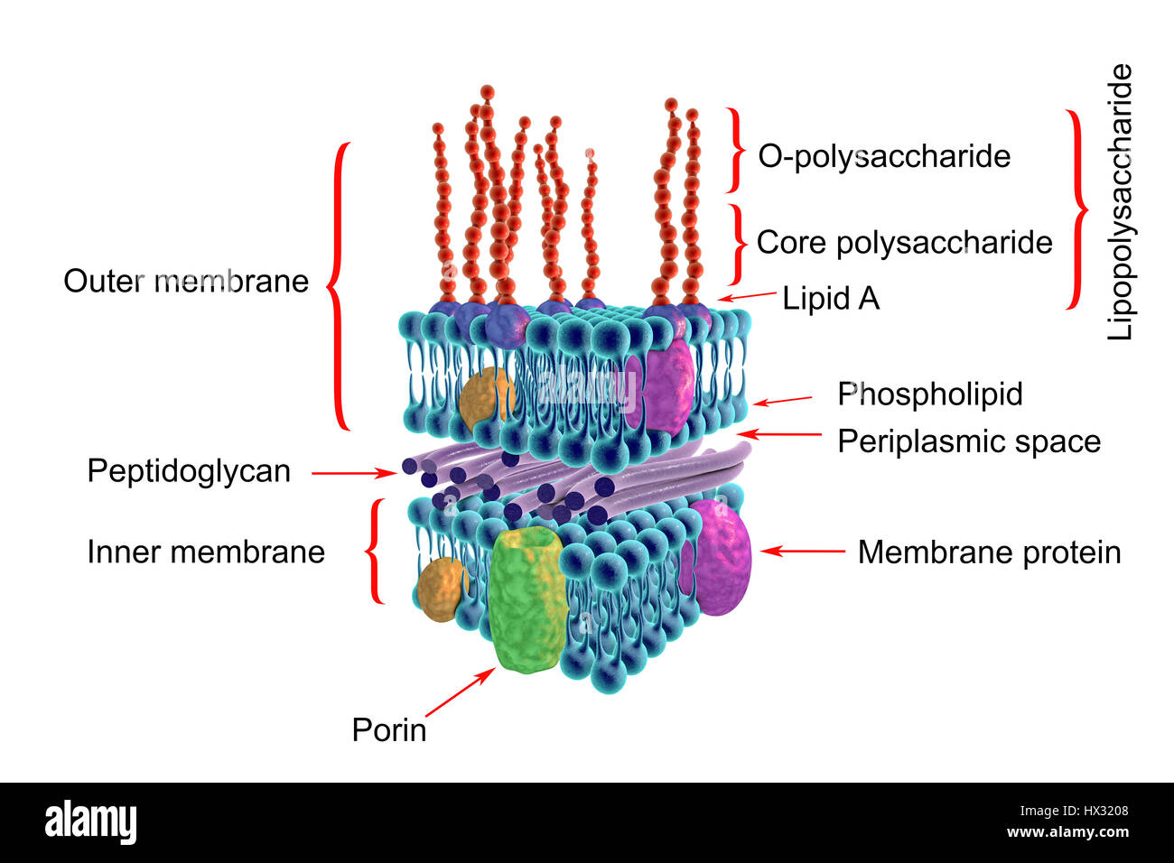
Gram-negative bacterial cell wall, artwork. The horizontal layers include both an external and an internal membrane, both containing transmembrane proteins (green, yellow and purple). The membranes are separated by a thin peptidoglycan layer. The outer surface of the external membrane is often a lipopolysaccharide layer with lipids (purple) in the membrane, and long saccharide side chains (red) extending out. This is termed a Gram-negative cell wall because it does not retain the Gram stain that helps identify microbial life.
RFID:Image ID:HX3208
Image details
Contributor:
Science Photo Library / Alamy Stock PhotoImage ID:
HX3208File size:
100 MB (1.9 MB Compressed download)Releases:
Model - no | Property - noDo I need a release?Dimensions:
7241 x 4827 px | 61.3 x 40.9 cm | 24.1 x 16.1 inches | 300dpiDate taken:
20 March 2017Photographer:
KATERYNA KON/SCIENCE PHOTO LIBRARY