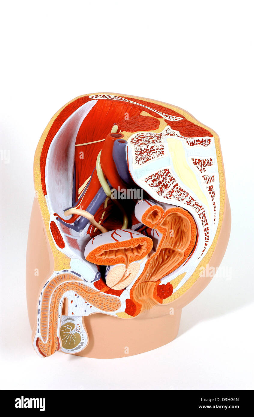
ANATOMY, MALE GENITALIA
Image details
Contributor:
BSIP SA / Alamy Stock PhotoImage ID:
D3HG6NFile size:
24.6 MB (947.7 KB Compressed download)Releases:
Model - no | Property - yesDo I need a release?Dimensions:
2365 x 3630 px | 20 x 30.7 cm | 7.9 x 12.1 inches | 300dpiDate taken:
23 June 2005Photographer:
GOUNOT/3B SCIENTIFIC / BSIPMore information:
Model of the internal anatomy of an adult male pelvis (median section). The pelvic musculature and vascular network, as well as the spinal column, are described in image no. 1149205. The ureter (in greenish yellow) carries the urine from the kidney to the bladder. The urinary bladder wall is mainly composed of the bladder muscle (in orange-red), the contraction of which allows urine evacuation through the urethra. This muscle is inwardly covered by a mucous membrane (in orange) with many folds and, outwardly, by the perimetrium (in white). The rectum is composed of two segments: the rectal ampulla in its upper part, storing the feces, and the anal canal, ending in the anus. The rectal ampulla forms curves, giving rise to folds of the inner rectal mucosa - the rectal valves. It also has vertical folds, the anal columns. The rectal wall includes three tissue layers: an outer muscularis (in orange-red), constituted by muscle fibers allowing defecation, a middle submucosa and an inner mucosa (in orange). The opening of the extremity of the anal canal, the anus, is controlled by an internal anal sphincter, composed of smooth muscle (in orange, reflex opening for defecation) and an external anal sphincter of striated muscle (in red, voluntary relaxation). Outside the pelvic cavity, the testicles (in khaki), located in the scrotum, produce spermatozoa and secrete sex hormones. The scrotum wall is inwardly covered by the Colles' fascia (in white) including a bundle of muscle fibers, the dartos, in contact with the abdominal wall. Each testicle is wrapped in the tunica vaginalis (in white) and in a fibrous capsule, the tunica albuginea (in white), the projections of which form walls separating the testicle into lobules. Spermatogenesis takes place in these lobules, then the spermatozoa are propelled towards the duct of the epididymis. The epididymis (in grey) is mainly made up of this duct, a long curled-up tube which performs sperm maturation; it is formed by three parts: