ALZHEIMER DISEASE, MRI
RMID:Image ID:CRYF8T
Image details
Contributor:
BSIP SA / Alamy Stock PhotoImage ID:
CRYF8TFile size:
25.1 MB (1.4 MB Compressed download)Releases:
Model - no | Property - noDo I need a release?Dimensions:
3630 x 2420 px | 30.7 x 20.5 cm | 12.1 x 8.1 inches | 300dpiDate taken:
13 June 2002Photographer:
DR CATH. OPPENHEIM / BSIPMore information:
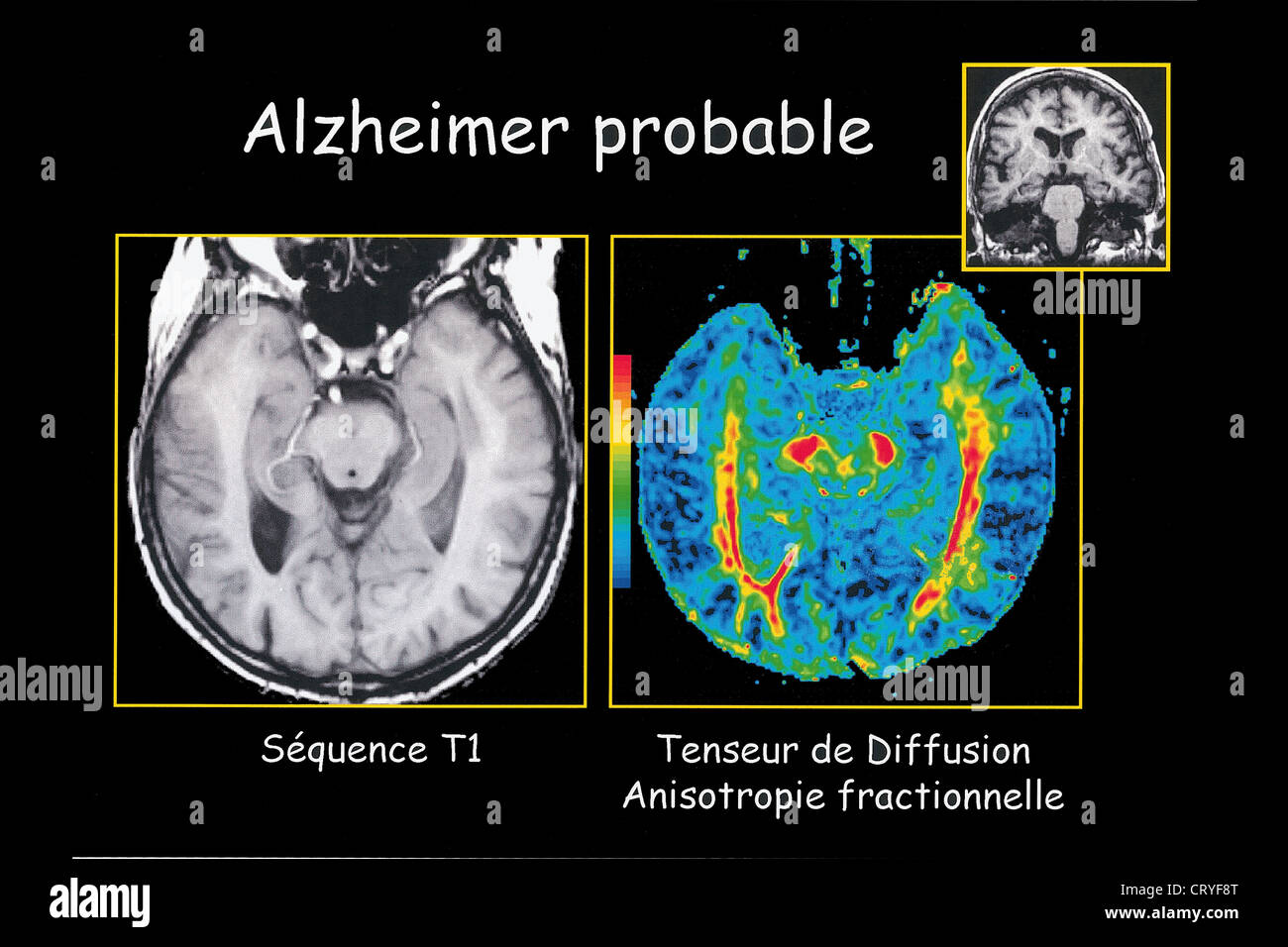
Department of morphological and functional imaging, led by Professor Fredy, at the Pitié Salpêtrière Hospital in Paris, France. At left, MRI-T1. Axial cut-away at the level of cerebral peduncles passing through the two temporal lobes. Temporal cerebellar tonsil apparently normal. At right, diffusion tensor MRI, which assesses the activity of the fibers connecting the basal ganglia, reveals the cerebellar tonsils' loss of function. Diagnosis of probable Alzheimer's disease.