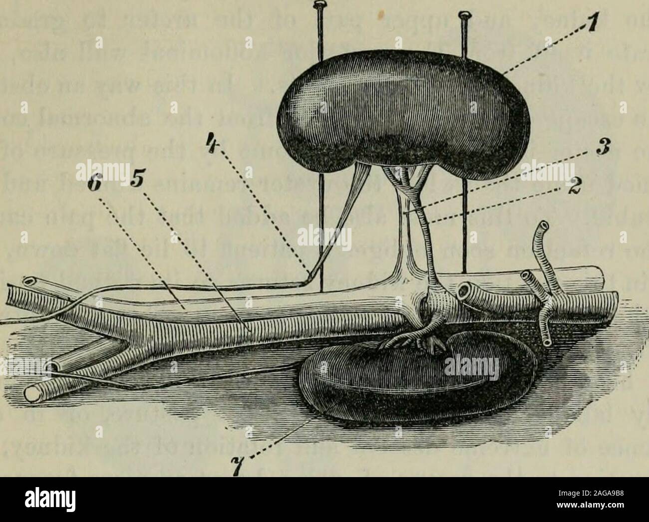
. Selected monographs. Right kidney, twisted on its hori-zontal axis, and with its ontcrborder looking downwards. Right renal artery. Right renal vein. 4. Right ureter, kinked and twisted. 5. Abdominal aorta.(). Vena cava inferior. 7. Left kidney in its normal situa-tion. numerous cxpeinmcnts involviuf^ tLo ligature and artificialnarrowing of the renal vein. In this relation however thedisturbance of the circulation, even when it occurs more orless contemporaneously with the disturbance of the circulation,is secondary, and caused by the alteration in the secretion.But I must affirm, contrary t
Image details
Contributor:
The Reading Room / Alamy Stock PhotoImage ID:
2AGA9B8File size:
7.2 MB (333.6 KB Compressed download)Releases:
Model - no | Property - noDo I need a release?Dimensions:
1843 x 1356 px | 31.2 x 23 cm | 12.3 x 9 inches | 150dpiMore information:
This image is a public domain image, which means either that copyright has expired in the image or the copyright holder has waived their copyright. Alamy charges you a fee for access to the high resolution copy of the image.
This image could have imperfections as it’s either historical or reportage.
. Selected monographs. Right kidney, twisted on its hori-zontal axis, and with its ontcrborder looking downwards. Right renal artery. Right renal vein. 4. Right ureter, kinked and twisted. 5. Abdominal aorta.(). Vena cava inferior. 7. Left kidney in its normal situa-tion. numerous cxpeinmcnts involviuf^ tLo ligature and artificialnarrowing of the renal vein. In this relation however thedisturbance of the circulation, even when it occurs more orless contemporaneously with the disturbance of the circulation, is secondary, and caused by the alteration in the secretion.But I must affirm, contrary to the general opinion, that thesecretion of the urine especially, may be primarily suppressedby narrowing or occlusion of the ureter. As soon as the kidney descends, the origin of the ureter, which is normally placed at the lowest point of the renalpelvis, and therefore at the point most favourable for the MOVEABLE KIDNEY IN WOMEN.Fig. 7. 303. The last dissection (Fig. 6) seen in profile. References the same as to Fig. 6. escape of the urine, moves to a liiglier position, and, if thekidney is very low down, to the very highest point of thepelvis. But as the kidney rotates (as it usually does) theureter will also become twisted on its axis. In the formercase kinking of the ureter results, in the latter torsion, ineither case obstruction to the flow of urine must result. Here, as in the case of the renal vessels, I have endeavouredto render the torsion, kinking and bending of the ureterintelligible by the help of drawings. Figs. 6 and 7, in whichthe ureter is bent and twisted, represent the same dissectionfrom two different views; Fig. 6 from the front. Fig. 7 inprofile. The very perceptible upward bend of the ureter, which we have already seen in Fig. 5, is repeated in Fig. 8below. If this is compared with Moslers post-mortem account, (see above p. 258) the resemblance between the course of theureter as there described and as