···
Red blood cell. Coloured scanning electron micrograph (SEM) of human red blood cells (erythrocytes). Red blood cells are biconcave, giving them a larg Image details File size:
51.1 MB (1.7 MB Compressed download)
Open your image file to the full size using image processing software.
Dimensions:
4550 x 3923 px | 38.5 x 33.2 cm | 15.2 x 13.1 inches | 300dpi
More information:
Red blood cell. Coloured scanning electron micrograph (SEM) of human red blood cells (erythrocytes). Red blood cells are biconcave, giving them a large surface area for gas exchange, and highly elastic, enabling them to pass through narrow capillary vessels. The main function of red blood cells is to distribute oxygen to body tissues and to carry waste carbon dioxide back to the lungs. Magnification: x7500 when printed at 10 centimetres wide.
Search stock photos by tags
Similar stock images Activated platelets, coloured scanning electron micrograph (SEM). Platelets are blood cell fragments that play an essential role in blood clotting and wound repair, and can also activate certain immune responses. They are formed in the red bone marrow, lungs, and spleen by fragmentation of very large cells known as megakaryocytes. Platelets in the blood are small oval disks and are termed non-activated platelets or thrombocytes. They are the body's first line of defence against excessive blood loss. Magnification: x2,000 at 10 cm wide. Stock Photo https://www.alamy.com/licenses-and-pricing/?v=1 https://www.alamy.com/activated-platelets-coloured-scanning-electron-micrograph-sem-platelets-are-blood-cell-fragments-that-play-an-essential-role-in-blood-clotting-and-wound-repair-and-can-also-activate-certain-immune-responses-they-are-formed-in-the-red-bone-marrow-lungs-and-spleen-by-fragmentation-of-very-large-cells-known-as-megakaryocytes-platelets-in-the-blood-are-small-oval-disks-and-are-termed-non-activated-platelets-or-thrombocytes-they-are-the-bodys-first-line-of-defence-against-excessive-blood-loss-magnification-x2000-at-10-cm-wide-image220702406.html RF PR1T4P – Activated platelets, coloured scanning electron micrograph (SEM). Platelets are blood cell fragments that play an essential role in blood clotting and wound repair, and can also activate certain immune responses. They are formed in the red bone marrow, lungs, and spleen by fragmentation of very large cells known as megakaryocytes. Platelets in the blood are small oval disks and are termed non-activated platelets or thrombocytes. They are the body's first line of defence against excessive blood loss. Magnification: x2,000 at 10 cm wide. SEM of sickle cell and normal red blood cells Stock Photo https://www.alamy.com/licenses-and-pricing/?v=1 https://www.alamy.com/sem-of-sickle-cell-and-normal-red-blood-cells-image65496031.html RF DPFGRY – SEM of sickle cell and normal red blood cells Coloured scanning electron micrograph (SEM) of red blood cells (RBCs, erythrocytes). Red blood cells are biconcave, disc-shaped cells that transport oxygen from the lungs to body cells. They circulate in the blood and also remove carbon dioxide to the lungs for exhalation. Their red colour is due to the oxygen-carrying protein haemoglobin. Red blood cells, the most abundant cell in the blood, have no nucleus and are about 7 micrometres across. Magnification: x3500 when printed at 10 centimetres across. Stock Photo https://www.alamy.com/licenses-and-pricing/?v=1 https://www.alamy.com/coloured-scanning-electron-micrograph-sem-of-red-blood-cells-rbcs-erythrocytes-red-blood-cells-are-biconcave-disc-shaped-cells-that-transport-oxygen-from-the-lungs-to-body-cells-they-circulate-in-the-blood-and-also-remove-carbon-dioxide-to-the-lungs-for-exhalation-their-red-colour-is-due-to-the-oxygen-carrying-protein-haemoglobin-red-blood-cells-the-most-abundant-cell-in-the-blood-have-no-nucleus-and-are-about-7-micrometres-across-magnification-x3500-when-printed-at-10-centimetres-across-image220702047.html RF PR1RKY – Coloured scanning electron micrograph (SEM) of red blood cells (RBCs, erythrocytes). Red blood cells are biconcave, disc-shaped cells that transport oxygen from the lungs to body cells. They circulate in the blood and also remove carbon dioxide to the lungs for exhalation. Their red colour is due to the oxygen-carrying protein haemoglobin. Red blood cells, the most abundant cell in the blood, have no nucleus and are about 7 micrometres across. Magnification: x3500 when printed at 10 centimetres across. SEM of a sickle cell red blood cell Stock Photo https://www.alamy.com/licenses-and-pricing/?v=1 https://www.alamy.com/sem-of-a-sickle-cell-red-blood-cell-image65496146.html RF DPFH02 – SEM of a sickle cell red blood cell Coloured scanning electron micrograph (SEM) of red blood cells (RBCs, erythrocytes). Red blood cells are biconcave, disc-shaped cells that transport oxygen from the lungs to body cells. They circulate in the blood and also remove carbon dioxide to the lungs for exhalation. Their red colour is due to the oxygen-carrying protein haemoglobin. Red blood cells, the most abundant cell in the blood, have no nucleus and are about 7 micrometres across. Magnification: x3500 when printed at 10 centimetres across. Stock Photo https://www.alamy.com/licenses-and-pricing/?v=1 https://www.alamy.com/coloured-scanning-electron-micrograph-sem-of-red-blood-cells-rbcs-erythrocytes-red-blood-cells-are-biconcave-disc-shaped-cells-that-transport-oxygen-from-the-lungs-to-body-cells-they-circulate-in-the-blood-and-also-remove-carbon-dioxide-to-the-lungs-for-exhalation-their-red-colour-is-due-to-the-oxygen-carrying-protein-haemoglobin-red-blood-cells-the-most-abundant-cell-in-the-blood-have-no-nucleus-and-are-about-7-micrometres-across-magnification-x3500-when-printed-at-10-centimetres-across-image220702034.html RF PR1RKE – Coloured scanning electron micrograph (SEM) of red blood cells (RBCs, erythrocytes). Red blood cells are biconcave, disc-shaped cells that transport oxygen from the lungs to body cells. They circulate in the blood and also remove carbon dioxide to the lungs for exhalation. Their red colour is due to the oxygen-carrying protein haemoglobin. Red blood cells, the most abundant cell in the blood, have no nucleus and are about 7 micrometres across. Magnification: x3500 when printed at 10 centimetres across. Scanning electron micrograph of HIV-1 virus Stock Photo https://www.alamy.com/licenses-and-pricing/?v=1 https://www.alamy.com/scanning-electron-micrograph-of-hiv-1-virus-image65495128.html RF DPFFKM – Scanning electron micrograph of HIV-1 virus Coloured scanning electron micrograph (SEM) of red blood cells (RBCs, erythrocytes). Red blood cells are biconcave, disc-shaped cells that transport oxygen from the lungs to body cells. They circulate in the blood and also remove carbon dioxide to the lungs for exhalation. Their red colour is due to the oxygen-carrying protein haemoglobin. Red blood cells, the most abundant cell in the blood, have no nucleus and are about 7 micrometres across. Magnification: x3500 when printed at 10 centimetres across. Stock Photo https://www.alamy.com/licenses-and-pricing/?v=1 https://www.alamy.com/coloured-scanning-electron-micrograph-sem-of-red-blood-cells-rbcs-erythrocytes-red-blood-cells-are-biconcave-disc-shaped-cells-that-transport-oxygen-from-the-lungs-to-body-cells-they-circulate-in-the-blood-and-also-remove-carbon-dioxide-to-the-lungs-for-exhalation-their-red-colour-is-due-to-the-oxygen-carrying-protein-haemoglobin-red-blood-cells-the-most-abundant-cell-in-the-blood-have-no-nucleus-and-are-about-7-micrometres-across-magnification-x3500-when-printed-at-10-centimetres-across-image220702009.html RF PR1RJH – Coloured scanning electron micrograph (SEM) of red blood cells (RBCs, erythrocytes). Red blood cells are biconcave, disc-shaped cells that transport oxygen from the lungs to body cells. They circulate in the blood and also remove carbon dioxide to the lungs for exhalation. Their red colour is due to the oxygen-carrying protein haemoglobin. Red blood cells, the most abundant cell in the blood, have no nucleus and are about 7 micrometres across. Magnification: x3500 when printed at 10 centimetres across. Red blood cells and platelets. Coloured scanning electon micrograph (SEM) of human erythrocytes (red blood cells) and a platelet aggregate (yellow). Platelets are fragments of white blood cells that under normal circumstances are small and biconcave in form. However, if there is a break in the surface of a blood vessel the platelets come into contact with molecules they are not used to and become activated. They become amorphous in form, with long projections (pseudopodia) that help them adhere to other cells and each other, forming a clot. Magnification: x3000 when printed at 10 centimetres Stock Photo https://www.alamy.com/licenses-and-pricing/?v=1 https://www.alamy.com/stock-photo-red-blood-cells-and-platelets-coloured-scanning-electon-micrograph-102520732.html RF FXP66M – Red blood cells and platelets. Coloured scanning electon micrograph (SEM) of human erythrocytes (red blood cells) and a platelet aggregate (yellow). Platelets are fragments of white blood cells that under normal circumstances are small and biconcave in form. However, if there is a break in the surface of a blood vessel the platelets come into contact with molecules they are not used to and become activated. They become amorphous in form, with long projections (pseudopodia) that help them adhere to other cells and each other, forming a clot. Magnification: x3000 when printed at 10 centimetres Red blood cells and platelets. Coloured scanning electon micrograph (SEM) of human erythrocytes (red blood cells) and a platelet aggregate (orange). Platelets are fragments of white blood cells that under normal circumstances are small and biconcave in form. However, if there is a break in the surface of a blood vessel the platelets come into contact with molecules they are not used to and become activated. They become amorphous in form, with long projections (pseudopodia) that help them adhere to other cells and each other, forming a clot. Magnification: x3000 when printed at 10 centimetres Stock Photo https://www.alamy.com/licenses-and-pricing/?v=1 https://www.alamy.com/stock-photo-red-blood-cells-and-platelets-coloured-scanning-electon-micrograph-102520725.html RF FXP66D – Red blood cells and platelets. Coloured scanning electon micrograph (SEM) of human erythrocytes (red blood cells) and a platelet aggregate (orange). Platelets are fragments of white blood cells that under normal circumstances are small and biconcave in form. However, if there is a break in the surface of a blood vessel the platelets come into contact with molecules they are not used to and become activated. They become amorphous in form, with long projections (pseudopodia) that help them adhere to other cells and each other, forming a clot. Magnification: x3000 when printed at 10 centimetres Red blood cells and platelets. Coloured scanning electon micrograph (SEM) of human erythrocytes (red blood cells) and a platelet aggregate (orange). Platelets are fragments of white blood cells that under normal circumstances are small and biconcave in form. However, if there is a break in the surface of a blood vessel the platelets come into contact with molecules they are not used to and become activated. They become amorphous in form, with long projections (pseudopodia) that help them adhere to other cells and each other, forming a clot. Magnification: x3000 when printed at 10 centimetres Stock Photo https://www.alamy.com/licenses-and-pricing/?v=1 https://www.alamy.com/stock-photo-red-blood-cells-and-platelets-coloured-scanning-electon-micrograph-102520727.html RF FXP66F – Red blood cells and platelets. Coloured scanning electon micrograph (SEM) of human erythrocytes (red blood cells) and a platelet aggregate (orange). Platelets are fragments of white blood cells that under normal circumstances are small and biconcave in form. However, if there is a break in the surface of a blood vessel the platelets come into contact with molecules they are not used to and become activated. They become amorphous in form, with long projections (pseudopodia) that help them adhere to other cells and each other, forming a clot. Magnification: x3000 when printed at 10 centimetres Red blood cells and platelets. Coloured scanning electon micrograph (SEM) of human erythrocytes (red blood cells) and a platelet aggregate (blue). Platelets are fragments of white blood cells that under normal circumstances are small and biconcave in form. However, if there is a break in the surface of a blood vessel the platelets come into contact with molecules they are not used to and become activated. They become amorphous in form, with long projections (pseudopodia) that help them adhere to other cells and each other, forming a clot. Magnification: x3000 when printed at 10 centimetres Stock Photo https://www.alamy.com/licenses-and-pricing/?v=1 https://www.alamy.com/stock-photo-red-blood-cells-and-platelets-coloured-scanning-electon-micrograph-102520722.html RF FXP66A – Red blood cells and platelets. Coloured scanning electon micrograph (SEM) of human erythrocytes (red blood cells) and a platelet aggregate (blue). Platelets are fragments of white blood cells that under normal circumstances are small and biconcave in form. However, if there is a break in the surface of a blood vessel the platelets come into contact with molecules they are not used to and become activated. They become amorphous in form, with long projections (pseudopodia) that help them adhere to other cells and each other, forming a clot. Magnification: x3000 when printed at 10 centimetres Activated T lymphocytes and red blood cells (RBCs). Coloured scanning electron micrograph (SEM) of activated T lymphocytes and RBCs from a human blood Stock Photo https://www.alamy.com/licenses-and-pricing/?v=1 https://www.alamy.com/activated-t-lymphocytes-and-red-blood-cells-rbcs-coloured-scanning-electron-micrograph-sem-of-activated-t-lymphocytes-and-rbcs-from-a-human-blood-image184718633.html RF MMEJE1 – Activated T lymphocytes and red blood cells (RBCs). Coloured scanning electron micrograph (SEM) of activated T lymphocytes and RBCs from a human blood Blood cells. Coloured scanning electron micrograph (SEM) of human red blood cells (erythrocytes, red), white blood cells (leukocytes, blue and cyan), and platelets (thrombocytes, yellow). The disc-shaped, biconcave erythrocytes transport oxygen to the body's cells and remove carbon dioxide to the lungs. Leukocytes are part of the immune system, defending the body against infection by ingesting pathogens by phagocytosis or by producing antibodies. Magnification: x5000 when printed 10 centimetres high. Stock Photo https://www.alamy.com/licenses-and-pricing/?v=1 https://www.alamy.com/blood-cells-coloured-scanning-electron-micrograph-sem-of-human-red-blood-cells-erythrocytes-red-white-blood-cells-leukocytes-blue-and-cyan-and-platelets-thrombocytes-yellow-the-disc-shaped-biconcave-erythrocytes-transport-oxygen-to-the-bodys-cells-and-remove-carbon-dioxide-to-the-lungs-leukocytes-are-part-of-the-immune-system-defending-the-body-against-infection-by-ingesting-pathogens-by-phagocytosis-or-by-producing-antibodies-magnification-x5000-when-printed-10-centimetres-high-image220702013.html RF PR1RJN – Blood cells. Coloured scanning electron micrograph (SEM) of human red blood cells (erythrocytes, red), white blood cells (leukocytes, blue and cyan), and platelets (thrombocytes, yellow). The disc-shaped, biconcave erythrocytes transport oxygen to the body's cells and remove carbon dioxide to the lungs. Leukocytes are part of the immune system, defending the body against infection by ingesting pathogens by phagocytosis or by producing antibodies. Magnification: x5000 when printed 10 centimetres high. 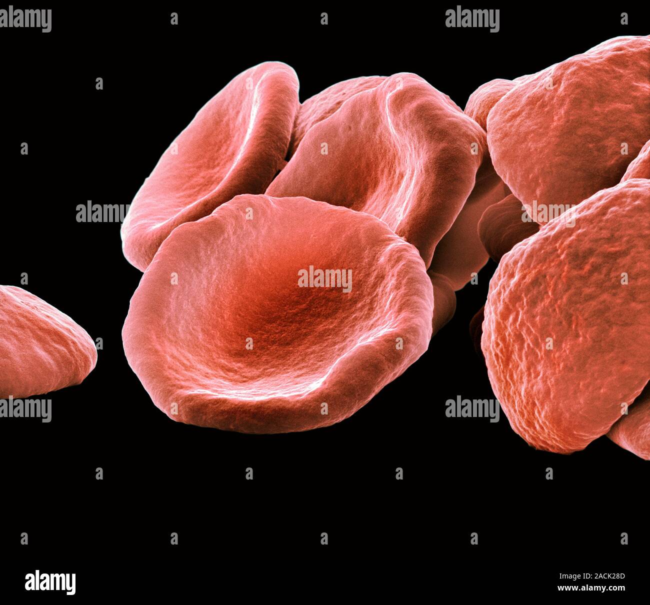