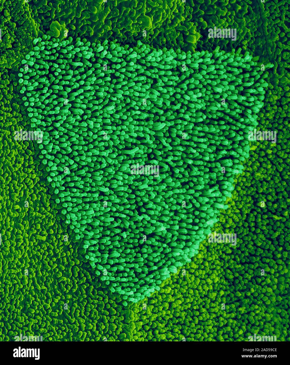
Nasopharynx surface that contains squamous nasal epithelial cells, coloured scanning electron micrograph (SEM). The nasopharynx (nasal part of the pha
Image details
Contributor:
Science Photo Library / Alamy Stock PhotoImage ID:
2AD59CEFile size:
25 MB (1.8 MB Compressed download)Releases:
Model - no | Property - noDo I need a release?Dimensions:
2726 x 3206 px | 23.1 x 27.1 cm | 9.1 x 10.7 inches | 300dpiDate taken:
22 November 2016Photographer:
DENNIS KUNKEL MICROSCOPY/SCIENCE PHOTO LIBRARYMore information:
Nasopharynx surface that contains squamous nasal epithelial cells, coloured scanning electron micrograph (SEM). The nasopharynx (nasal part of the pharynx) lies behind the nose and above the level of the soft palate. Stratified squamous nasal epithelial cells line the surface of the nasopharynx. A prominent triangular shaped nasal epithelial cell is seen in this image. The epithelial cell surfaces are covered with tiny microvilli that increase the cell surface area. The microvilli likely aid in localization of foreign debris (coming from the nose) and detection by immune cells. Mucus, secreted by cells in the epithelial lining (not seen), traps foreign objects, such as bacteria, preventing them from entering the lungs. Other sensory cells and features (not seen) also occur in the nasal epithelial layer. Magnification: x2, 200 when shortest axis printed at 25 millimetres.