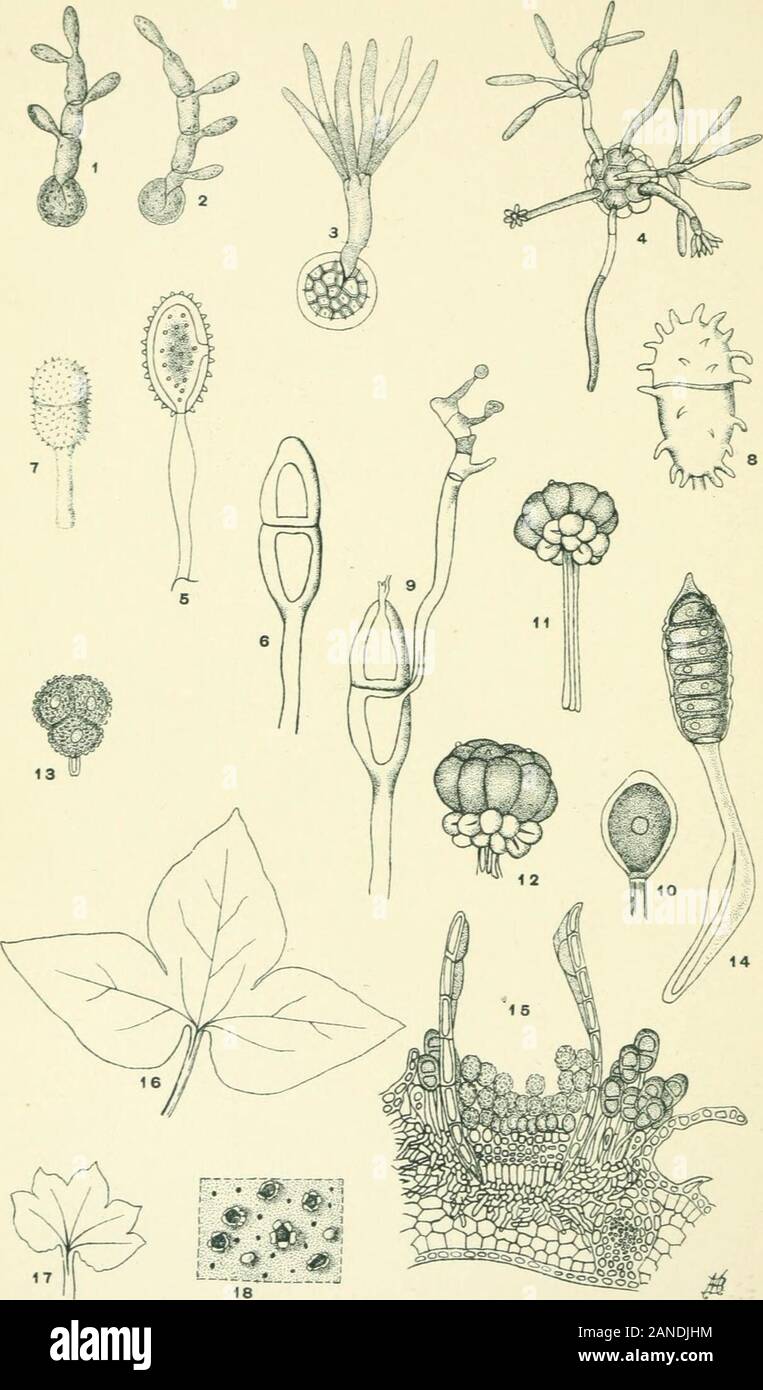
Moulds, mildews, and mushrooms; a guide to the systematic study of the Fungi and Mycetozoa and their literature . the: MELIOTyPE PRINTING CO., BOSTON. Pl. 6.. THt MELIOTYPE PRINTING CO., BOSTON. EXPLANATION OF PLATE VI Basidiomycetes Figs, i, 2. Ustilago avenae {J?,Ti-LA.Giy.A.i.Y.s). Germinating chlamyd-ospores producing spores laterally and terminally. X 350. (Redrawnfrom Brefeld.) Fig. 3. Tilletia zonata (USTILAGINALES). Germinating chlamyd-ospore producing a cluster of spores at the apex. X 300. (Redrawnfrom Brefeld.) Fig. 4. Urocystis violae (Ustilaginales). Germination of a chlamyd-ospo
Image details
Contributor:
The Reading Room / Alamy Stock PhotoImage ID:
2ANDJHMFile size:
7.1 MB (262.1 KB Compressed download)Releases:
Model - no | Property - noDo I need a release?Dimensions:
1211 x 2063 px | 20.5 x 34.9 cm | 8.1 x 13.8 inches | 150dpiMore information:
This image is a public domain image, which means either that copyright has expired in the image or the copyright holder has waived their copyright. Alamy charges you a fee for access to the high resolution copy of the image.
This image could have imperfections as it’s either historical or reportage.
Moulds, mildews, and mushrooms; a guide to the systematic study of the Fungi and Mycetozoa and their literature . the: MELIOTyPE PRINTING CO., BOSTON. Pl. 6.. THt MELIOTYPE PRINTING CO., BOSTON. EXPLANATION OF PLATE VI Basidiomycetes Figs, i, 2. Ustilago avenae {J?, Ti-LA.Giy.A.i.Y.s). Germinating chlamyd-ospores producing spores laterally and terminally. X 350. (Redrawnfrom Brefeld.) Fig. 3. Tilletia zonata (USTILAGINALES). Germinating chlamyd-ospore producing a cluster of spores at the apex. X 300. (Redrawnfrom Brefeld.) Fig. 4. Urocystis violae (Ustilaginales). Germination of a chlamyd-ospore. X 180. (Redrawn from Brefeld.) Fig. 5. Puccinia graminis (Uredinales). Uredospore with separ-able pedicel. X 200. (Redrawn from Sachs.) Fig. 6. Puccinia graniinis (Uredinales). Teleutospore. X 330.Redrawn from Peck). Fig. 7. Puccinia anemones (Uredinales). Teleutospore. X 300.(Redrawn from Peck.) Fig. 8. Puccinia podophylli (Uredinales). Teleutospore. X 300.(Redrawn from Peck.) Fig 9. Puccinia graminis [UrediNales). Germinating teleutosporeproducing the basidiospores from the upper part of the promycelium, X330. (Redrawn from Sachs.) Fig. 10. Uromy