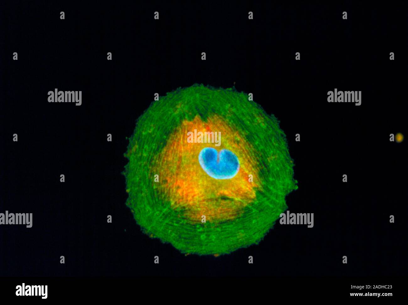
Melanoma cell. Immunofluorescent Light Micrograph of a melanoma cancer cell, cultured from a human tumour. The cell nucleus (blue) is in the process o
RMID:Image ID:2ADHC23
Image details
Contributor:
Science Photo Library / Alamy Stock PhotoImage ID:
2ADHC23File size:
26.6 MB (420.9 KB Compressed download)Releases:
Model - no | Property - noDo I need a release?Dimensions:
3700 x 2516 px | 31.3 x 21.3 cm | 12.3 x 8.4 inches | 300dpiDate taken:
21 April 1993Photographer:
NANCY KEDERSHA/IMMUNOGEN/SCIENCE PHOTO LIBRARYMore information:
Melanoma cell. Immunofluorescent Light Micrograph of a melanoma cancer cell, cultured from a human tumour. The cell nucleus (blue) is in the process of division; cytoplasm stains orange; actin stress fibres (green, protein) surround the cell & serve as a supportive network. This cancer is typically derived from melanin-forming skin cells. Melanoma is a highly malignant cancer consisting of large undifferentiated cells that divide rapidly, and invade healthy tissue. Immunofluorescence is a technique which uses antibodies to attach fluorescent dyes to specific tissues and molecules within the cell. Magnification: x125 at 35mm size.