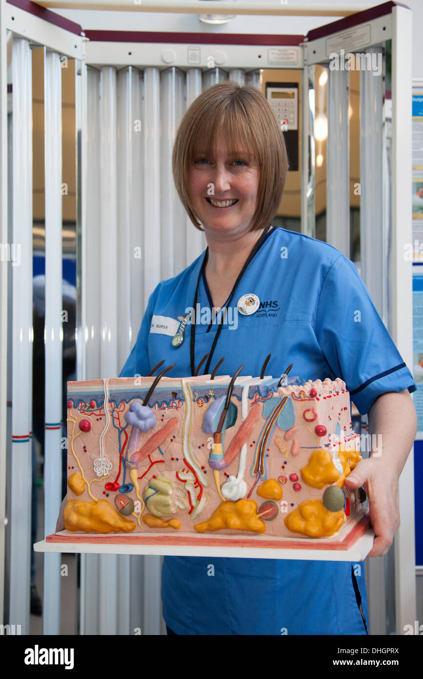
Medically accurate 3D model of skin in Dundee, Scotland, UK. November, 2013. Ms Susan Yule NHS nurse with a 3D cross-section of human skin, dermis showing micronucleus, multi-layered, highly differentiated layers, and Ultra Violet UVB patient treatment machine UVB, Ninewells University Hospital Human Genetics research labs. A city-wide celebration of anatomy medical science, biology, and curiosity for all ages.
RMID:Image ID:DHGPRX
Image details
Contributor:
MediaWorldImages / Alamy Stock PhotoImage ID:
DHGPRXFile size:
24.7 MB (997.5 KB Compressed download)Releases:
Model - yes | Property - noDo I need a release?Dimensions:
2400 x 3600 px | 20.3 x 30.5 cm | 8 x 12 inches | 300dpiDate taken:
9 November 2013Location:
Dundee, Scotland, UKMore information:
This image could have imperfections as it’s either historical or reportage.
"The epidermis is composed of the outermost layers of cells in the skin, "epi" in Greek meaning "over" or "upon", which together with the dermis forms the cutis. The epidermis is a stratified squamous epithelium, composed of proliferating basal and differentiated suprabasal keratinocytes which acts as the body's major barrier against an inhospitable environment, by preventing pathogens from entering, making the skin a natural barrier to infection. It also regulates the amount of water released from the body into the atmosphere through transepidermal water loss."