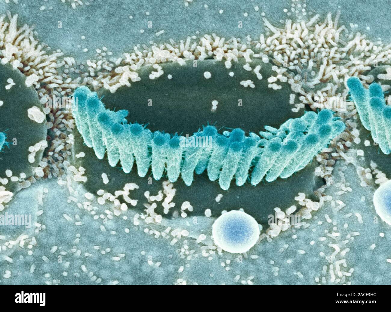
Inner ear hair cells. Coloured scanning electron micrograph (SEM) of sensory hair cells from the cochlea of the inner ear. The hairs are surrounded by
RMID:Image ID:2ACF3HC
Image details
Contributor:
Science Photo Library / Alamy Stock PhotoImage ID:
2ACF3HCFile size:
50.6 MB (3.2 MB Compressed download)Releases:
Model - no | Property - noDo I need a release?Dimensions:
4926 x 3591 px | 41.7 x 30.4 cm | 16.4 x 12 inches | 300dpiDate taken:
8 January 2009More information:
Inner ear hair cells. Coloured scanning electron micrograph (SEM) of sensory hair cells from the cochlea of the inner ear. The hairs are surrounded by a fluid called the endolymph. As sound enters the ear it causes waves to form in the endolymph, which in turn cause these hairs to move. The movement is converted into an electrical signal, which is passed to the brain. Each crescent-shaped arrangement of hairs lies on the top of a single cell.