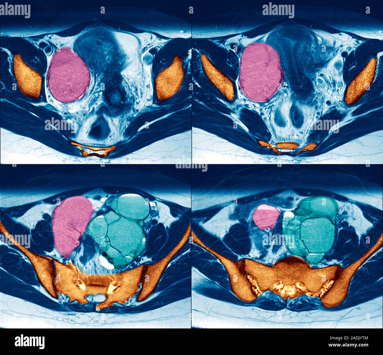
Endometriosis and ovarian cyst, sequence of axial magnetic resonance imaging (MRI) scans through the abdomen of a twenty-seven year old woman. The fro
Image details
Contributor:
Science Photo Library / Alamy Stock PhotoImage ID:
2ADJYTMFile size:
31.5 MB (1 MB Compressed download)Releases:
Model - no | Property - noDo I need a release?Dimensions:
3583 x 3071 px | 30.3 x 26 cm | 11.9 x 10.2 inches | 300dpiDate taken:
9 September 2004Photographer:
ZEPHYR/SCIENCE PHOTO LIBRARYMore information:
Endometriosis and ovarian cyst, sequence of axial magnetic resonance imaging (MRI) scans through the abdomen of a twenty-seven year old woman. The front of the body is at the top of the image. The pink mass at centre left is a cyst, a fluid-filled sac, of an ovary. The light blue masses at centre right are endometriomas, or chocolate cysts, a result of endometriosis. Endometriosis is the movement of cells from the lining of the uterus to the abdominal cavity. The cells still respond to the menstrual cycle and so bleed once a month. However, the blood has no way of leaving the body and so causes pain, inflammation and scar tissue, and can lead to infertility.