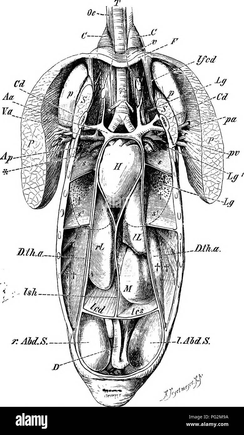
. Elements of the comparative anatomy of vertebrates. Anatomy, Comparative. r s.ijiM^. ' Ish— r.AM.S.— 1.AM.S. Fig. 237.—Abdominal Viscera and Air-Sacs of a Duck ai-teb the Re- moval OF THE Ventral Body-Wall. (From a drawing by H. Strasser.) T, trachea; H, heart, enclosed within the pericardium; rL,lL, right and left lobes of liver; Ish, suspensory (falciform) ligament, and led, Ics, right and left coronary ligament of the liver ; D, intestine ; P, pectoralis major; pa, pv, pectoral artery and vein ; S, subclavius muscle ; Od, coracoid ; F, furcula ; Ifcd, coraco-furcular ligament; Lg, Lg^, lu
Image details
Contributor:
Central Historic Books / Alamy Stock PhotoImage ID:
PG2M9AFile size:
7.2 MB (515.9 KB Compressed download)Releases:
Model - no | Property - noDo I need a release?Dimensions:
1225 x 2041 px | 20.7 x 34.6 cm | 8.2 x 13.6 inches | 150dpiMore information:
This image is a public domain image, which means either that copyright has expired in the image or the copyright holder has waived their copyright. Alamy charges you a fee for access to the high resolution copy of the image.
This image could have imperfections as it’s either historical or reportage.
. Elements of the comparative anatomy of vertebrates. Anatomy, Comparative. r s.ijiM^. ' Ish— r.AM.S.— 1.AM.S. Fig. 237.—Abdominal Viscera and Air-Sacs of a Duck ai-teb the Re- moval OF THE Ventral Body-Wall. (From a drawing by H. Strasser.) T, trachea; H, heart, enclosed within the pericardium; rL, lL, right and left lobes of liver; Ish, suspensory (falciform) ligament, and led, Ics, right and left coronary ligament of the liver ; D, intestine ; P, pectoralis major; pa, pv, pectoral artery and vein ; S, subclavius muscle ; Od, coracoid ; F, furcula ; Ifcd, coraco-furcular ligament; Lg, Lg^, lung; r.abd.S, I.abd.S, right and left abdominal (posterior) air-sac ; D.tji.a, oblique septum; tt> posterior intermediate air sac ; t, anterior intermediate air-sac ; s^, s^, partition-walls between these sacs; s, s, partition walls between the anterior intermediate air-sacs and the unpaired sub-bronchial sac, lying in the anterior part of the body-cavity; v, portion of anterior wall of latter; p, axillary sac lying between the coracoid, scapula, and the anterior ribs, and communicating with the sub-bronchial air-sac; G, C, prebronchial sacs; *, point of entrance of the bronchi into the lung ; Ap, pulmonary artery ; Aa, Va, innominate artery and vein with their branches.. Please note that these images are extracted from scanned page images that may have been digitally enhanced for readability - coloration and appearance of these illustrations may not perfectly resemble the original work.. Wiedersheim, Robert, 1848-1923; Parker, William Newton, 1857-1923. London, Macmillan