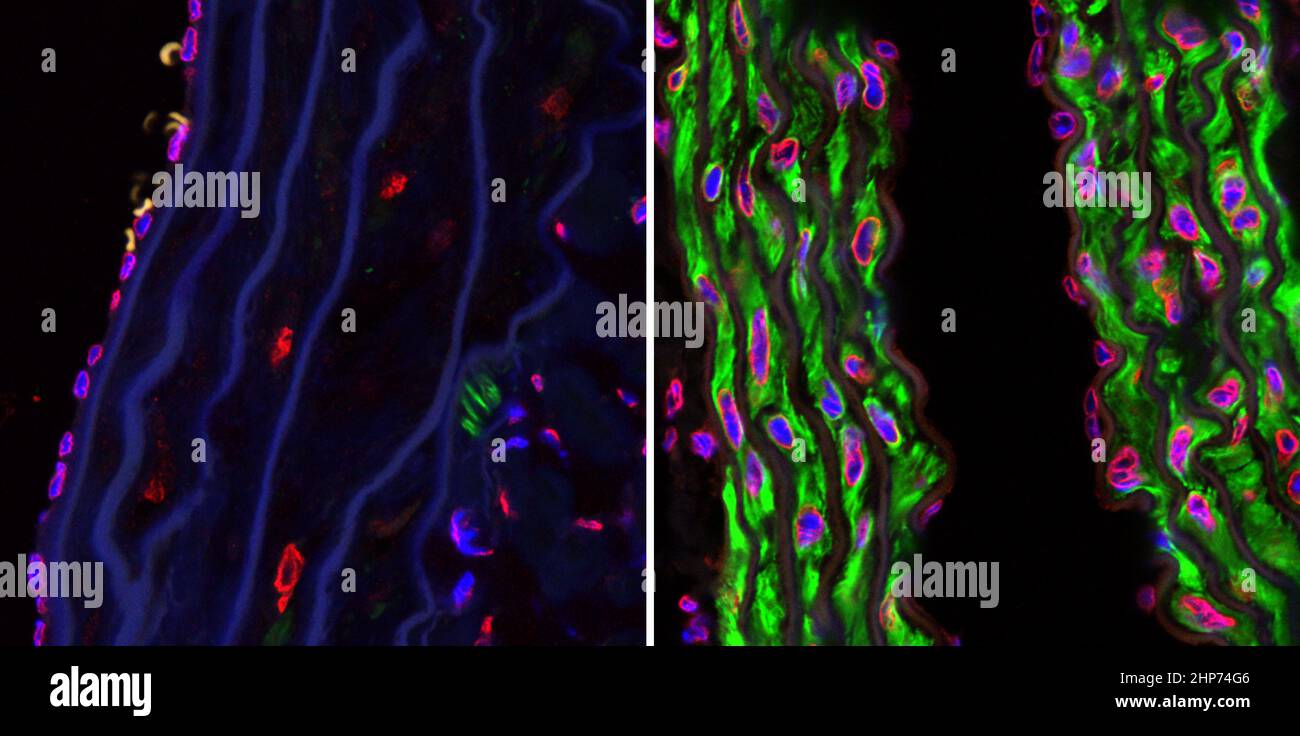
Confocal microscopy photographs of the descending aortas of two 15-month-old progeria mice, one untreated (left picture) and the other treated with the farnsyltransferase inhibitor drug tipifarnib (right picture). The microphotographs show prevention of the vascular smooth muscle cell loss that is otherwise rampant by this age. Staining was smooth muscle alpha-actin (green), lamins A/C (red) and DAPI (blue). (Original magnification, x 40) ca. 2009
RMID:Image ID:2HP74G6
Image details
Contributor:
PBH Images / Alamy Stock PhotoImage ID:
2HP74G6File size:
26.2 MB (599.4 KB Compressed download)Releases:
Model - no | Property - noDo I need a release?Dimensions:
4290 x 2133 px | 36.3 x 18.1 cm | 14.3 x 7.1 inches | 300dpiMore information:
This image could have imperfections as it’s either historical or reportage.