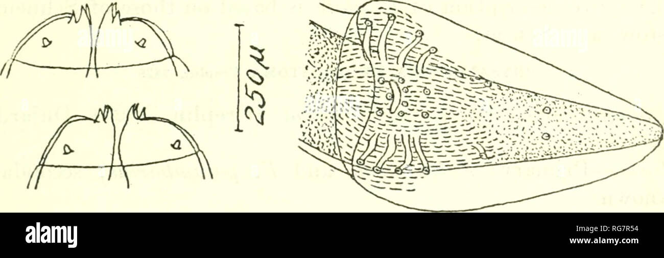
. Bulletin - United States National Museum. Science. NEMATODE PARASITES OF BIRDS 299 tris, F. pygargm^ F. 7*ufus, F. rutilans, F. species, F. mbbutt us, F. swainsonii, F. unicincttts, F. urubutinga, Tinnwnculus alaudarius; secondary: Unknown. Location.—Gizzard and intestine. Morphology.—Physaloptera (p. 295) : Cuticle may be completely reflected over the head, with a large crater-like opening anteriorly, or in young specimens this may be entirely lacking. Cervical papil- lae situated at level of terminal region of muscular esophagus. Mouth (fig. 360a) with 2 lateral lips (fig. 360Z>), each
Image details
Contributor:
Book Worm / Alamy Stock PhotoImage ID:
RG7R54File size:
7.1 MB (207.9 KB Compressed download)Releases:
Model - no | Property - noDo I need a release?Dimensions:
2792 x 895 px | 23.6 x 7.6 cm | 9.3 x 3 inches | 300dpiMore information:
This image is a public domain image, which means either that copyright has expired in the image or the copyright holder has waived their copyright. Alamy charges you a fee for access to the high resolution copy of the image.
This image could have imperfections as it’s either historical or reportage.
. Bulletin - United States National Museum. Science. NEMATODE PARASITES OF BIRDS 299 tris, F. pygargm^ F. 7*ufus, F. rutilans, F. species, F. mbbutt us, F. swainsonii, F. unicincttts, F. urubutinga, Tinnwnculus alaudarius; secondary: Unknown. Location.—Gizzard and intestine. Morphology.—Physaloptera (p. 295) : Cuticle may be completely reflected over the head, with a large crater-like opening anteriorly, or in young specimens this may be entirely lacking. Cervical papil- lae situated at level of terminal region of muscular esophagus. Mouth (fig. 360a) with 2 lateral lips (fig. 360Z>), each with a triangular external tooth and 3 smaller, blunter internal teeth; in addition 3 papillae, near the insertion of each lip. Male 17 to 20 mm. long. Entire esophagus 1/4 of body length. Cloacal aperture 650/* from caudal extremity. Circumcloacal region covered with small tubercules. Caudal alae (fig. 360<?) A'ery elon- gated. Five pairs of long pedunculated papillae, of which 2 are preanal. 1 ad anal and 2 postanal. Three papillae on the anterior edge of cloacal aperture. Four pairs of postanal ventral papillae, of which 2 pairs form a transverse row on the posterior edge of. Fig. 361.—Physaloptera alata Rudolphi of Ortlepp, 1922. Possibly equivalent to p. galinieri seuuat. see text for discussion. after ort- lepp, 1922. the cloacal aperture, the third pair is at the level of the most pos- terior pair of lateral pedunculated papillae, and the fourth pair is situated about midway from cloacal aperture to caudal extremity (Seurat says 5 pairs of postanal papillae but in describing their position only lists 4 pairs). Spicules 420/* long, according to Lin- stow; 280 and 265/t long, according to Seurat. Female 19 to 27 mm. long. Entire esophagus 1/5 of body length. Tail 1/21 of body length, conical, attenuated. Schneider states that the vulva is 7 mm. from the anterior extremity of a 19 mm. long specimen, but Seurat describes its position as more anterior (250/* posterio