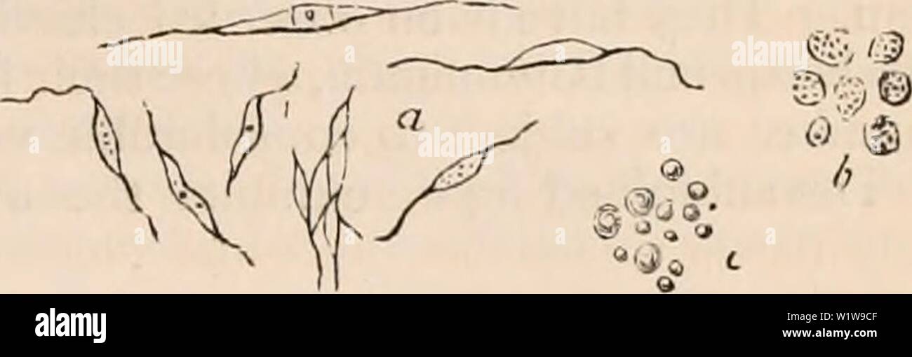
Archive image from page 626 of The cyclopædia of anatomy and. The cyclopædia of anatomy and physiology cyclopdiaofana05todd Year: 1859 Graafian vesicle of the rabbit x 100 (?) diameters. (After Barry.) a, outer coat or tunic of the ovisac; b, ovisac; c, epithelial lining or membrana granulosa, a por- tion of which has been removed in order to display del, retinacula (here too distinctly marked) ; e, tunica granulosa of Barry immediately surrounding the ovum, consisting of, /, zona pellucida, within which is the yelk and germinal vesicle and macula. in its spherical figure ; it carries numerou
Image details
Contributor:
Actep Burstov / Alamy Stock PhotoImage ID:
W1W9CFFile size:
5.7 MB (116.1 KB Compressed download)Releases:
Model - no | Property - noDo I need a release?Dimensions:
2488 x 804 px | 21.1 x 6.8 cm | 8.3 x 2.7 inches | 300dpiMore information:
This image is a public domain image, which means either that copyright has expired in the image or the copyright holder has waived their copyright. Alamy charges you a fee for access to the high resolution copy of the image.
This image could have imperfections as it’s either historical or reportage.
Archive image from page 626 of The cyclopædia of anatomy and. The cyclopædia of anatomy and physiology cyclopdiaofana05todd Year: 1859 Graafian vesicle of the rabbit x 100 (?) diameters. (After Barry.) a, outer coat or tunic of the ovisac; b, ovisac; c, epithelial lining or membrana granulosa, a por- tion of which has been removed in order to display del, retinacula (here too distinctly marked) ; e, tunica granulosa of Barry immediately surrounding the ovum, consisting of, /, zona pellucida, within which is the yelk and germinal vesicle and macula. in its spherical figure ; it carries numerous blood-vessels, which pass from the ovarian stroma to become expanded in a vascular net- work over its walls (fig. 371. D). Examined by the microscope, this membrane is seen to be highly vascular. It is composed of a fine membrane, containing few fibres, but everywhere abundantly studded with oval nuclei, visible without the aid of acetic acid, and probably, in part at least, due to the pre- sence of so many blood-vessels in its tissue. This coat contains no oil globules. Its chief use appears to be to give increased support and protection to the true ovisac which it sur- rounds, and to convey blood-vessels from the ovary for its nutrition, and for the supply of the fluids which the ovisac contains. The second or internal coat, as it is com- monly termed, of the Graafian follicle is the ovisac itself. It constitutes at first an inde- pendent structure; but receiving afterwards the before mentioned investment from the ovarian parenchyma, the two coats unite to form the Graafian follicle. The ovisac is. Fig. 375. Structure of ovisac. (Ad J'at. x 3.30.) composed of embryonic fibres of connective tissue (fig. 375. a), of rounded cells or granules, b; and of a large proportion of minute oil globules, c. The embryonic fibre-cells lie parallel with each other, and together with the granules form the bulk of the tissue in nearly equal proportions. The oil drops are very numerous