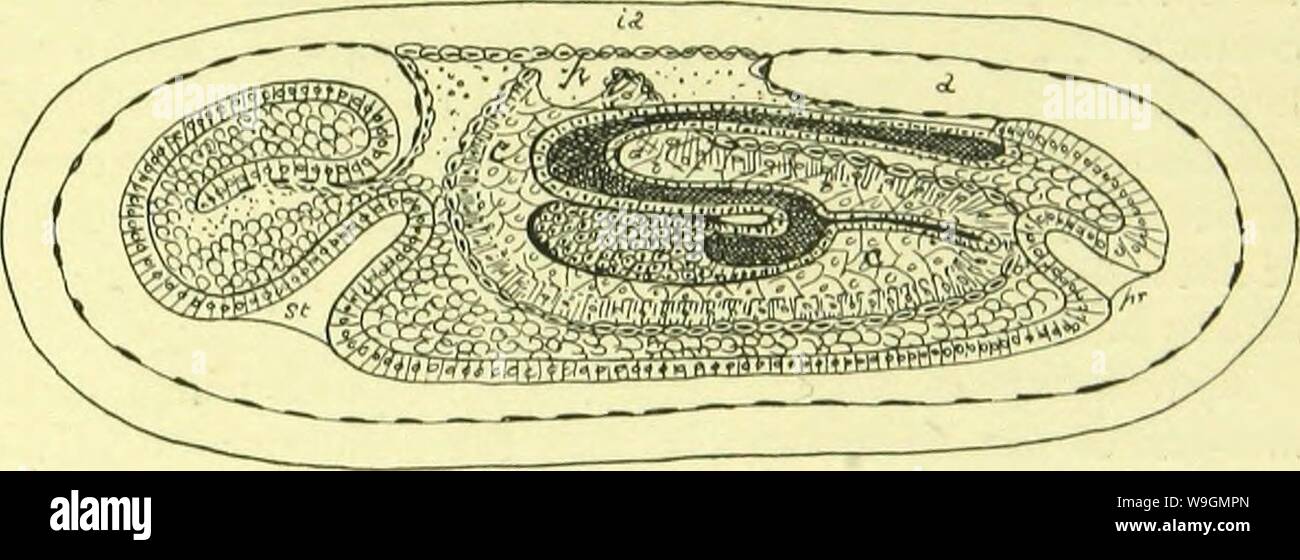
Archive image from page 290 of The anatomy, physiology, morphology and. The anatomy, physiology, morphology and development of the blow-fly (Calliphora erythrocephala.) A study in the comparative anatomy and morphology of insects; with plates and illustrations executed directly from the drawings of the author; CUbiodiversity4765349-9885 Year: 1890 ( ORIGIN OF THE ARCHENTERON. 249 veloped, which ultimately closes above and forms the metenteron. The metenteron then sinks into the yelk, and its blind posterior extremity, as I believe, eventually unites with the ventral proctodaeal involution; bu
Image details
Contributor:
Bookive / Alamy Stock PhotoImage ID:
W9GMPNFile size:
5.7 MB (254.5 KB Compressed download)Releases:
Model - no | Property - noDo I need a release?Dimensions:
2349 x 851 px | 39.8 x 14.4 cm | 15.7 x 5.7 inches | 150dpiMore information:
This image is a public domain image, which means either that copyright has expired in the image or the copyright holder has waived their copyright. Alamy charges you a fee for access to the high resolution copy of the image.
This image could have imperfections as it’s either historical or reportage.
Archive image from page 290 of The anatomy, physiology, morphology and. The anatomy, physiology, morphology and development of the blow-fly (Calliphora erythrocephala.) A study in the comparative anatomy and morphology of insects; with plates and illustrations executed directly from the drawings of the author; CUbiodiversity4765349-9885 Year: 1890 ( ORIGIN OF THE ARCHENTERON. 249 veloped, which ultimately closes above and forms the metenteron. The metenteron then sinks into the yelk, and its blind posterior extremity, as I believe, eventually unites with the ventral proctodaeal involution; but on this point I have no actual evidence, as I have been unable to trace the union of the hind gut and proctodaeum, although it is clear that if I am right in the interpretation of the observations of myself and others, such a union must occur. The mid-gut, that is, the part of the intestine in front of the blastopore, first turns backwards beneath the metenteron (Fig. 3), but its hinder blind extremity afterwards turns forwards Fk;. 39.—An ideal section of an embryo constructed from transverse sections, showing the relations of the archenteron, stomod.-eum, proctodeum, and cct'loniic sacs. The parablast of the ca'loniic sacs and pericardial (segmentation) cavity, is repre- sented l)y a chain of cells : a, amnial sac ; c c, cuelomic sac of the right side ; i a, space between the amnion and serosa ; p, pericardial cavity ; pr, proctoditum ; st, stomodix;uni. and forms a solid cellular growth, which penetrates the yelk and ultimately becomes the mid-gut (Fig. 39). The Malpighian tubes are developed as sacculi of the arch- enteron, one on either side of the blastopore, but subsequently travel back as far as the posterior extremity of the archenteric invagination, so that they open into it immediately behind its solid cellular portion. If we compare the development of the archenteron of the fly embryo, as I have described it above, with that of the crayfish, as described by H