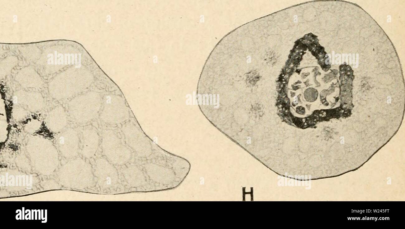
Archive image from page 207 of Cytology, with special reference to. Cytology, with special reference to the metazoan nucleus cytologywithspec00agar Year: 1920 Fig. 81. ' Illustrating the supposed emission of chromidia from various Metazoan cells. A, B, C, oocyte of Aricia foetida (alter Schaxel, Z.J.A., 1912). A, young oocyte, chromosomes still filamentar, cytoplasm destitute of chromidia ; B, older oocyte, still no chromidia in the cytoplasm ; C, still older oocyte, showing emission of chromidia from the nucleus into the cytoplasm. D, oocyte of Antedon bifida (after Chubb, Phil. Trans., 190
Image details
Contributor:
Bookive / Alamy Stock PhotoImage ID:
W245FTFile size:
5.7 MB (261.5 KB Compressed download)Releases:
Model - no | Property - noDo I need a release?Dimensions:
2007 x 997 px | 34 x 16.9 cm | 13.4 x 6.6 inches | 150dpiMore information:
This image is a public domain image, which means either that copyright has expired in the image or the copyright holder has waived their copyright. Alamy charges you a fee for access to the high resolution copy of the image.
This image could have imperfections as it’s either historical or reportage.
Archive image from page 207 of Cytology, with special reference to. Cytology, with special reference to the metazoan nucleus cytologywithspec00agar Year: 1920 Fig. 81. ' Illustrating the supposed emission of chromidia from various Metazoan cells. A, B, C, oocyte of Aricia foetida (alter Schaxel, Z.J.A., 1912). A, young oocyte, chromosomes still filamentar, cytoplasm destitute of chromidia ; B, older oocyte, still no chromidia in the cytoplasm ; C, still older oocyte, showing emission of chromidia from the nucleus into the cytoplasm. D, oocyte of Antedon bifida (after Chubb, Phil. Trans., 1906). Discharge of comparatively large masses of chromatin from the nucleolus. E, spermatocyte I. of Blatta ger- manica (after Wassilieff, A.m.A., 1907). F, oocyte of Proteus anguineus (after jorgensen, F.H., 1910). G, H, somatic cells of Musca (after Popoff, F.H., 1910). G, emission stage ; H, the chromidia congregated into a band round the nucleus. are divided as to whether they pass as formed bodies through deficiencies in the nuclear membrane (Buchner, 1910), or whether they are passed