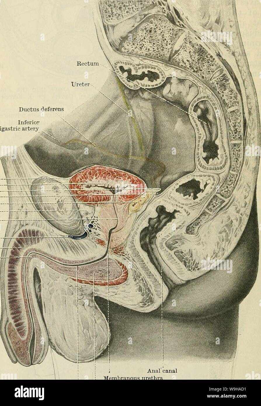
Archive image from page 1315 of Cunningham's Text-book of anatomy (1914). Cunningham's Text-book of anatomy cunninghamstextb00cunn Year: 1914 ( 1282 THE URINO-GENITAL SYSTEM. basal surface of the contracted and empty bladder receives a covering from the peritoneum, since the seminal vesicles and terminal portions of the ductus de- ferentes intervene as they lie in the anterior wall of the recto-vesical or recto- o-enital pouch. When the bladder is distended the posterior border, separating the upper and basal surfaces, is rounded out, and the peritoneum forming the horizontal shelf, just desc
Image details
Contributor:
Bookive / Alamy Stock PhotoImage ID:
W9HAD1File size:
5.7 MB (360.8 KB Compressed download)Releases:
Model - no | Property - noDo I need a release?Dimensions:
1173 x 1705 px | 19.9 x 28.9 cm | 7.8 x 11.4 inches | 150dpiMore information:
This image is a public domain image, which means either that copyright has expired in the image or the copyright holder has waived their copyright. Alamy charges you a fee for access to the high resolution copy of the image.
This image could have imperfections as it’s either historical or reportage.
Archive image from page 1315 of Cunningham's Text-book of anatomy (1914). Cunningham's Text-book of anatomy cunninghamstextb00cunn Year: 1914 ( 1282 THE URINO-GENITAL SYSTEM. basal surface of the contracted and empty bladder receives a covering from the peritoneum, since the seminal vesicles and terminal portions of the ductus de- ferentes intervene as they lie in the anterior wall of the recto-vesical or recto- o-enital pouch. When the bladder is distended the posterior border, separating the upper and basal surfaces, is rounded out, and the peritoneum forming the horizontal shelf, just described, is taken up (compare Eigs. 989 and 990). It is Inferior epigastric artery Superior peritoneal lig. of bladder Urinary bladder. Sacro-genital fold Recto-vesical pouch Ductus deferens Retro-pubic pad of fat Prostatic urethra Dorsal vein of penis Corpus cavernosum penis--„ Corpus cavernosum, hL urethrse T i Anal canal , i Membranous urethra Cavernous portion of urethra Bulb of urethra Fig. 1001.—Median Section of the Pelvis in an Adult Male. The coils of small intestine which Iky within the pelvis have been lifted out in order to give a view of the lateral wall of the pelvic cavity. to be specially noted that the level of the peritoneal reflection, forming the bottom of the recto-vesical pouch; does not vary much, as regards its relationship to the prostate, during distension and contraction of the bladder (Figs. 989 and 990). An examination of median Sections of the pelvis shows the great danger run by the ampullae of the ductus deferentes in any operation for reaching the bladder through the anterior wall of the rectum, and the difficulty in avoiding injury to the peritoneum. The term ' posterior false (or peritoneal) ligament' is often applied to the some- what variable crescentic fold of peritoneum which bounds on each side the entrance to the recto-vesical or recto-genital pouch, and which often unites with