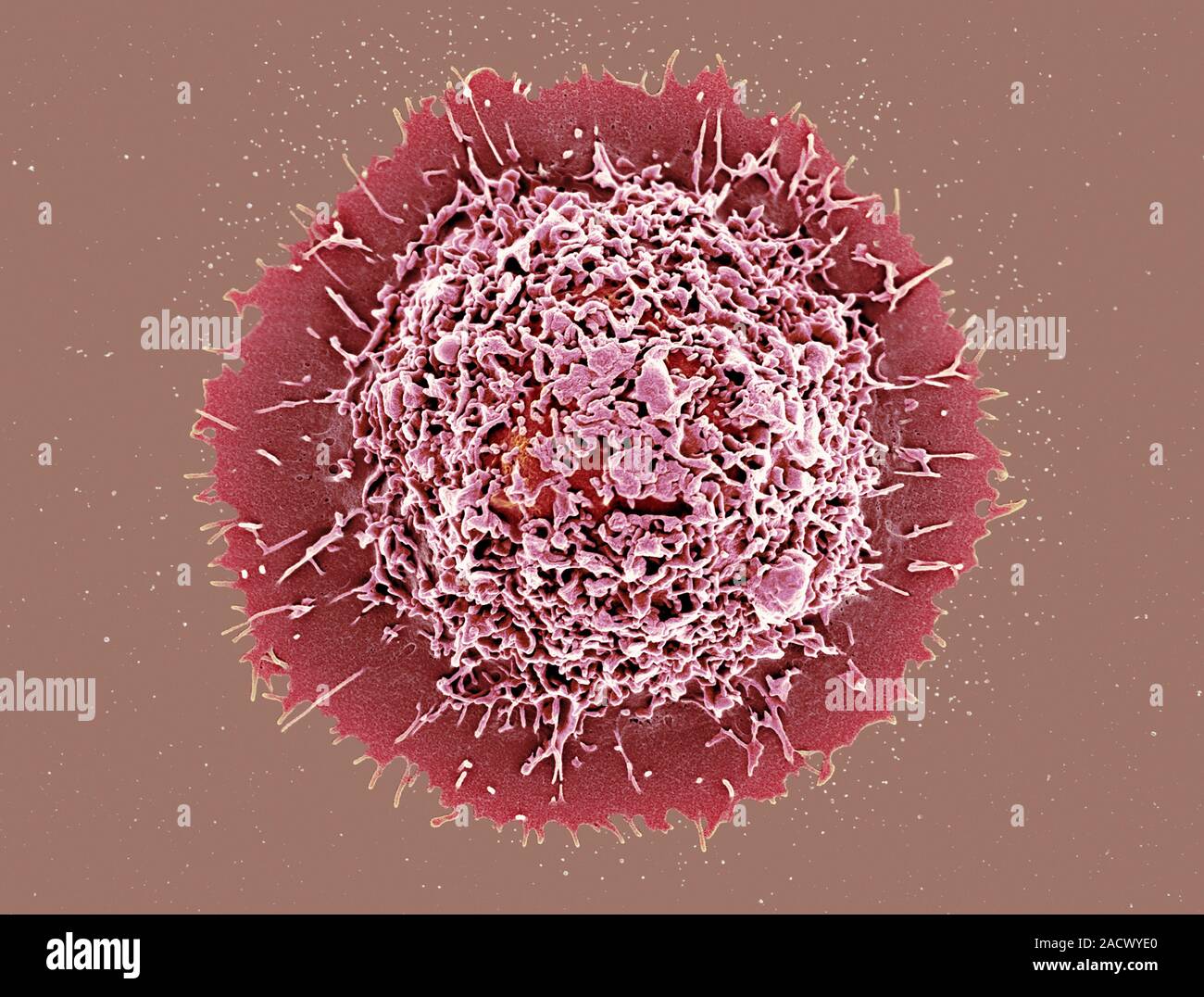
Alveolar macrophage. Coloured scanning electron micrograph (SEM) of an alveolar macrophage white blood cell from lung tissue. Macrophages are cells of
RMID:Image ID:2ACWYE0
Image details
Contributor:
Science Photo Library / Alamy Stock PhotoImage ID:
2ACWYE0File size:
54.3 MB (1.9 MB Compressed download)Releases:
Model - no | Property - noDo I need a release?Dimensions:
5000 x 3796 px | 42.3 x 32.1 cm | 16.7 x 12.7 inches | 300dpiDate taken:
31 March 2014More information:
Alveolar macrophage. Coloured scanning electron micrograph (SEM) of an alveolar macrophage white blood cell from lung tissue. Macrophages are cells of the body's immune system that are found in the tissues rather than in the circulating blood. They recognise foreign particles, including bacteria, pollen and dust, and phagocytose (engulf) and digest them. Alveolar macrophages are associated with lung alveoli (air sacs) and epithelium (lining tissue). Their role is primarily to remove dust, hence their other name of dust cell.