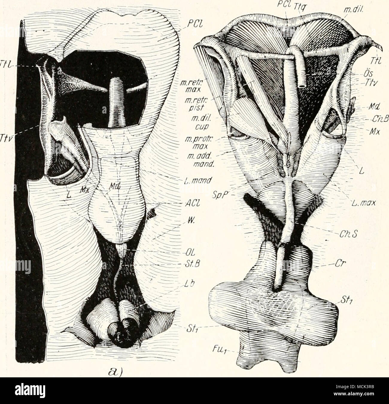
. Abb. 11. Imago von Psylla mali, Vorderkopf, Labiuin und Prosternum. «) Ventral- (Außen-)ansicht. Aus dem Postclypeus ist ein Fenster ausgeschnitten. Muskeln ent- fernt, b) Dorsal- (Innen-) Ansieht. Muskeln belassen. — ACl= Anteclypeus: ChB = seh- nenartige Brücke; Ch S = Chitinstab; Cr = Crumena; Fu^^ = Furca 1; L ^ maxillarer Protractorarm; Lb = Labium; L'))ia)id= Lamina mandibularis; Lmax=^ Lamina maxillaris; LV = Vorderrand des Postclypeus; Md = Mandibel; Mx= Maxille; m dil = Musculus dilatator; m retr max= Musculus retractor niax.; M retr pist = Musculus retractor pistilli; w dil cup = M
Image details
Contributor:
The Bookworm Collection / Alamy Stock PhotoImage ID:
MCK3RBFile size:
14.3 MB (747.6 KB Compressed download)Releases:
Model - no | Property - noDo I need a release?Dimensions:
2271 x 2201 px | 38.5 x 37.3 cm | 15.1 x 14.7 inches | 150dpiMore information:
This image is a public domain image, which means either that copyright has expired in the image or the copyright holder has waived their copyright. Alamy charges you a fee for access to the high resolution copy of the image.
This image could have imperfections as it’s either historical or reportage.
. Abb. 11. Imago von Psylla mali, Vorderkopf, Labiuin und Prosternum. «) Ventral- (Außen-)ansicht. Aus dem Postclypeus ist ein Fenster ausgeschnitten. Muskeln ent- fernt, b) Dorsal- (Innen-) Ansieht. Muskeln belassen. — ACl= Anteclypeus: ChB = seh- nenartige Brücke; Ch S = Chitinstab; Cr = Crumena; Fu^^ = Furca 1; L ^ maxillarer Protractorarm; Lb = Labium; L'))ia)id= Lamina mandibularis; Lmax=^ Lamina maxillaris; LV = Vorderrand des Postclypeus; Md = Mandibel; Mx= Maxille; m dil = Musculus dilatator; m retr max= Musculus retractor niax.; M retr pist = Musculus retractor pistilli; w dil cup = Musculus dilatator cui)ulae: '»i prutr mnx = Musculus protractor maxill; m add menu/= Musculus adductor mandibulae; OL = Oberlippe; Ös = Oesophagus; PCL^ Postclypeus; Sj) P = Speichelpunipe; StB= Stechborsten; Stj^= Sternum; Ttl. Ttv, Ttr = Tentorium; W = Wulst. — Nach Weber. Hypopharynx. Laminae maxillares und Anteclypeus legen sicli eng zur Bildung einerstarren Mundhöhle zusannnen^. An der Übergangsstelle zum Pharynx ist das Mundhöhlendach durch ein Paar drucldaiopfartige Gebilde, die Mundknöpfe (M Kn, Abb. 18) an der Ventralwand des Hypo- pharynx befestigt. 1 Diese Höhle ist, morphologisch betrachtet, in Wirklichkeit ein Teil der Präoral- höhle, während die eigentliche Mundhöhle mit dem Cibarium (also ebenfalls der Pväoral- höhle in eler Mundpumpe aufgegangen ist. Näheres im allgemeinen Teil. Der Herausgeber..