Quick filters:
Muscle tendon Stock Photos and Images
 Young teen asian woman muscle tendon joint wrist pain from using laptop computer massage for pain relief. Stock Photohttps://www.alamy.com/image-license-details/?v=1https://www.alamy.com/young-teen-asian-woman-muscle-tendon-joint-wrist-pain-from-using-laptop-computer-massage-for-pain-relief-image557022613.html
RF2RA6G0N–Young teen asian woman muscle tendon joint wrist pain from using laptop computer massage for pain relief.
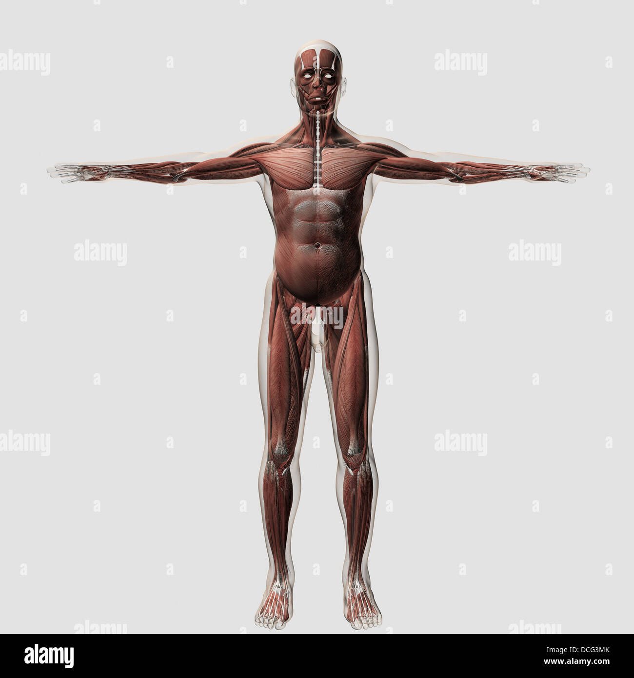 Anatomy of male muscular system, front view. Stock Photohttps://www.alamy.com/image-license-details/?v=1https://www.alamy.com/stock-photo-anatomy-of-male-muscular-system-front-view-59361139.html
RFDCG3MK–Anatomy of male muscular system, front view.
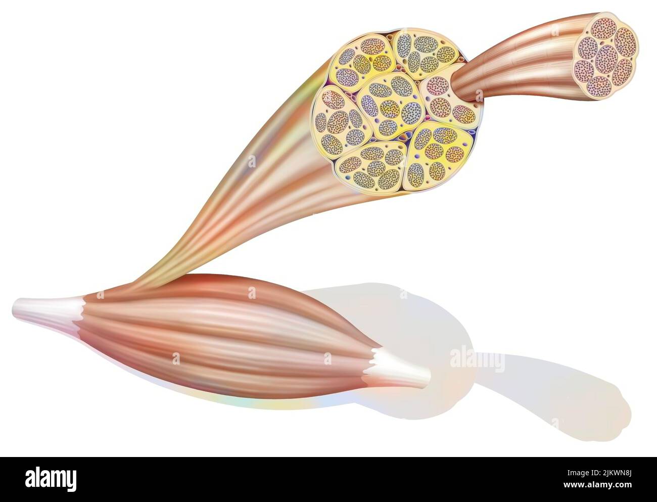 From muscle to muscle fiber: tendon, muscle, muscle fiber. Stock Photohttps://www.alamy.com/image-license-details/?v=1https://www.alamy.com/from-muscle-to-muscle-fiber-tendon-muscle-muscle-fiber-image476923906.html
RF2JKWN8J–From muscle to muscle fiber: tendon, muscle, muscle fiber.
 Diagnosis - Sprain. Medical Concept. 3D Render. Stock Photohttps://www.alamy.com/image-license-details/?v=1https://www.alamy.com/stock-photo-diagnosis-sprain-medical-concept-3d-render-83508219.html
RFERT3GB–Diagnosis - Sprain. Medical Concept. 3D Render.
 Tendons on the Back of the Left Hand, vintage line drawing or engraving illustration. Stock Vectorhttps://www.alamy.com/image-license-details/?v=1https://www.alamy.com/tendons-on-the-back-of-the-left-hand-vintage-line-drawing-or-engraving-illustration-image348651098.html
RF2B76C0X–Tendons on the Back of the Left Hand, vintage line drawing or engraving illustration.
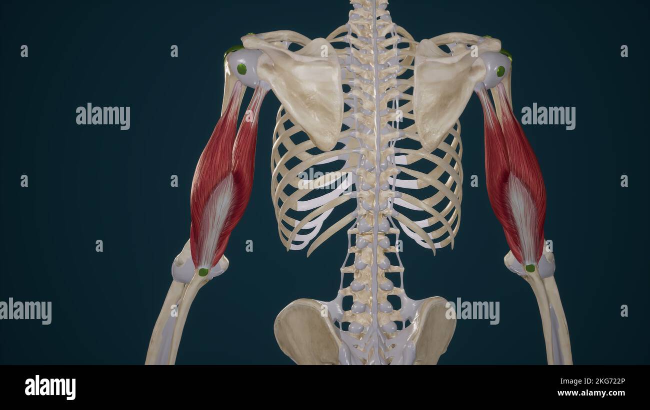 Triceps Brachii Muscle Stock Photohttps://www.alamy.com/image-license-details/?v=1https://www.alamy.com/triceps-brachii-muscle-image491880110.html
RF2KG722P–Triceps Brachii Muscle
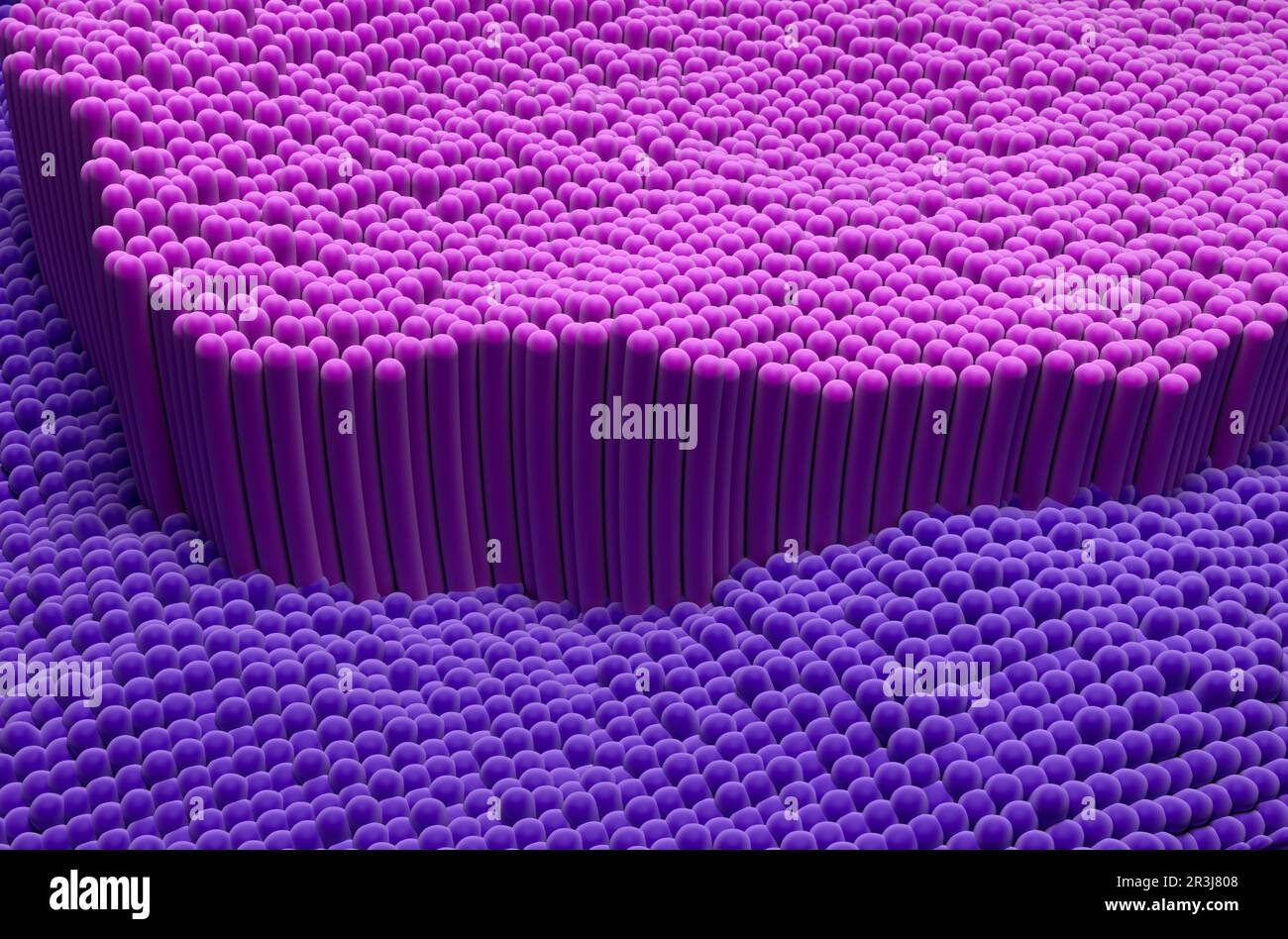 Tendon structure in the human body - 3d illustration isometric view Stock Photohttps://www.alamy.com/image-license-details/?v=1https://www.alamy.com/tendon-structure-in-the-human-body-3d-illustration-isometric-view-image552977160.html
RF2R3J808–Tendon structure in the human body - 3d illustration isometric view
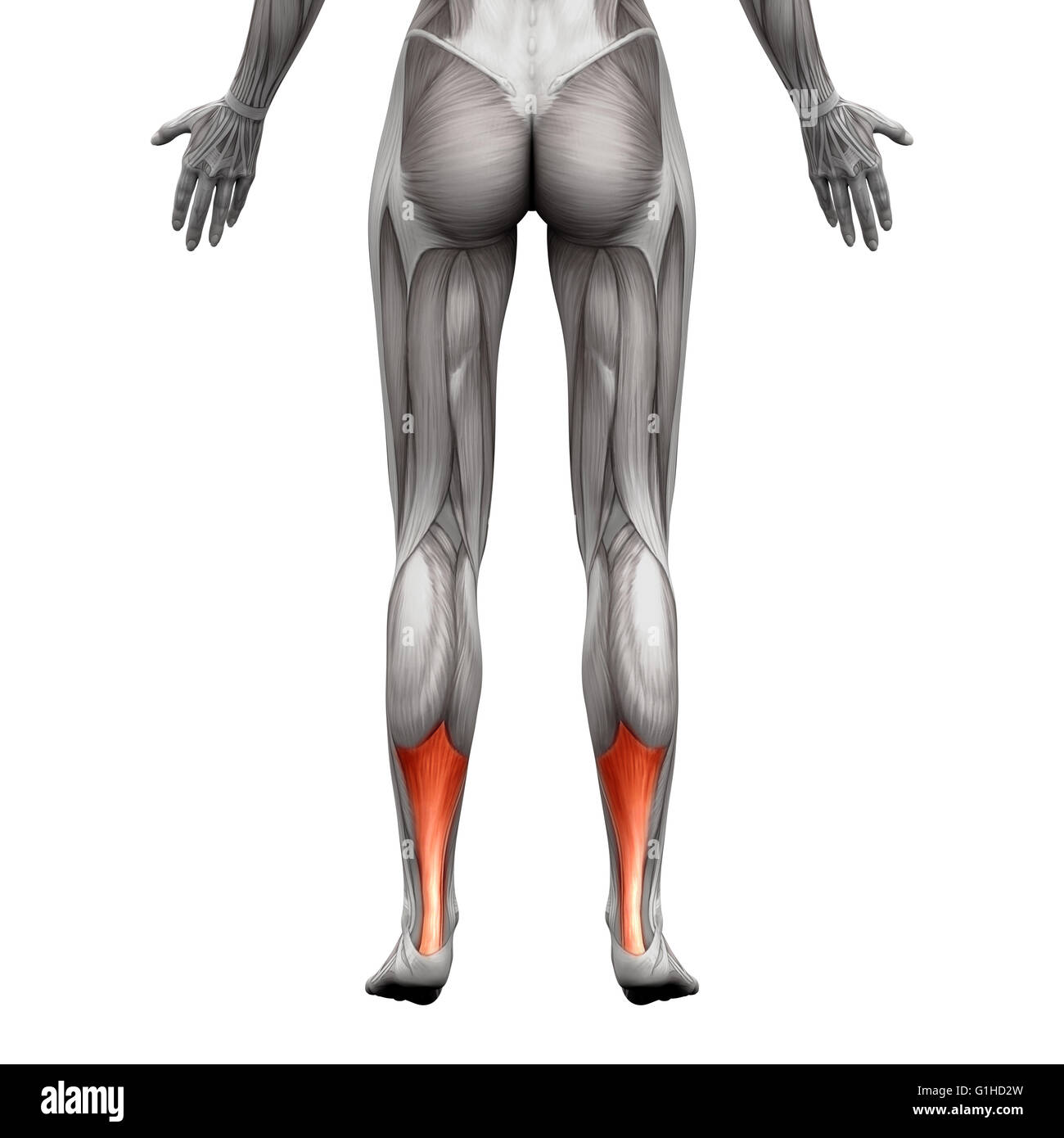 Achilles Tendon - Anatomy Muscle - isolated on white - 3D illustration Stock Photohttps://www.alamy.com/image-license-details/?v=1https://www.alamy.com/stock-photo-achilles-tendon-anatomy-muscle-isolated-on-white-3d-illustration-104260321.html
RFG1HD2W–Achilles Tendon - Anatomy Muscle - isolated on white - 3D illustration
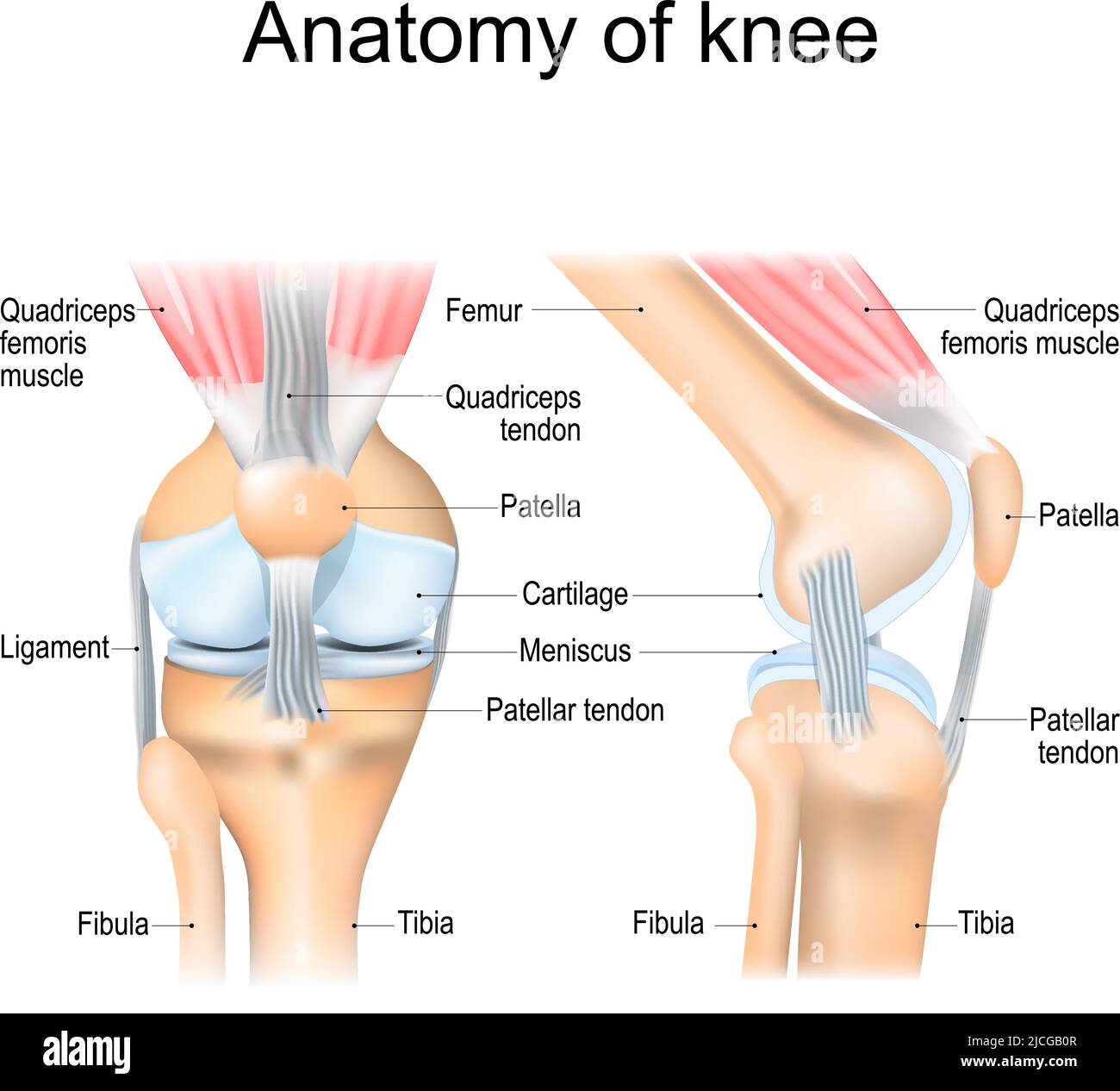 Knee anatomy. Structure of leg joint. Major parts. Vector poster with text label for medical education Stock Vectorhttps://www.alamy.com/image-license-details/?v=1https://www.alamy.com/knee-anatomy-structure-of-leg-joint-major-parts-vector-poster-with-text-label-for-medical-education-image472415687.html
RF2JCGB0R–Knee anatomy. Structure of leg joint. Major parts. Vector poster with text label for medical education
 Ultrasound examination of wrist Stock Photohttps://www.alamy.com/image-license-details/?v=1https://www.alamy.com/stock-photo-ultrasound-examination-of-wrist-102868817.html
RMFYA269–Ultrasound examination of wrist
 Muscle-tendon conection in a long bone. X25 at 10 cm wide. Stock Photohttps://www.alamy.com/image-license-details/?v=1https://www.alamy.com/muscle-tendon-conection-in-a-long-bone-x25-at-10-cm-wide-image591999403.html
RF2WB3W7R–Muscle-tendon conection in a long bone. X25 at 10 cm wide.
 Golgi Tendon Organ. It is a sensory organ that perceives tendon or muscle strength. Stock Photohttps://www.alamy.com/image-license-details/?v=1https://www.alamy.com/golgi-tendon-organ-it-is-a-sensory-organ-that-perceives-tendon-or-muscle-strength-image626642104.html
RF2YBE0B4–Golgi Tendon Organ. It is a sensory organ that perceives tendon or muscle strength.
 medically accurate illustration of the achilles tendon Stock Photohttps://www.alamy.com/image-license-details/?v=1https://www.alamy.com/stock-photo-medically-accurate-illustration-of-the-achilles-tendon-89760723.html
RFF60XM3–medically accurate illustration of the achilles tendon
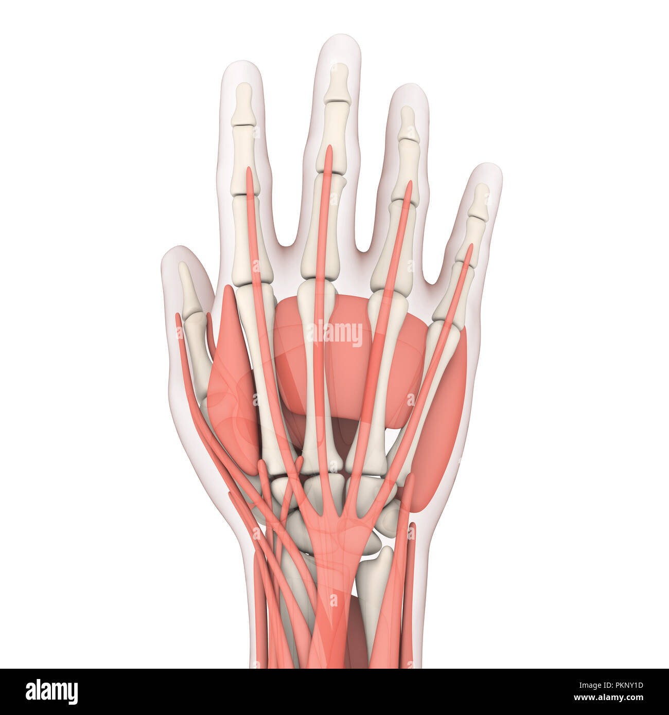 Human Hand Anatomy Illustration Stock Photohttps://www.alamy.com/image-license-details/?v=1https://www.alamy.com/human-hand-anatomy-illustration-image218685081.html
RFPKNY1D–Human Hand Anatomy Illustration
 Transition of the striated muscle fibers into the tendon. Illustration of the 19th century. Germany. White background. Stock Photohttps://www.alamy.com/image-license-details/?v=1https://www.alamy.com/transition-of-the-striated-muscle-fibers-into-the-tendon-illustration-of-the-19th-century-germany-white-background-image402543575.html
RF2EAWCC7–Transition of the striated muscle fibers into the tendon. Illustration of the 19th century. Germany. White background.
 anatomy back without skin isolated on white. 3D rendering Stock Photohttps://www.alamy.com/image-license-details/?v=1https://www.alamy.com/stock-photo-anatomy-back-without-skin-isolated-on-white-3d-rendering-122816267.html
RFH3PNB7–anatomy back without skin isolated on white. 3D rendering
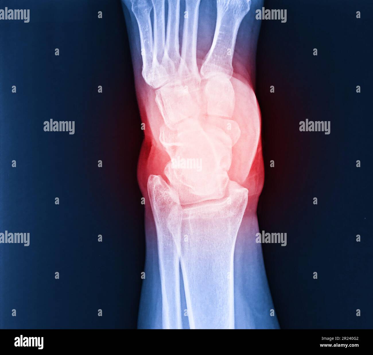 Arthritis at ankle joint (Gout , Rheumatoid arthritis) Stock Photohttps://www.alamy.com/image-license-details/?v=1https://www.alamy.com/arthritis-at-ankle-joint-gout-rheumatoid-arthritis-image552049346.html
RF2R240G2–Arthritis at ankle joint (Gout , Rheumatoid arthritis)
 Back view of woman sit at desk at home have webcam conference on laptop computer with Orthopedic Surgeons about bone, joint, tendon and muscle. Stock Photohttps://www.alamy.com/image-license-details/?v=1https://www.alamy.com/back-view-of-woman-sit-at-desk-at-home-have-webcam-conference-on-laptop-computer-with-orthopedic-surgeons-about-bone-joint-tendon-and-muscle-image547902598.html
RF2PRB39X–Back view of woman sit at desk at home have webcam conference on laptop computer with Orthopedic Surgeons about bone, joint, tendon and muscle.
 Heel pain, joint inflammation from arthritis or neuropathy. Inflamed feet with numbness, callus, or bruise. Bone, muscle, tendon issues, and sprain. M Stock Photohttps://www.alamy.com/image-license-details/?v=1https://www.alamy.com/heel-pain-joint-inflammation-from-arthritis-or-neuropathy-inflamed-feet-with-numbness-callus-or-bruise-bone-muscle-tendon-issues-and-sprain-m-image632779935.html
RF2YNDH7B–Heel pain, joint inflammation from arthritis or neuropathy. Inflamed feet with numbness, callus, or bruise. Bone, muscle, tendon issues, and sprain. M
 ARCHIVE - An archival image dated 18 October 2016 shows the German Bundesliga soccer club Bayern Munich player Jerome Boateng during a training session in Munich, Germany. The central defender will not be able to take part in the club's scheduled training session in Qatar between the 03.01.17 and 11.01.17 due to a chest muscle tendon injury. The team will be in the Middle Eastern in order to prepare for the remainder of the Bundesliga season. Photo: Sven Hoppe/dpa Stock Photohttps://www.alamy.com/image-license-details/?v=1https://www.alamy.com/stock-photo-archive-an-archival-image-dated-18-october-2016-shows-the-german-bundesliga-130329536.html
RMHG10J8–ARCHIVE - An archival image dated 18 October 2016 shows the German Bundesliga soccer club Bayern Munich player Jerome Boateng during a training session in Munich, Germany. The central defender will not be able to take part in the club's scheduled training session in Qatar between the 03.01.17 and 11.01.17 due to a chest muscle tendon injury. The team will be in the Middle Eastern in order to prepare for the remainder of the Bundesliga season. Photo: Sven Hoppe/dpa
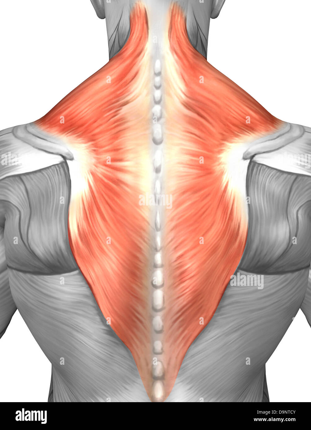 Muscles of the back and neck (splenius capitis muscle, trapezius muscle). Stock Photohttps://www.alamy.com/image-license-details/?v=1https://www.alamy.com/stock-photo-muscles-of-the-back-and-neck-splenius-capitis-muscle-trapezius-muscle-57643179.html
RFD9NTCY–Muscles of the back and neck (splenius capitis muscle, trapezius muscle).
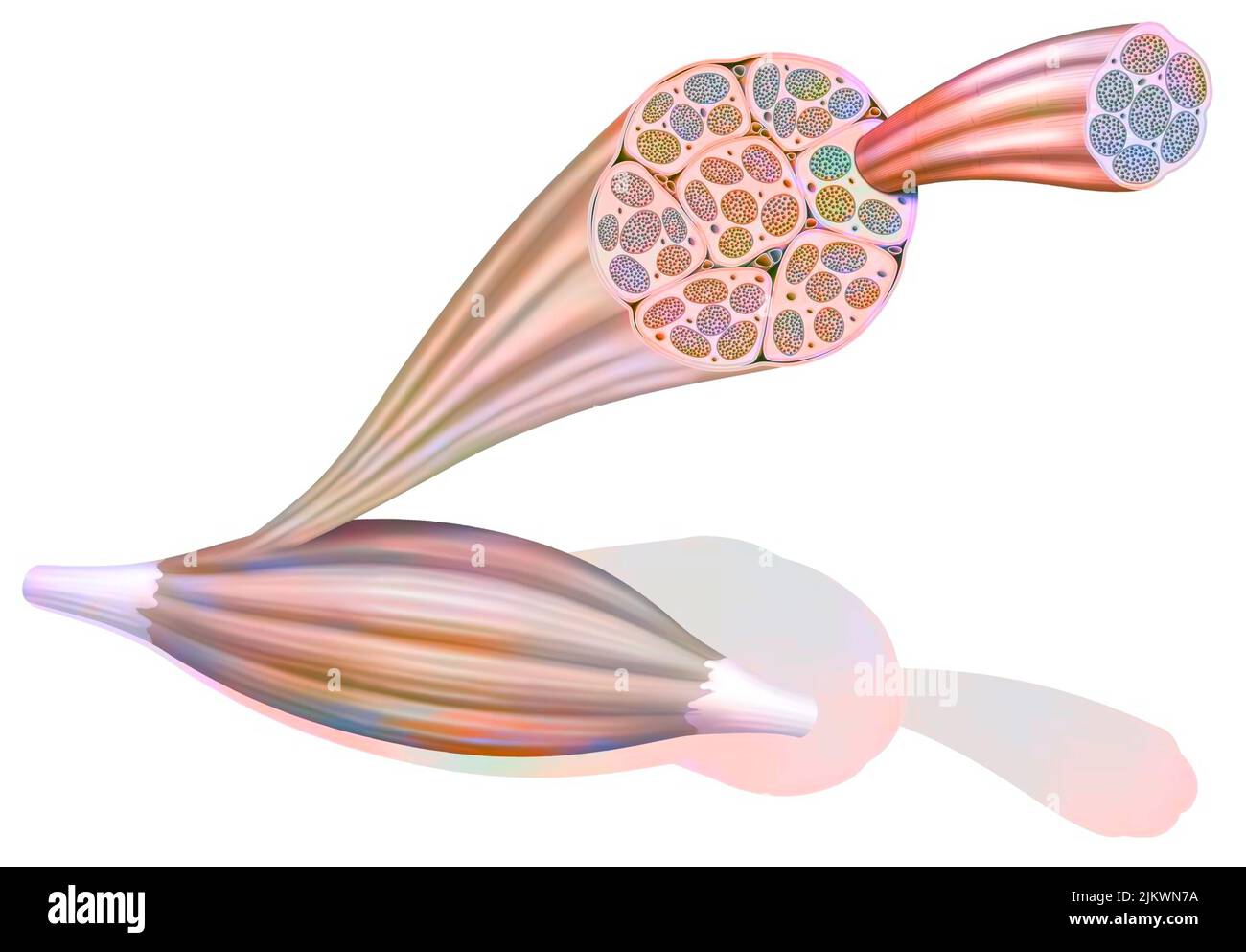 From muscle to muscle fiber: tendon, muscle, muscle fiber. Stock Photohttps://www.alamy.com/image-license-details/?v=1https://www.alamy.com/from-muscle-to-muscle-fiber-tendon-muscle-muscle-fiber-image476923870.html
RF2JKWN7A–From muscle to muscle fiber: tendon, muscle, muscle fiber.
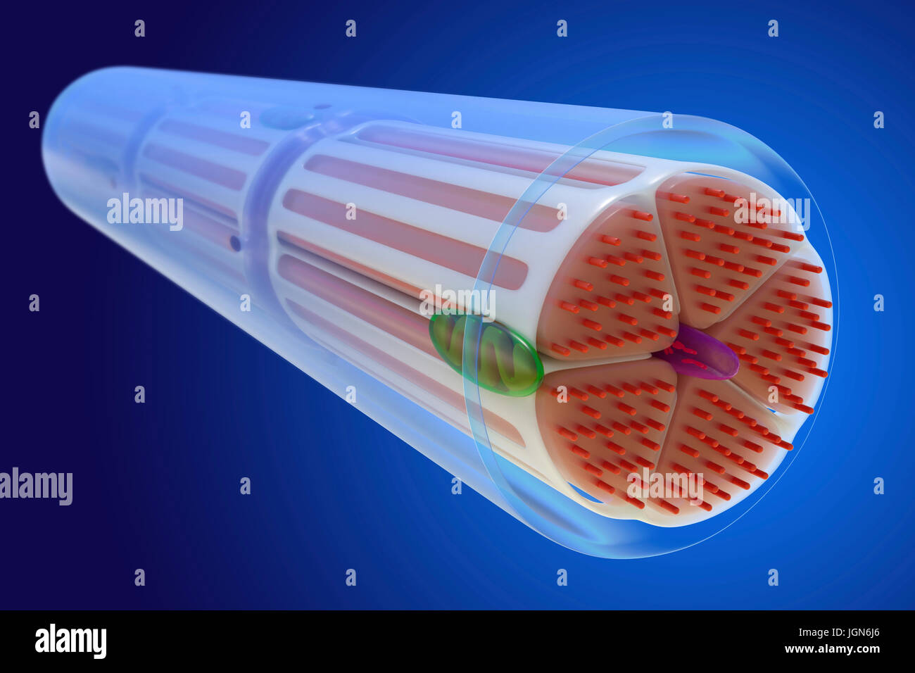 Skeletal muscle, illustration. The muscle is surrounded by epimysium connective tissue (transparent), which forms the tendon that attaches the muscle to bone. Bundles of muscle fibres (red) are surrounded by perimysium connective tissue (yellow). Stock Photohttps://www.alamy.com/image-license-details/?v=1https://www.alamy.com/stock-photo-skeletal-muscle-illustration-the-muscle-is-surrounded-by-epimysium-147983646.html
RFJGN6J6–Skeletal muscle, illustration. The muscle is surrounded by epimysium connective tissue (transparent), which forms the tendon that attaches the muscle to bone. Bundles of muscle fibres (red) are surrounded by perimysium connective tissue (yellow).
 A typical representation of the muscles of the leg showing how they pass into tendons at the ankle, vintage line drawing or engraving illustration. Stock Vectorhttps://www.alamy.com/image-license-details/?v=1https://www.alamy.com/a-typical-representation-of-the-muscles-of-the-leg-showing-how-they-pass-into-tendons-at-the-ankle-vintage-line-drawing-or-engraving-illustration-image348666179.html
RF2B7737F–A typical representation of the muscles of the leg showing how they pass into tendons at the ankle, vintage line drawing or engraving illustration.
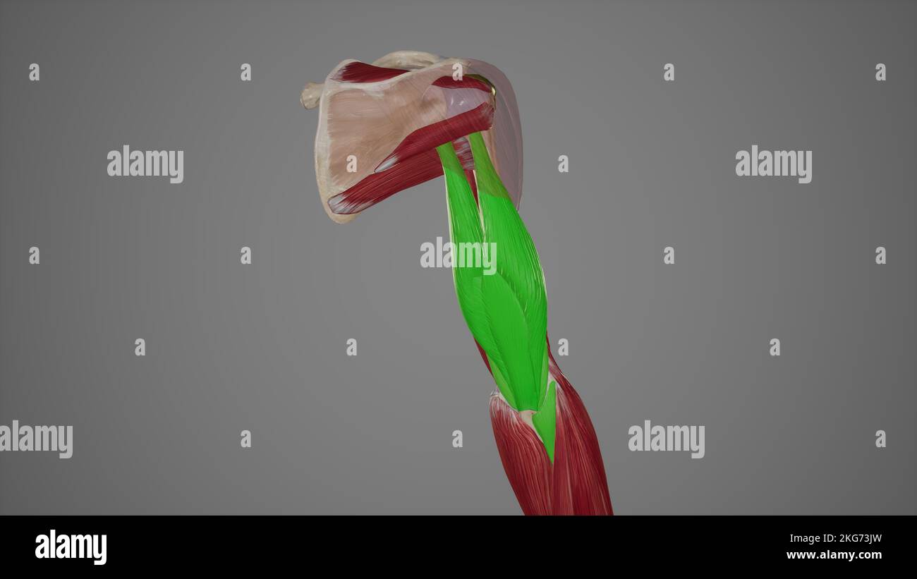 Posterior Muscles of Upper Arm Stock Photohttps://www.alamy.com/image-license-details/?v=1https://www.alamy.com/posterior-muscles-of-upper-arm-image491881345.html
RF2KG73JW–Posterior Muscles of Upper Arm
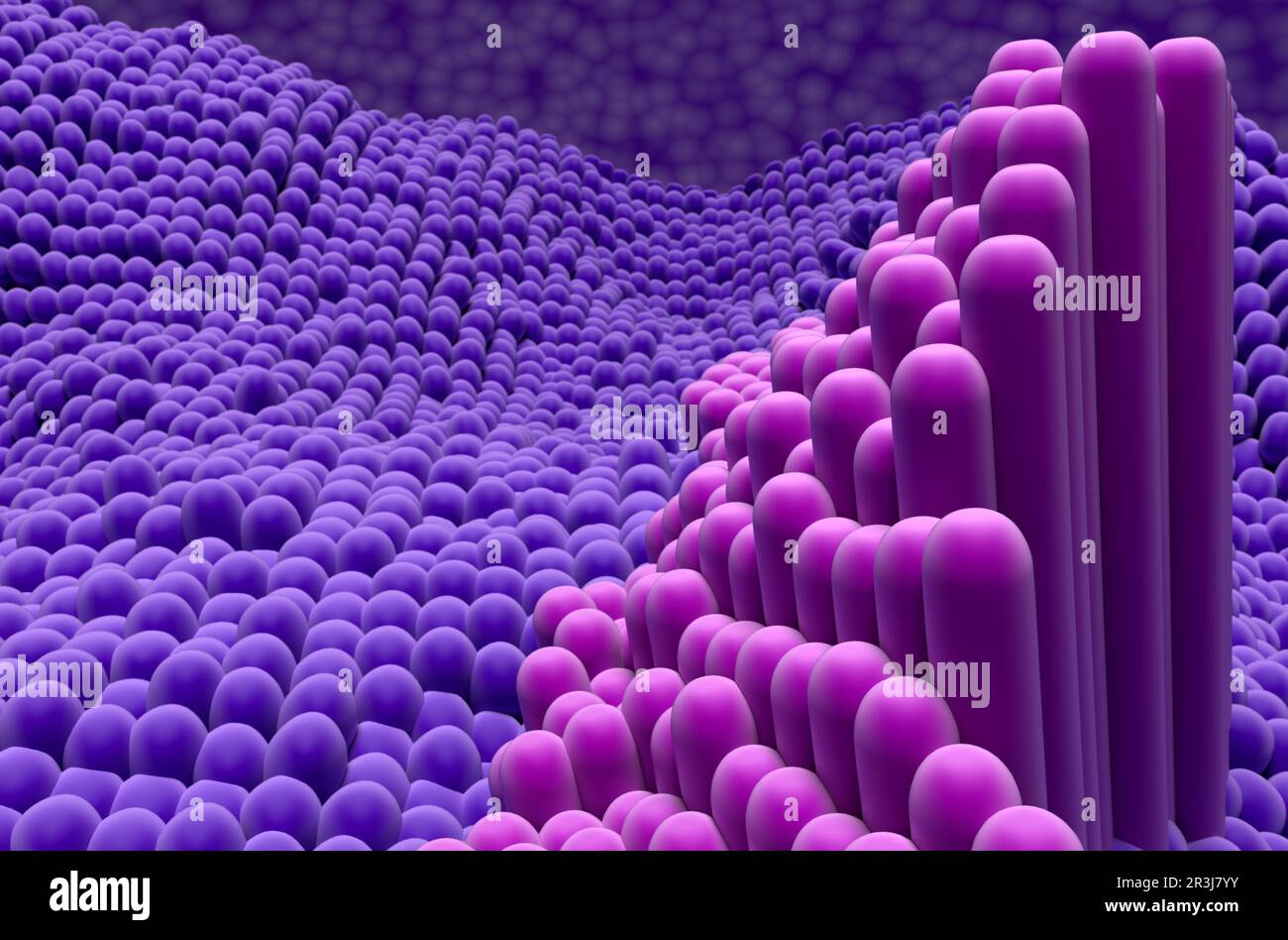 Tendon structure in the human body - 3d illustration closeup view Stock Photohttps://www.alamy.com/image-license-details/?v=1https://www.alamy.com/tendon-structure-in-the-human-body-3d-illustration-closeup-view-image552977151.html
RF2R3J7YY–Tendon structure in the human body - 3d illustration closeup view
 Foot of statue showing achilles tendon, close-up Stock Photohttps://www.alamy.com/image-license-details/?v=1https://www.alamy.com/foot-of-statue-showing-achilles-tendon-close-up-image566321748.html
RF2RWA54M–Foot of statue showing achilles tendon, close-up
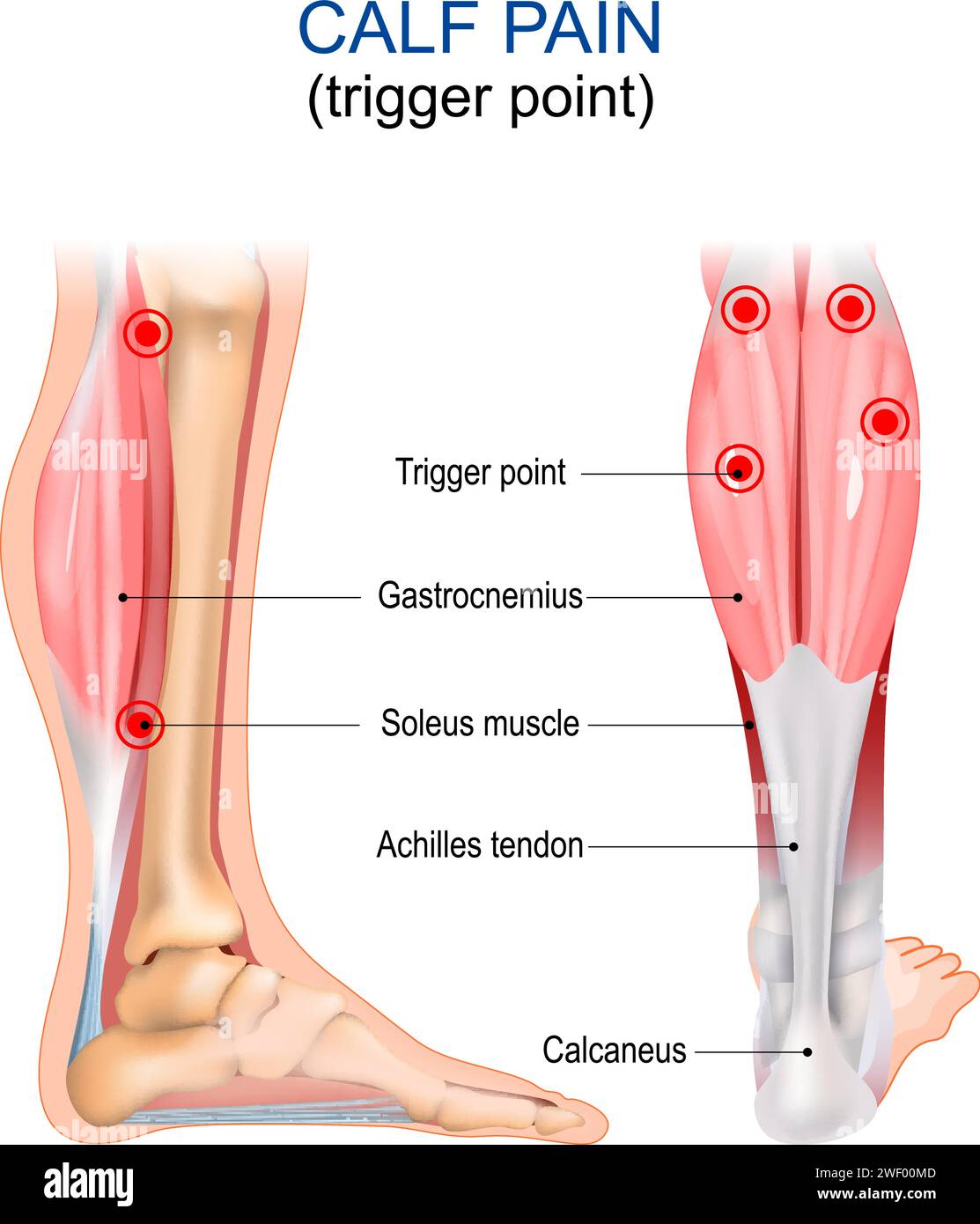 Calf pain. Trigger point. Gastrocnemius, Soleus muscle, Achilles tendon and Calcaneus. Myofascial pain syndrome. Human body anatomy. Vector illustrati Stock Vectorhttps://www.alamy.com/image-license-details/?v=1https://www.alamy.com/calf-pain-trigger-point-gastrocnemius-soleus-muscle-achilles-tendon-and-calcaneus-myofascial-pain-syndrome-human-body-anatomy-vector-illustrati-image594372925.html
RF2WF00MD–Calf pain. Trigger point. Gastrocnemius, Soleus muscle, Achilles tendon and Calcaneus. Myofascial pain syndrome. Human body anatomy. Vector illustrati
 Concept of leg tendon injury of fat man - obese person holding leg suffering muscle pain - overweight man fitness concept at outdoor park. Stock Photohttps://www.alamy.com/image-license-details/?v=1https://www.alamy.com/concept-of-leg-tendon-injury-of-fat-man-obese-person-holding-leg-suffering-muscle-pain-overweight-man-fitness-concept-at-outdoor-park-image332770546.html
RF2A9B06A–Concept of leg tendon injury of fat man - obese person holding leg suffering muscle pain - overweight man fitness concept at outdoor park.
 Muscle-tendon conection in a long bone. X75 at 10 cm wide. Stock Photohttps://www.alamy.com/image-license-details/?v=1https://www.alamy.com/muscle-tendon-conection-in-a-long-bone-x75-at-10-cm-wide-image591999407.html
RF2WB3W7Y–Muscle-tendon conection in a long bone. X75 at 10 cm wide.
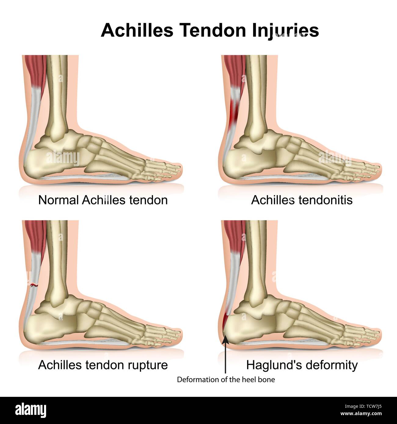 Achilles tendon injures medical vector illustration isolated on white background with english description eps 10 infographic Stock Vectorhttps://www.alamy.com/image-license-details/?v=1https://www.alamy.com/achilles-tendon-injures-medical-vector-illustration-isolated-on-white-background-with-english-description-eps-10-infographic-image248875821.html
RFTCW7J5–Achilles tendon injures medical vector illustration isolated on white background with english description eps 10 infographic
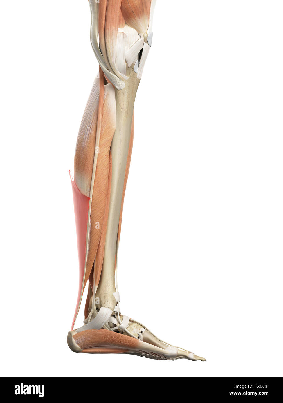 medically accurate illustration of the achilles tendon Stock Photohttps://www.alamy.com/image-license-details/?v=1https://www.alamy.com/stock-photo-medically-accurate-illustration-of-the-achilles-tendon-89760714.html
RFF60XKP–medically accurate illustration of the achilles tendon
 1185 Pg 203 Muscles and Tendons Stock Photohttps://www.alamy.com/image-license-details/?v=1https://www.alamy.com/1185-pg-203-muscles-and-tendons-image213440219.html
RMPB714Y–1185 Pg 203 Muscles and Tendons
 a young caucasian man, wearing sports clothes, massaging the muscles of his neck with a massage gun Stock Photohttps://www.alamy.com/image-license-details/?v=1https://www.alamy.com/a-young-caucasian-man-wearing-sports-clothes-massaging-the-muscles-of-his-neck-with-a-massage-gun-image484900380.html
RF2K4W3AM–a young caucasian man, wearing sports clothes, massaging the muscles of his neck with a massage gun
 Human male anatomy, limbs and hip muscular and skeletal systems, with internal muscle layers. Front and back views. black background. 3d ilustration. Stock Photohttps://www.alamy.com/image-license-details/?v=1https://www.alamy.com/human-male-anatomy-limbs-and-hip-muscular-and-skeletal-systems-with-internal-muscle-layers-front-and-back-views-black-background-3d-ilustration-image273575845.html
RFWW2CNW–Human male anatomy, limbs and hip muscular and skeletal systems, with internal muscle layers. Front and back views. black background. 3d ilustration.
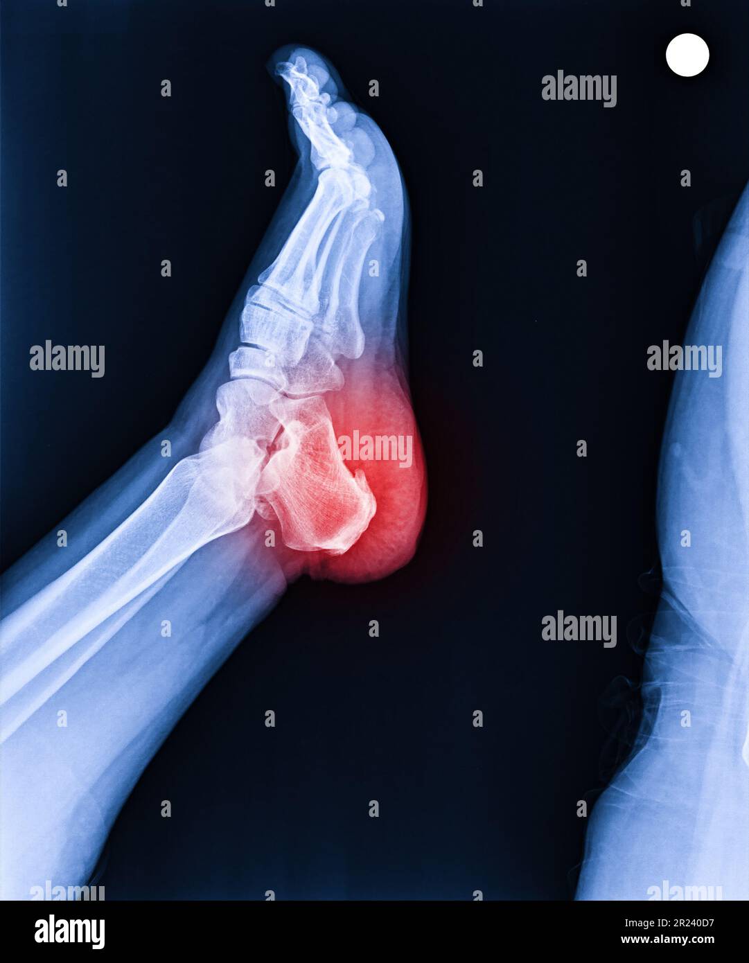 Foot and ankle pain on x-ray, isolated on black background, heel pain, heel spur Stock Photohttps://www.alamy.com/image-license-details/?v=1https://www.alamy.com/foot-and-ankle-pain-on-x-ray-isolated-on-black-background-heel-pain-heel-spur-image552049267.html
RF2R240D7–Foot and ankle pain on x-ray, isolated on black background, heel pain, heel spur
 Back view of woman in white gown have webcam conference on tablet computer with young asian doctor about bone, joint, tendon and muscle. Stock Photohttps://www.alamy.com/image-license-details/?v=1https://www.alamy.com/back-view-of-woman-in-white-gown-have-webcam-conference-on-tablet-computer-with-young-asian-doctor-about-bone-joint-tendon-and-muscle-image550241250.html
RF2PY5J96–Back view of woman in white gown have webcam conference on tablet computer with young asian doctor about bone, joint, tendon and muscle.
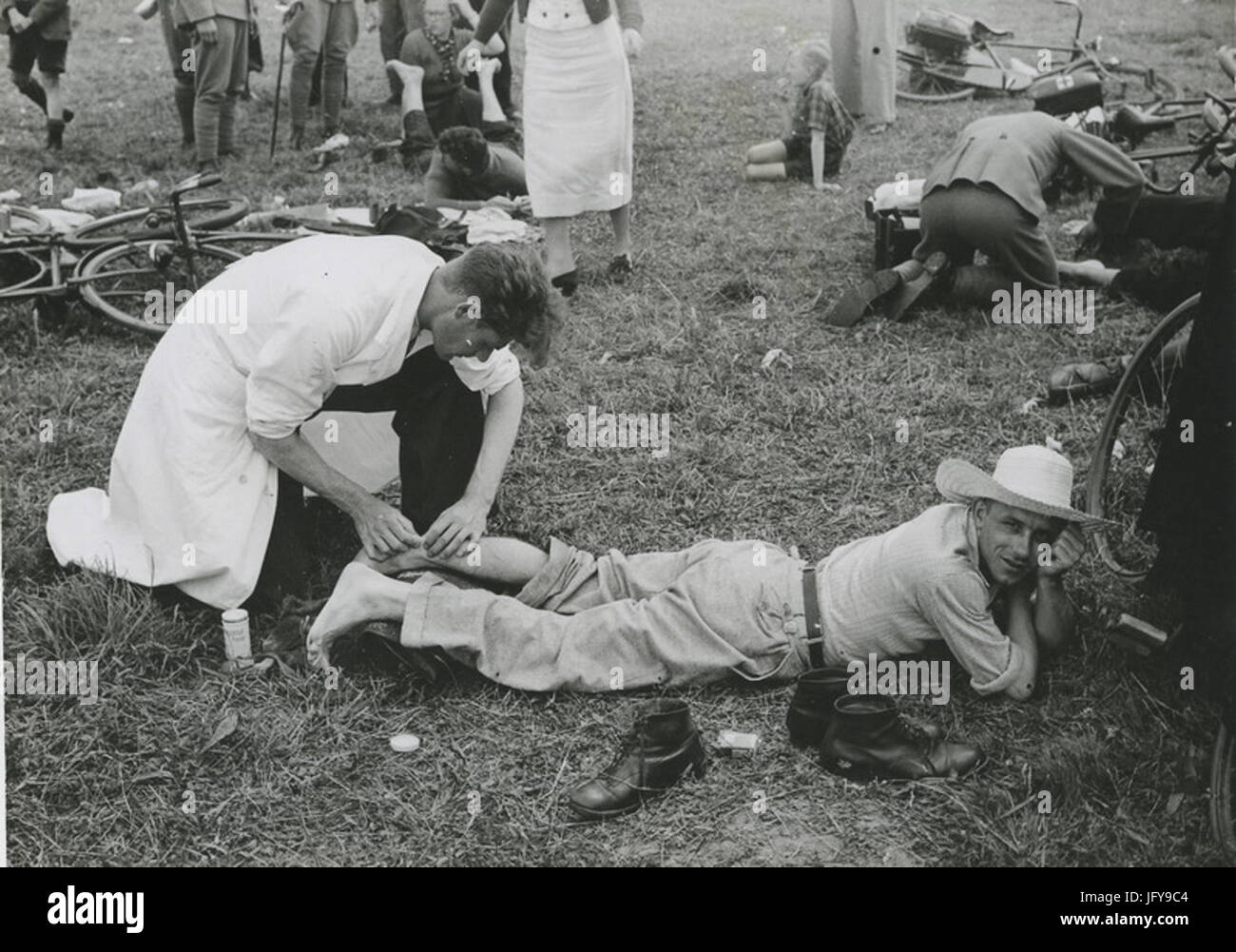 Eerste hulp bij verkramping aan de achillespees op de vierde dag van de 28e Vier - F40966 - KNBLO Stock Photohttps://www.alamy.com/image-license-details/?v=1https://www.alamy.com/stock-photo-eerste-hulp-bij-verkramping-aan-de-achillespees-op-de-vierde-dag-van-147502884.html
RMJFY9C4–Eerste hulp bij verkramping aan de achillespees op de vierde dag van de 28e Vier - F40966 - KNBLO
 ARCHIVE - An archival image dated 26 October 2016 shows the German Bundesliga soccer club Bayern Munich player Jerome Boateng in action during a German Soccer Association Cup (DFB-Pokal) match between FC Bayern Munich and FC Augsburg in the Allianz Arena in Munich, Germany. The central defender will not be able to take part in the club's scheduled training session in Qatar between the 03.01.17 and 11.01.17 due to a chest muscle tendon injury. The team will be in the Middle Eastern in order to prepare for the remainder of the Bundesliga season. Photo: Matthias Balk/dpa Stock Photohttps://www.alamy.com/image-license-details/?v=1https://www.alamy.com/stock-photo-archive-an-archival-image-dated-26-october-2016-shows-the-german-bundesliga-130329534.html
RMHG10J6–ARCHIVE - An archival image dated 26 October 2016 shows the German Bundesliga soccer club Bayern Munich player Jerome Boateng in action during a German Soccer Association Cup (DFB-Pokal) match between FC Bayern Munich and FC Augsburg in the Allianz Arena in Munich, Germany. The central defender will not be able to take part in the club's scheduled training session in Qatar between the 03.01.17 and 11.01.17 due to a chest muscle tendon injury. The team will be in the Middle Eastern in order to prepare for the remainder of the Bundesliga season. Photo: Matthias Balk/dpa
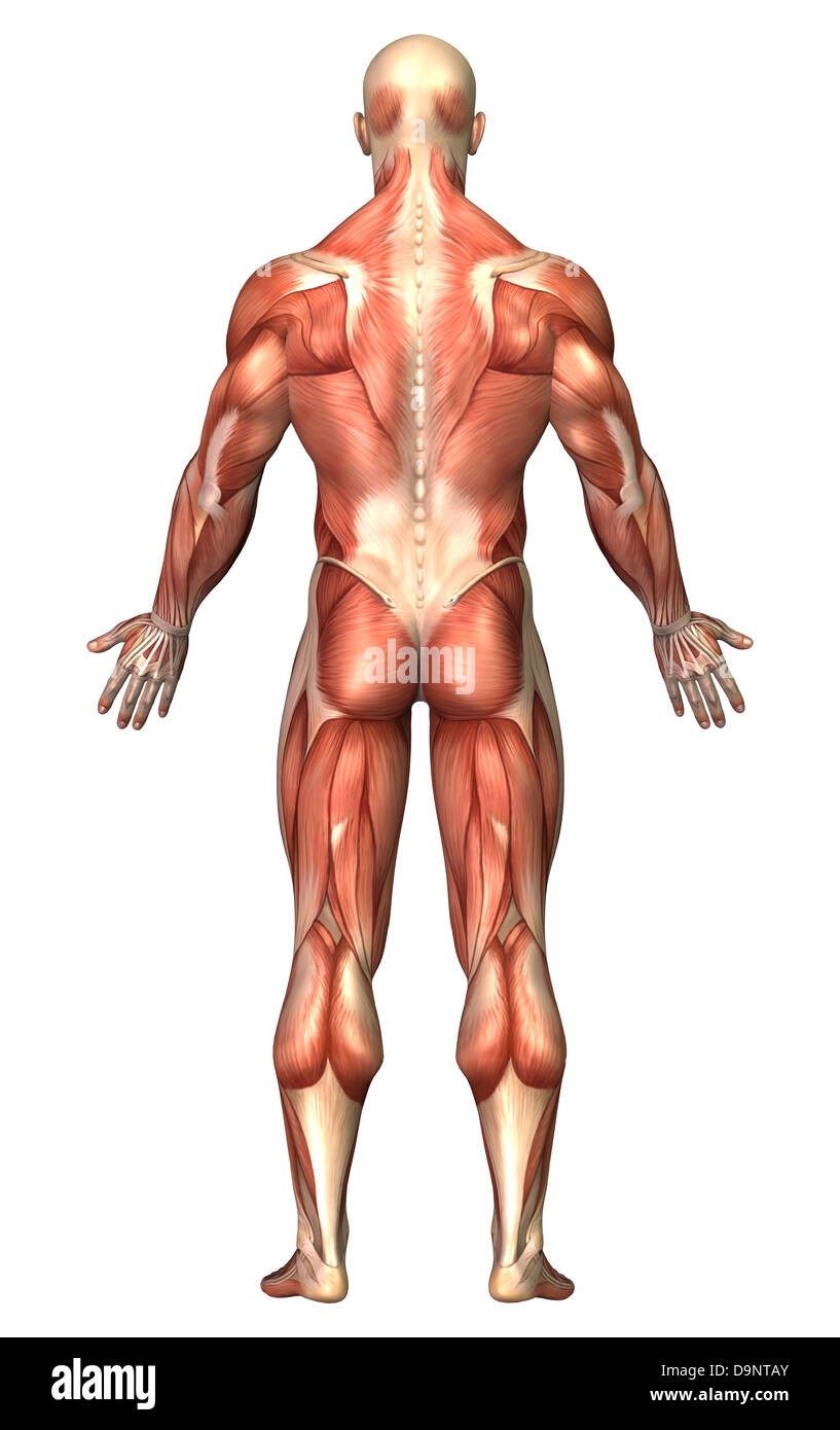 Anatomy of male muscular system, back view. Stock Photohttps://www.alamy.com/image-license-details/?v=1https://www.alamy.com/stock-photo-anatomy-of-male-muscular-system-back-view-57643123.html
RFD9NTAY–Anatomy of male muscular system, back view.
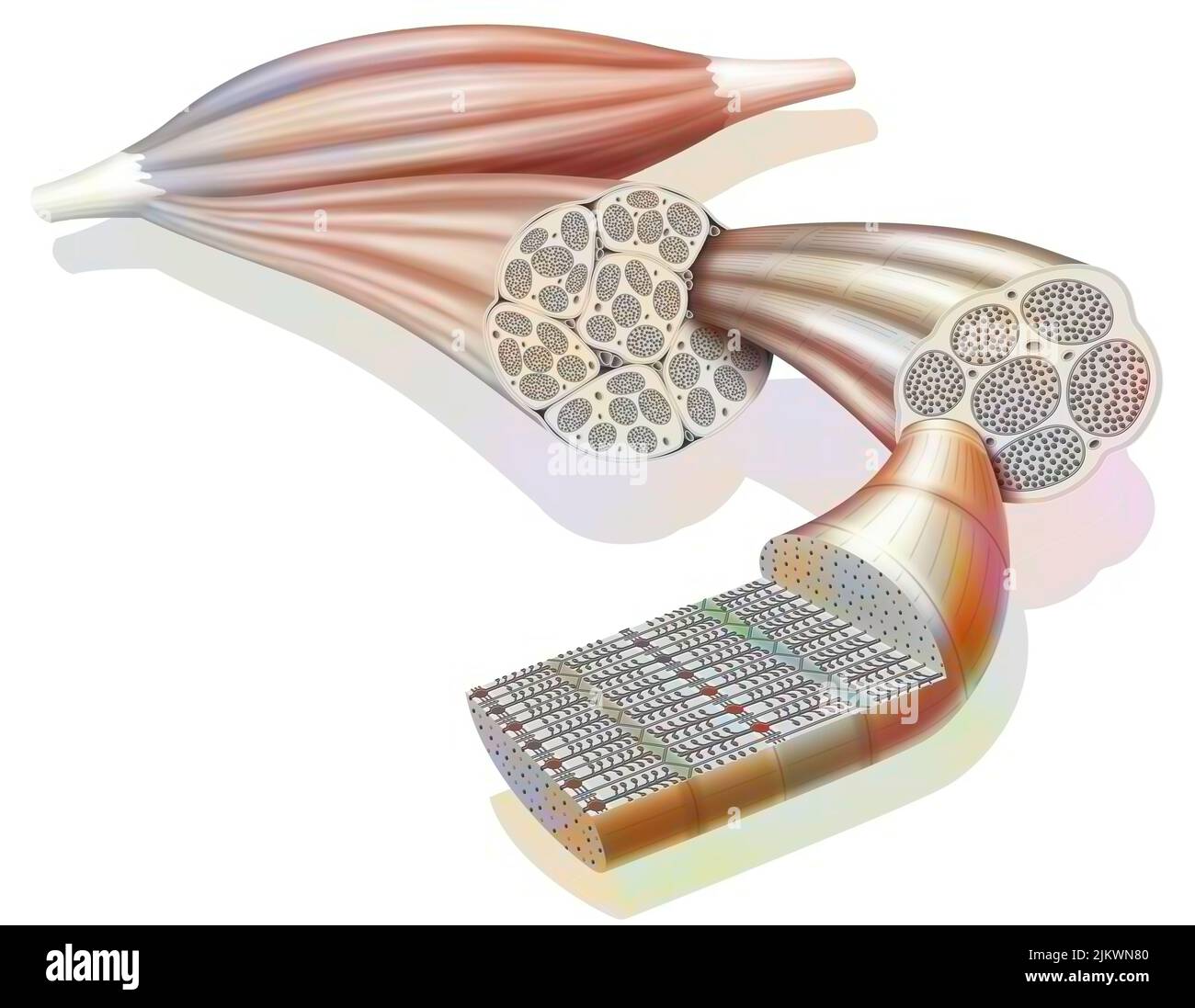 From muscle to muscle fiber: tendon, muscle, muscle fiber. Stock Photohttps://www.alamy.com/image-license-details/?v=1https://www.alamy.com/from-muscle-to-muscle-fiber-tendon-muscle-muscle-fiber-image476923888.html
RF2JKWN80–From muscle to muscle fiber: tendon, muscle, muscle fiber.
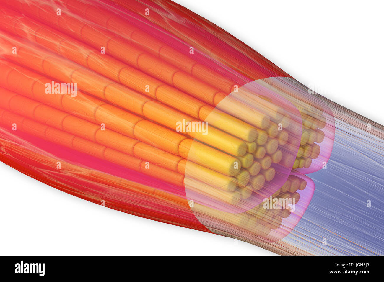 Skeletal muscle, illustration. The muscle is surrounded by epimysium connective tissue (transparent), which forms the tendon that attaches the muscle to bone. Bundles of muscle fibres (yellow) are surrounded by perimysium connective tissue (pink). Stock Photohttps://www.alamy.com/image-license-details/?v=1https://www.alamy.com/stock-photo-skeletal-muscle-illustration-the-muscle-is-surrounded-by-epimysium-147983643.html
RFJGN6J3–Skeletal muscle, illustration. The muscle is surrounded by epimysium connective tissue (transparent), which forms the tendon that attaches the muscle to bone. Bundles of muscle fibres (yellow) are surrounded by perimysium connective tissue (pink).
 Tendons on the Top of the Right Foot, vintage line drawing or engraving illustration. Stock Vectorhttps://www.alamy.com/image-license-details/?v=1https://www.alamy.com/tendons-on-the-top-of-the-right-foot-vintage-line-drawing-or-engraving-illustration-image348632545.html
RF2B75GA9–Tendons on the Top of the Right Foot, vintage line drawing or engraving illustration.
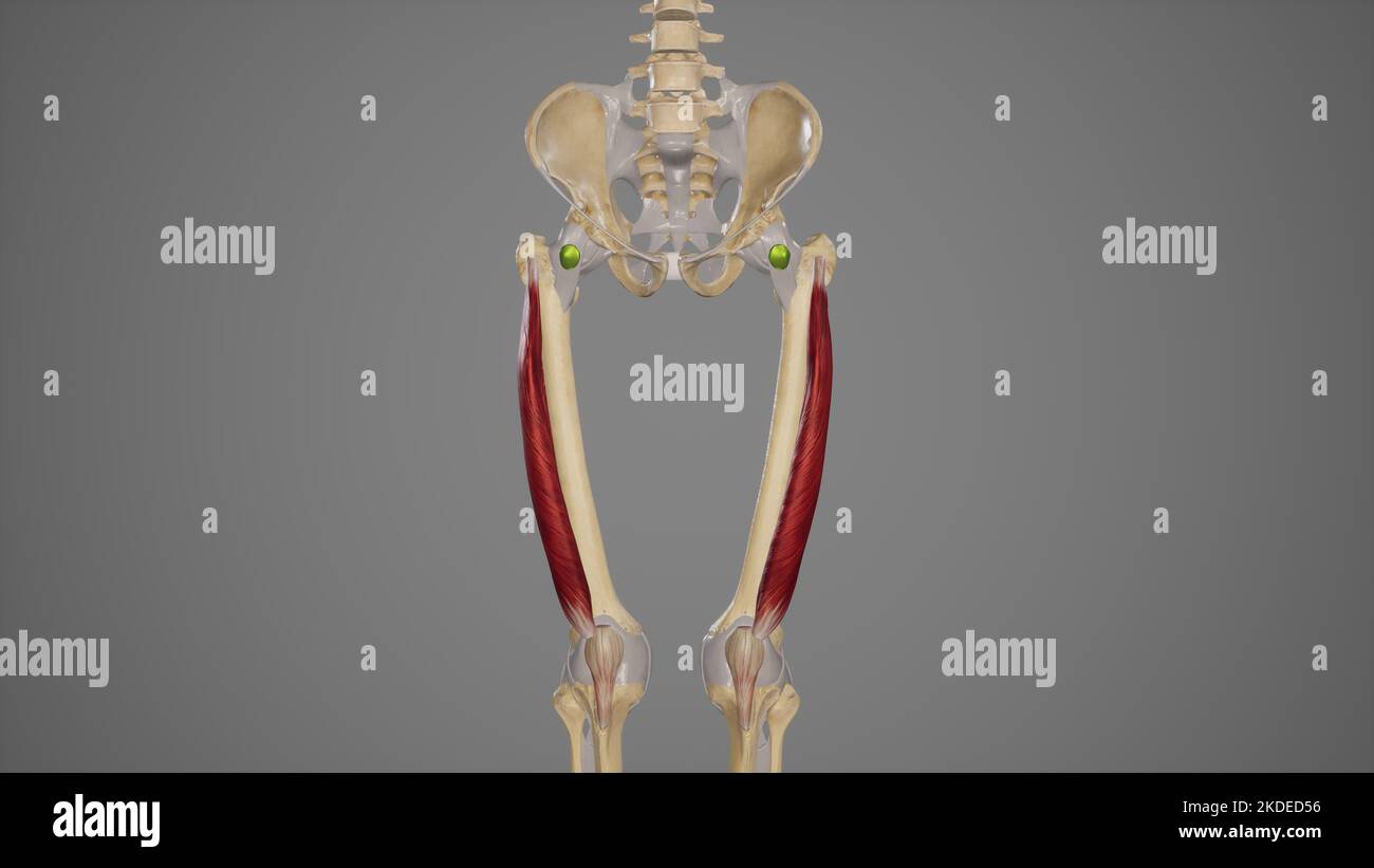 Medical Illustration of Vastus Lateralis Muscle Stock Photohttps://www.alamy.com/image-license-details/?v=1https://www.alamy.com/medical-illustration-of-vastus-lateralis-muscle-image490198498.html
RF2KDED56–Medical Illustration of Vastus Lateralis Muscle
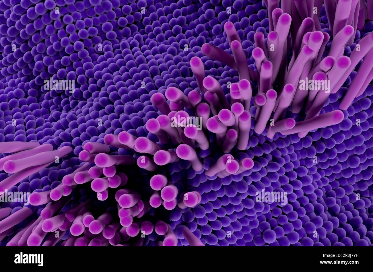 Tendon structure in the human body - 3d illustration top view Stock Photohttps://www.alamy.com/image-license-details/?v=1https://www.alamy.com/tendon-structure-in-the-human-body-3d-illustration-top-view-image552977141.html
RF2R3J7YH–Tendon structure in the human body - 3d illustration top view
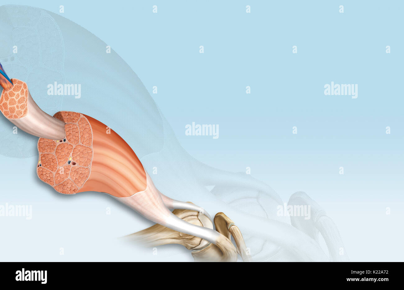 This image shows the structure of a skeletal muscle, revealing the muscle fibers bundle, the motor neuron, the muscle fiber and the myofibril. Stock Photohttps://www.alamy.com/image-license-details/?v=1https://www.alamy.com/this-image-shows-the-structure-of-a-skeletal-muscle-revealing-the-image156174566.html
RMK22A72–This image shows the structure of a skeletal muscle, revealing the muscle fibers bundle, the motor neuron, the muscle fiber and the myofibril.
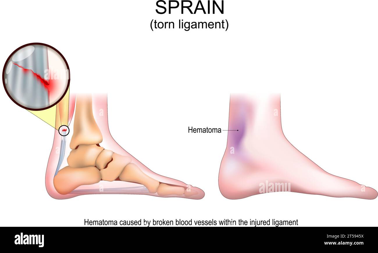 Sprain. Torn ligament after Painful foot twist. Signs and symptoms of a Soft tissue injury. Muscle damage, tendon tear, ligament problem description. Stock Vectorhttps://www.alamy.com/image-license-details/?v=1https://www.alamy.com/sprain-torn-ligament-after-painful-foot-twist-signs-and-symptoms-of-a-soft-tissue-injury-muscle-damage-tendon-tear-ligament-problem-description-image571216294.html
RF2T5945X–Sprain. Torn ligament after Painful foot twist. Signs and symptoms of a Soft tissue injury. Muscle damage, tendon tear, ligament problem description.
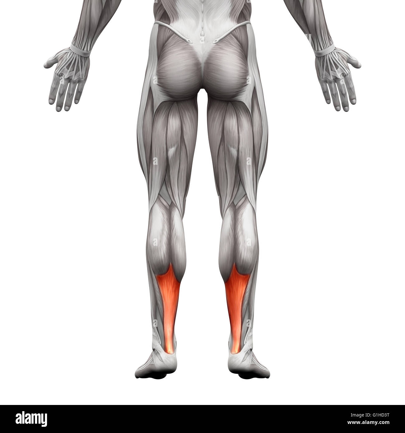 Achilles Tendon - Anatomy Muscle - isolated on white - 3D illustration Stock Photohttps://www.alamy.com/image-license-details/?v=1https://www.alamy.com/stock-photo-achilles-tendon-anatomy-muscle-isolated-on-white-3d-illustration-104260348.html
RFG1HD3T–Achilles Tendon - Anatomy Muscle - isolated on white - 3D illustration
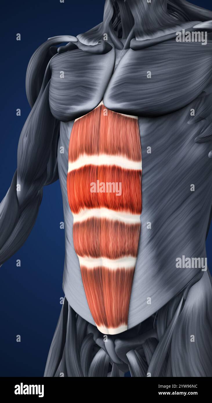 Anatomy of Highlighted Abdominal Muscle Sections Stock Photohttps://www.alamy.com/image-license-details/?v=1https://www.alamy.com/anatomy-of-highlighted-abdominal-muscle-sections-image635142520.html
RF2YW96NC–Anatomy of Highlighted Abdominal Muscle Sections
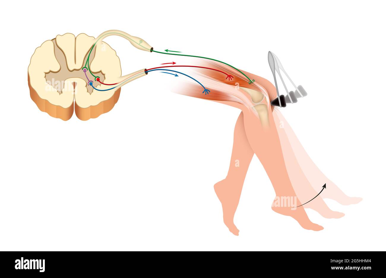 Knee jerk or patellar tendon reflex Stock Photohttps://www.alamy.com/image-license-details/?v=1https://www.alamy.com/knee-jerk-or-patellar-tendon-reflex-image433719556.html
RF2G5HHM4–Knee jerk or patellar tendon reflex
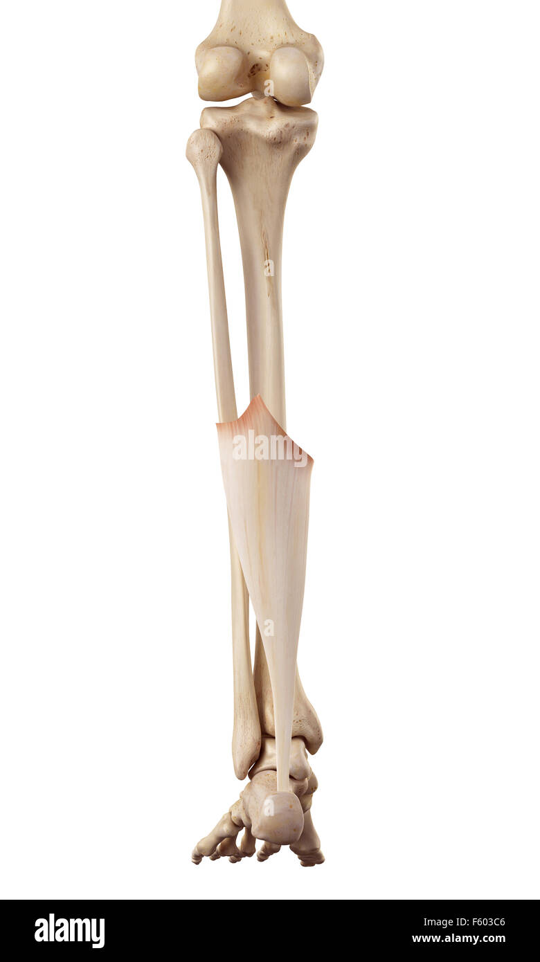 medical accurate illustration of the achilles tendon Stock Photohttps://www.alamy.com/image-license-details/?v=1https://www.alamy.com/stock-photo-medical-accurate-illustration-of-the-achilles-tendon-89742470.html
RFF603C6–medical accurate illustration of the achilles tendon
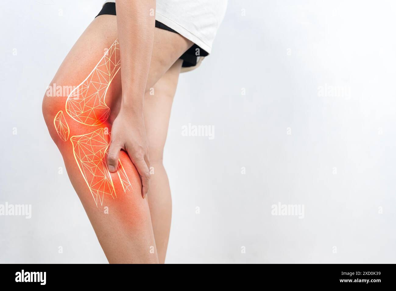 Elderly woman suffering from pain in knee. Tendon problems and Joint inflammation on white background. Stock Photohttps://www.alamy.com/image-license-details/?v=1https://www.alamy.com/elderly-woman-suffering-from-pain-in-knee-tendon-problems-and-joint-inflammation-on-white-background-image610368397.html
RF2XD0K39–Elderly woman suffering from pain in knee. Tendon problems and Joint inflammation on white background.
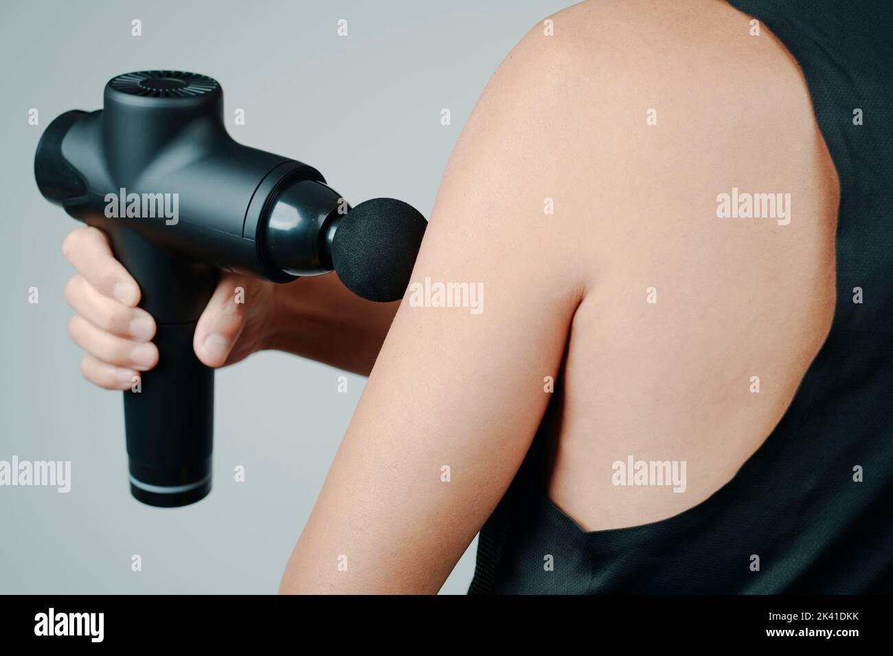 closeup of a young caucasian man, wearing sports clothes, using a massage gun to massage the muscles of his arm Stock Photohttps://www.alamy.com/image-license-details/?v=1https://www.alamy.com/closeup-of-a-young-caucasian-man-wearing-sports-clothes-using-a-massage-gun-to-massage-the-muscles-of-his-arm-image484381623.html
RF2K41DKK–closeup of a young caucasian man, wearing sports clothes, using a massage gun to massage the muscles of his arm
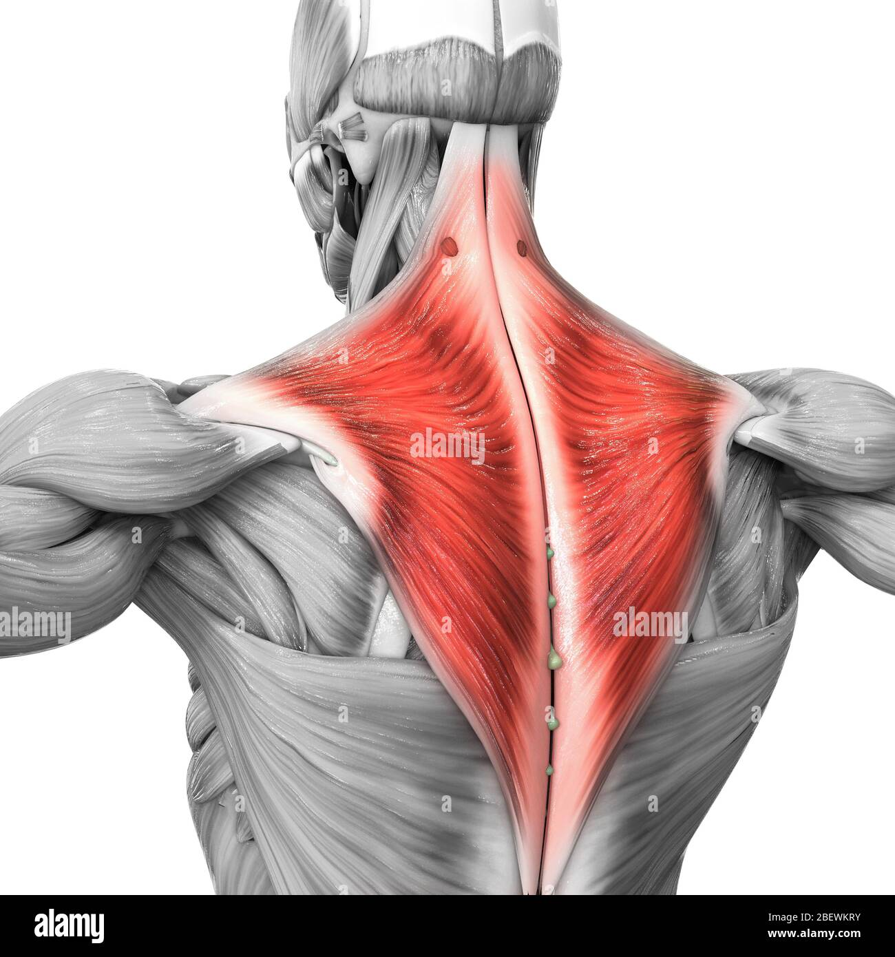 Human Muscular System Parts Trapezius Muscle Anatomy Stock Photohttps://www.alamy.com/image-license-details/?v=1https://www.alamy.com/human-muscular-system-parts-trapezius-muscle-anatomy-image353376911.html
RF2BEWKRY–Human Muscular System Parts Trapezius Muscle Anatomy
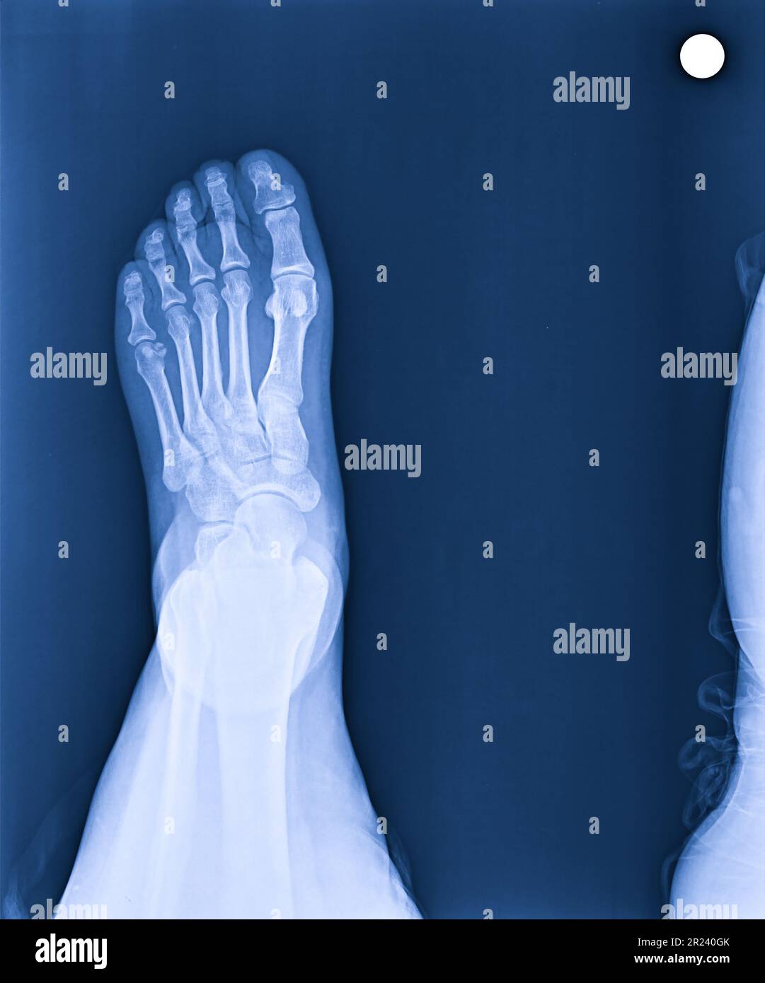 X-ray normal human's foot lateral Stock Photohttps://www.alamy.com/image-license-details/?v=1https://www.alamy.com/x-ray-normal-humans-foot-lateral-image552049363.html
RF2R240GK–X-ray normal human's foot lateral
 Young asian Orthopedic Surgeons in white gown and stethoscope asking for the professor advice about bone, joint, tendon and muscle. Working atmosphere in hospital. Stock Photohttps://www.alamy.com/image-license-details/?v=1https://www.alamy.com/young-asian-orthopedic-surgeons-in-white-gown-and-stethoscope-asking-for-the-professor-advice-about-bone-joint-tendon-and-muscle-working-atmosphere-in-hospital-image547835502.html
RF2PR81NJ–Young asian Orthopedic Surgeons in white gown and stethoscope asking for the professor advice about bone, joint, tendon and muscle. Working atmosphere in hospital.
 Achilles tendon 02 Stock Photohttps://www.alamy.com/image-license-details/?v=1https://www.alamy.com/achilles-tendon-02-image155513277.html
RMK106NH–Achilles tendon 02
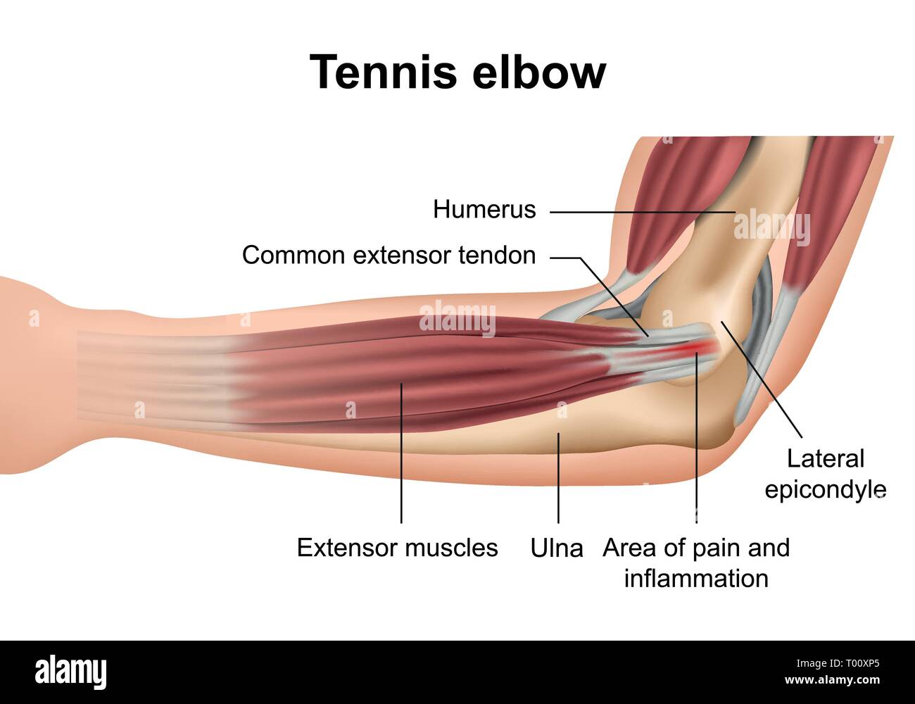 Tennis elbow injury medical vector illustration on white background Stock Vectorhttps://www.alamy.com/image-license-details/?v=1https://www.alamy.com/tennis-elbow-injury-medical-vector-illustration-on-white-background-image240966157.html
RFT00XP5–Tennis elbow injury medical vector illustration on white background
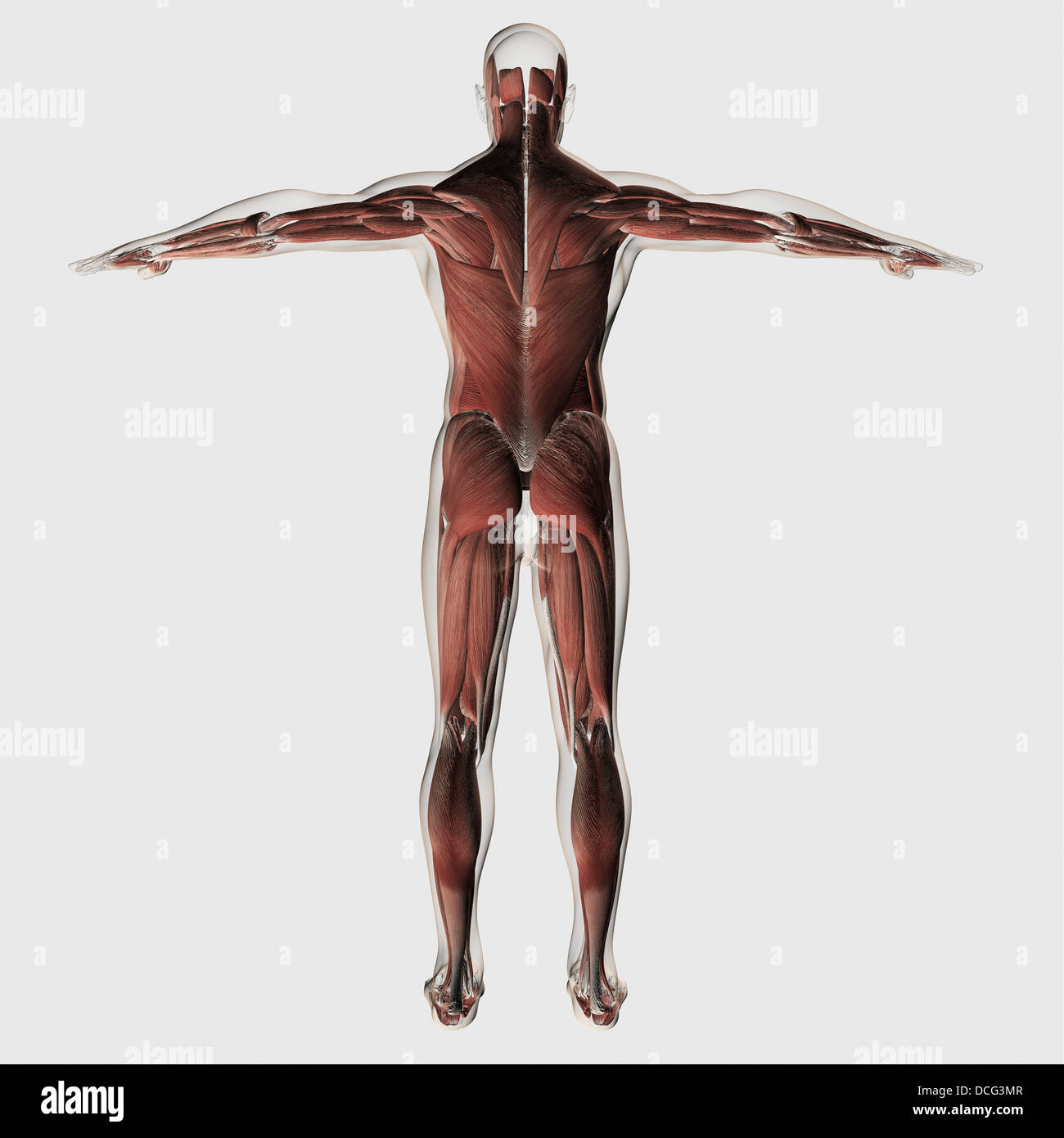 Anatomy of male muscular system, posterior view. Stock Photohttps://www.alamy.com/image-license-details/?v=1https://www.alamy.com/stock-photo-anatomy-of-male-muscular-system-posterior-view-59361143.html
RFDCG3MR–Anatomy of male muscular system, posterior view.
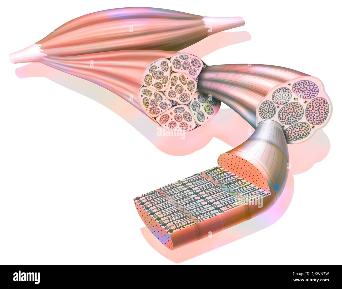 From muscle to muscle fiber: tendon, muscle, muscle fiber. Stock Photohttps://www.alamy.com/image-license-details/?v=1https://www.alamy.com/from-muscle-to-muscle-fiber-tendon-muscle-muscle-fiber-image476923885.html
RF2JKWN7W–From muscle to muscle fiber: tendon, muscle, muscle fiber.
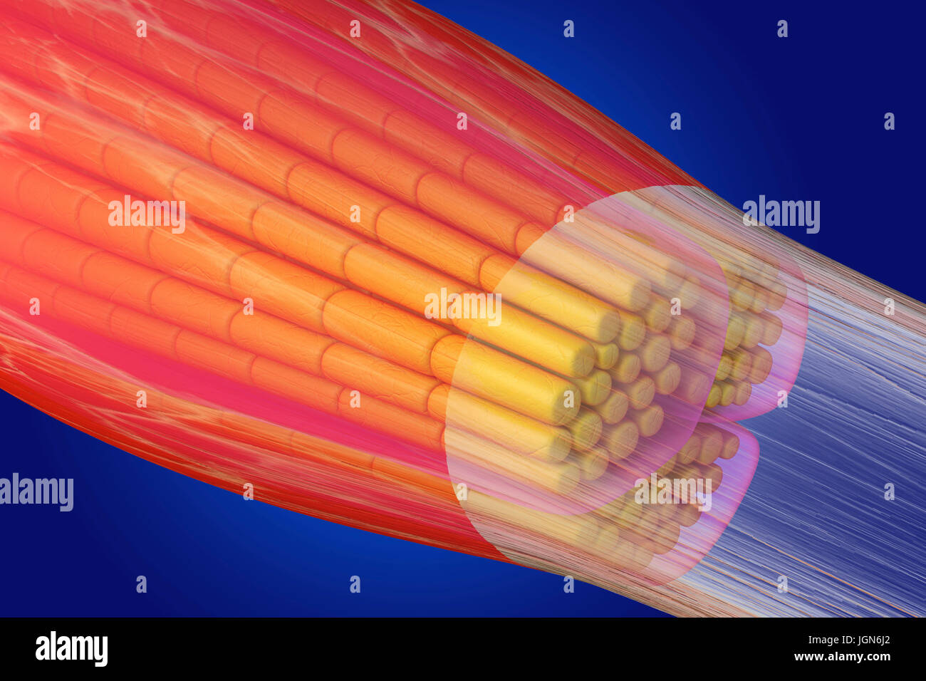 Skeletal muscle, illustration. The muscle is surrounded by epimysium connective tissue (transparent), which forms the tendon that attaches the muscle to bone. Bundles of muscle fibres (yellow) are surrounded by perimysium connective tissue (pink). Stock Photohttps://www.alamy.com/image-license-details/?v=1https://www.alamy.com/stock-photo-skeletal-muscle-illustration-the-muscle-is-surrounded-by-epimysium-147983642.html
RFJGN6J2–Skeletal muscle, illustration. The muscle is surrounded by epimysium connective tissue (transparent), which forms the tendon that attaches the muscle to bone. Bundles of muscle fibres (yellow) are surrounded by perimysium connective tissue (pink).
 The thigh has three sets of strong muscles. Shown here is some of the human larger muscles on the back of the thigh. Powerful tendons at the hip and o Stock Vectorhttps://www.alamy.com/image-license-details/?v=1https://www.alamy.com/the-thigh-has-three-sets-of-strong-muscles-shown-here-is-some-of-the-human-larger-muscles-on-the-back-of-the-thigh-powerful-tendons-at-the-hip-and-o-image367217623.html
RF2C9C5R3–The thigh has three sets of strong muscles. Shown here is some of the human larger muscles on the back of the thigh. Powerful tendons at the hip and o
 Medical Illustration of Plantaris Muscle Stock Photohttps://www.alamy.com/image-license-details/?v=1https://www.alamy.com/medical-illustration-of-plantaris-muscle-image490198380.html
RF2KDED10–Medical Illustration of Plantaris Muscle
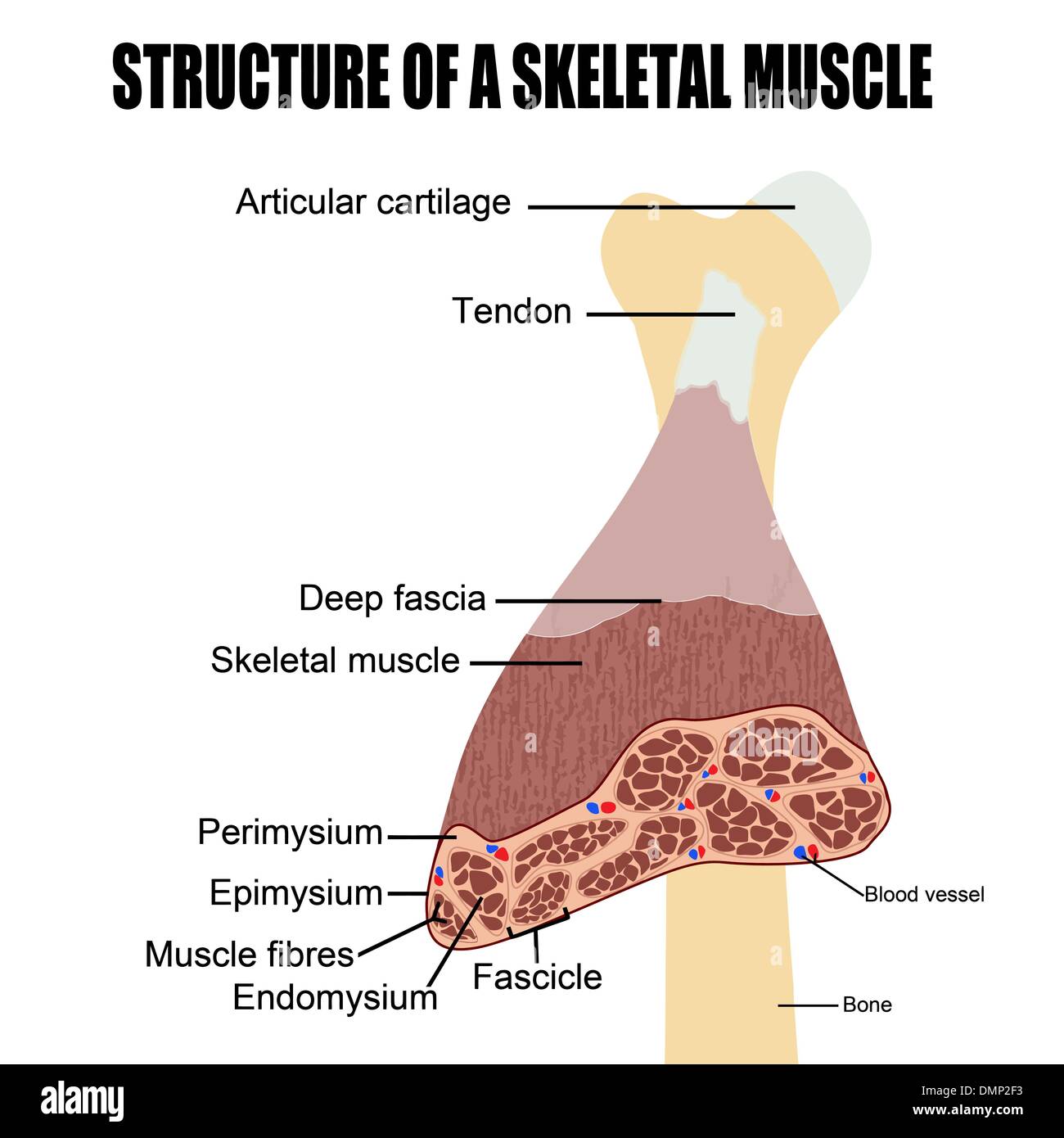 Structure of a skeletal muscle Stock Vectorhttps://www.alamy.com/image-license-details/?v=1https://www.alamy.com/structure-of-a-skeletal-muscle-image64409159.html
RFDMP2F3–Structure of a skeletal muscle
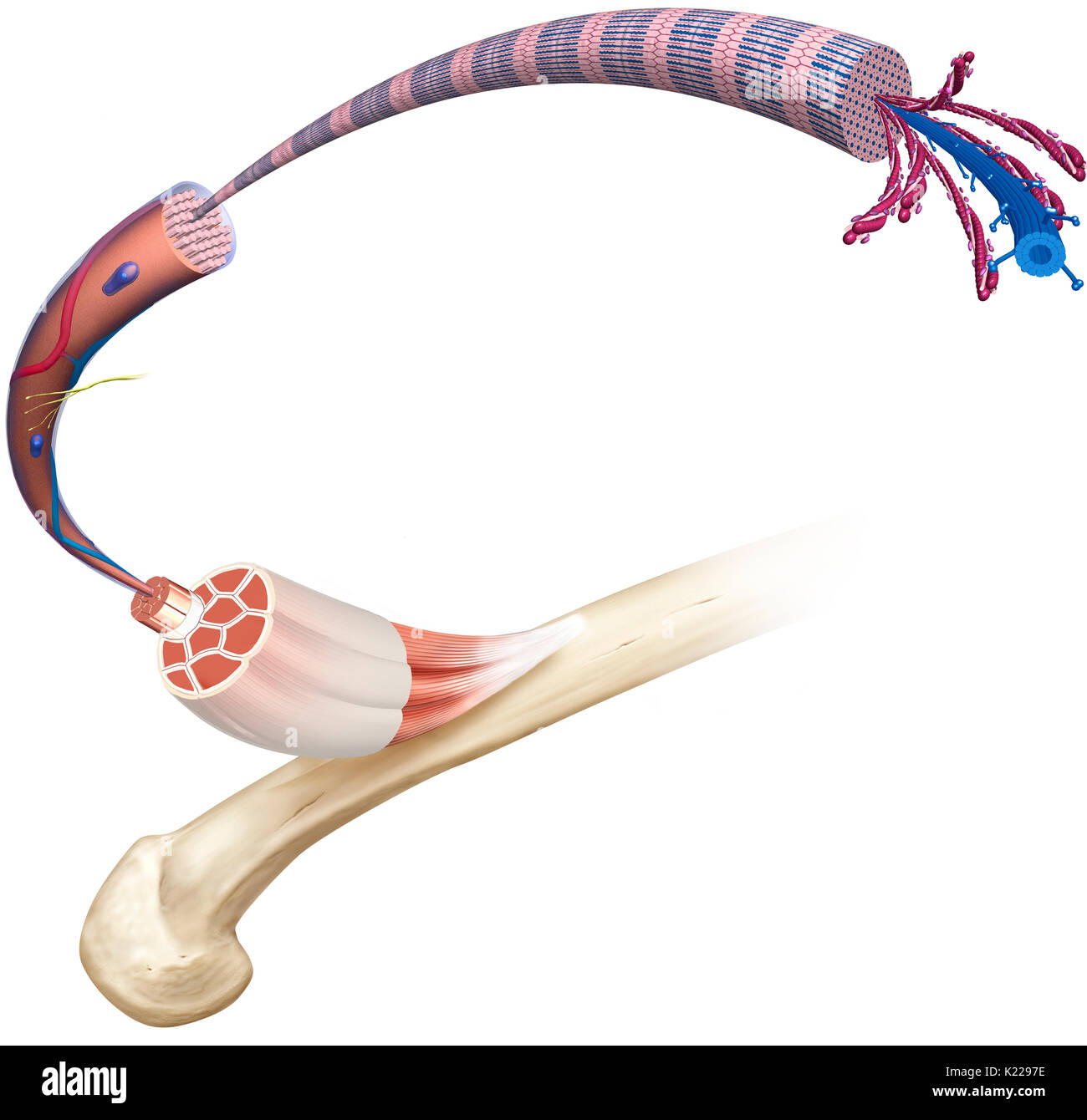 This image shows the structure of a skeletal muscle, revealing the muscle fibers bundle, the motor neuron, the muscle fiber and the myofibril. Stock Photohttps://www.alamy.com/image-license-details/?v=1https://www.alamy.com/this-image-shows-the-structure-of-a-skeletal-muscle-revealing-the-image156173794.html
RMK2297E–This image shows the structure of a skeletal muscle, revealing the muscle fibers bundle, the motor neuron, the muscle fiber and the myofibril.
 Calf pain. Calf muscle anatomy. Muscular structure of a human lerg. Medical infographic. Vector illustration Stock Vectorhttps://www.alamy.com/image-license-details/?v=1https://www.alamy.com/calf-pain-calf-muscle-anatomy-muscular-structure-of-a-human-lerg-medical-infographic-vector-illustration-image569293247.html
RF2T25F9K–Calf pain. Calf muscle anatomy. Muscular structure of a human lerg. Medical infographic. Vector illustration
 Elbow joint pain symptom. Woman holding sore arm. Inflammation or injury concept. Muscle or tendon strain. Medical problem with upper limb. Stock Photohttps://www.alamy.com/image-license-details/?v=1https://www.alamy.com/elbow-joint-pain-symptom-woman-holding-sore-arm-inflammation-or-injury-concept-muscle-or-tendon-strain-medical-problem-with-upper-limb-image677324051.html
RF3B9XNMK–Elbow joint pain symptom. Woman holding sore arm. Inflammation or injury concept. Muscle or tendon strain. Medical problem with upper limb.
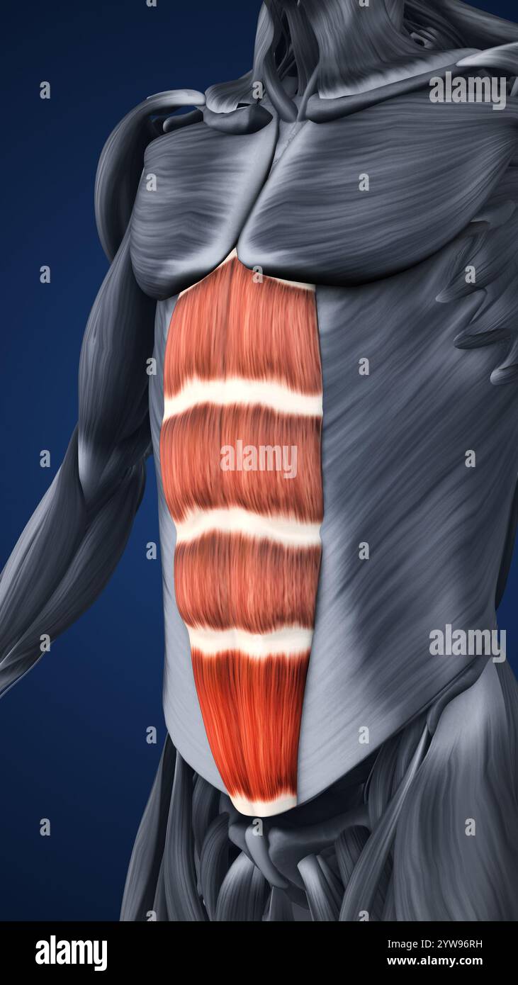 Anatomy of Highlighted Abdominal Muscle Sections Stock Photohttps://www.alamy.com/image-license-details/?v=1https://www.alamy.com/anatomy-of-highlighted-abdominal-muscle-sections-image635142581.html
RF2YW96RH–Anatomy of Highlighted Abdominal Muscle Sections
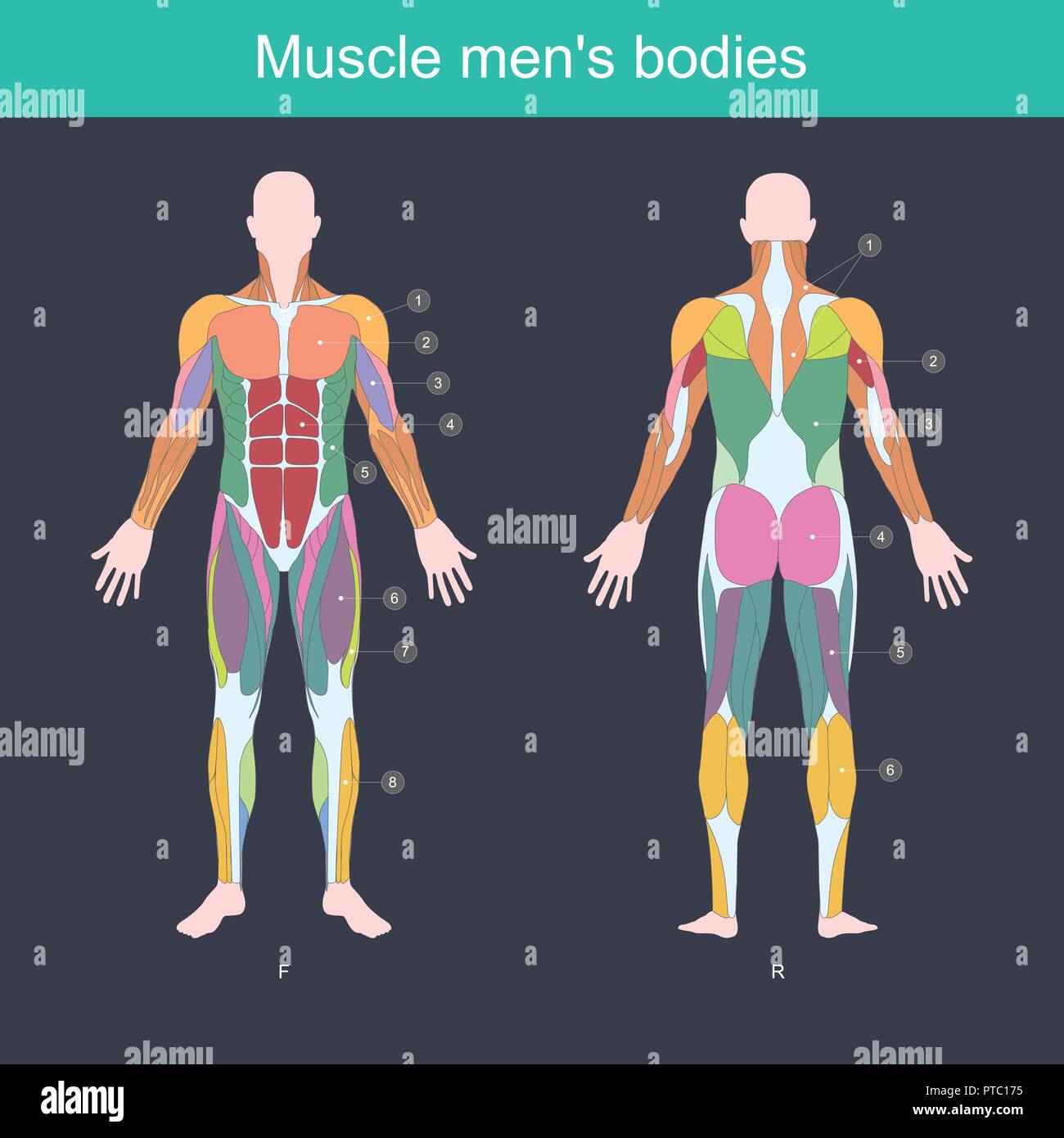 Muscle is the part of the body that exerts, And control the movement of the internal organs. Illustration front and back side. Stock Vectorhttps://www.alamy.com/image-license-details/?v=1https://www.alamy.com/muscle-is-the-part-of-the-body-that-exerts-and-control-the-movement-of-the-internal-organs-illustration-front-and-back-side-image221540569.html
RFPTC175–Muscle is the part of the body that exerts, And control the movement of the internal organs. Illustration front and back side.
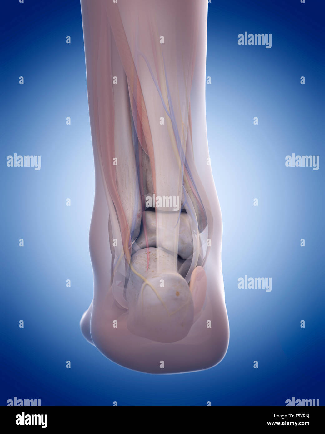 medically accurate illustration of the achilles tendon Stock Photohttps://www.alamy.com/image-license-details/?v=1https://www.alamy.com/stock-photo-medically-accurate-illustration-of-the-achilles-tendon-89736042.html
RFF5YR6J–medically accurate illustration of the achilles tendon
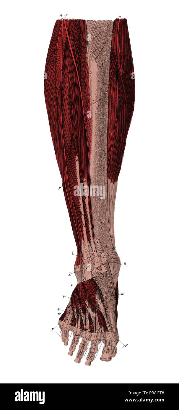 19th century illustration of right lower leg from the front to the distance of the top muscle layer. Published in Systematischer Bilder-Atlas zum Conv Stock Photohttps://www.alamy.com/image-license-details/?v=1https://www.alamy.com/19th-century-illustration-of-right-lower-leg-from-the-front-to-the-distance-of-the-top-muscle-layer-published-in-systematischer-bilder-atlas-zum-conv-image220850344.html
RFPR8GT8–19th century illustration of right lower leg from the front to the distance of the top muscle layer. Published in Systematischer Bilder-Atlas zum Conv
 a young caucasian man is using a massage gun to massage the muscles of his arm next to his elbow, sitting on a couch Stock Photohttps://www.alamy.com/image-license-details/?v=1https://www.alamy.com/a-young-caucasian-man-is-using-a-massage-gun-to-massage-the-muscles-of-his-arm-next-to-his-elbow-sitting-on-a-couch-image484381666.html
RF2K41DN6–a young caucasian man is using a massage gun to massage the muscles of his arm next to his elbow, sitting on a couch
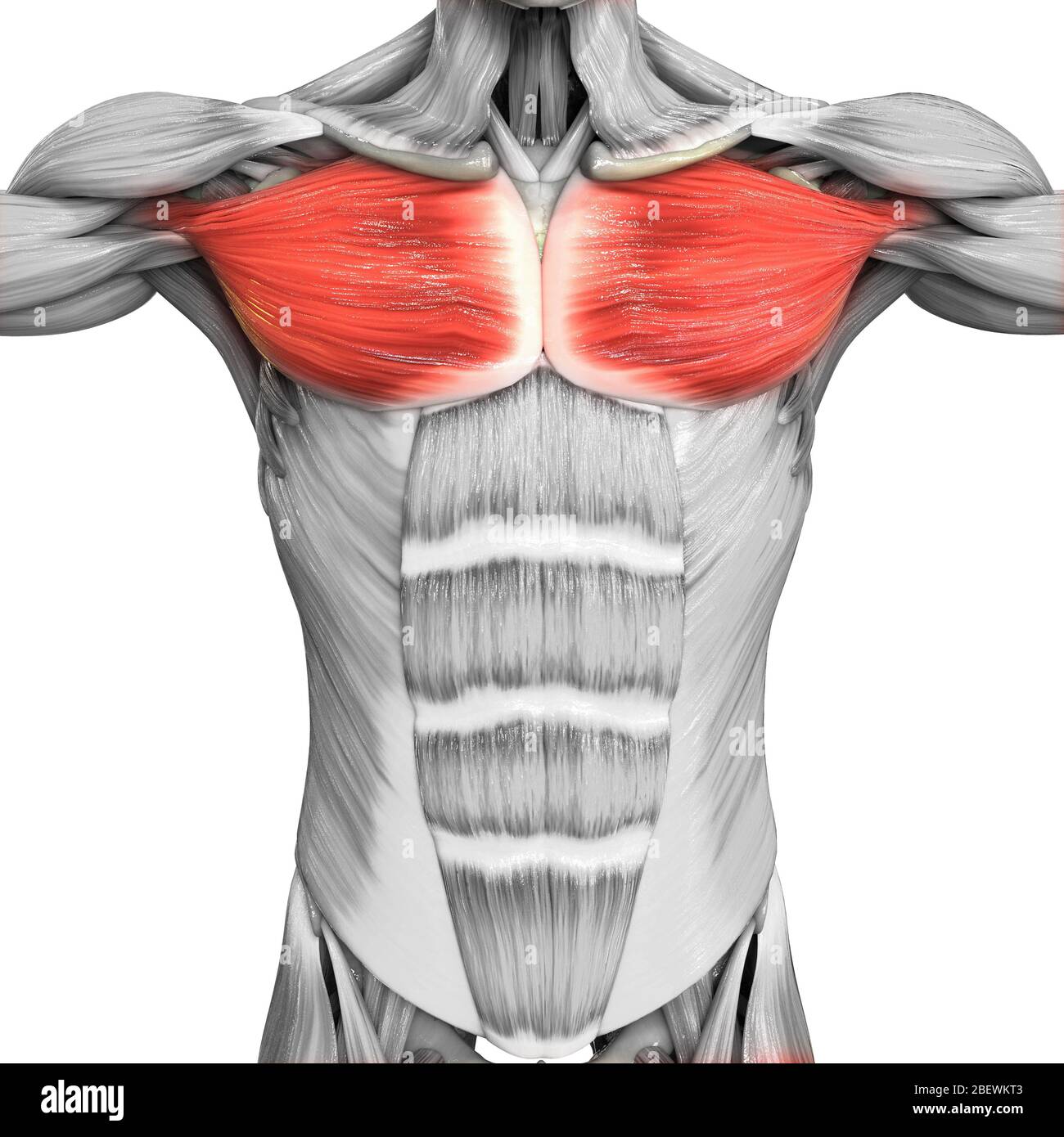 Human Muscular System Parts Pectoral Muscle Anatomy Stock Photohttps://www.alamy.com/image-license-details/?v=1https://www.alamy.com/human-muscular-system-parts-pectoral-muscle-anatomy-image353376915.html
RF2BEWKT3–Human Muscular System Parts Pectoral Muscle Anatomy
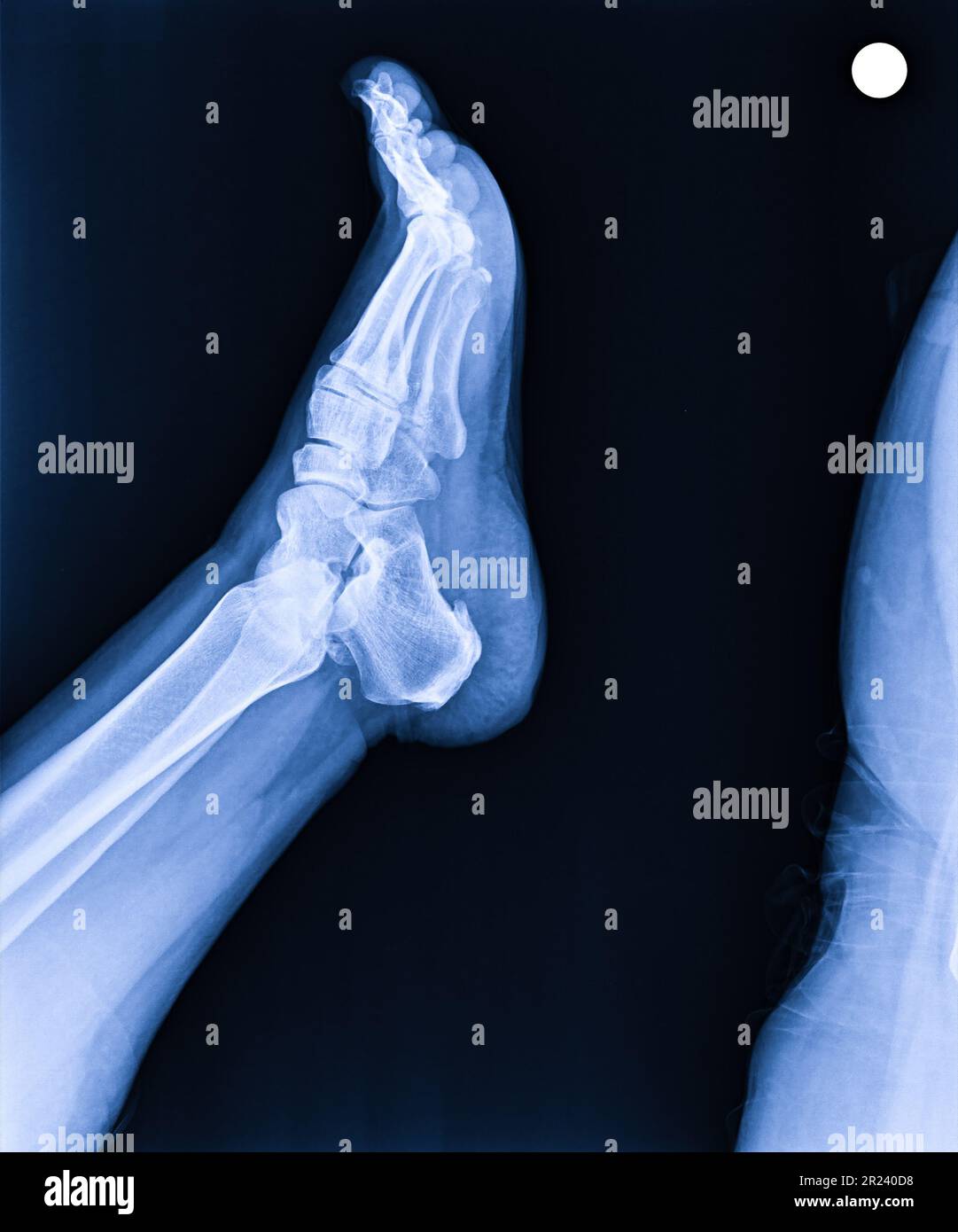 X-ray normal human's foot lateral Stock Photohttps://www.alamy.com/image-license-details/?v=1https://www.alamy.com/x-ray-normal-humans-foot-lateral-image552049268.html
RF2R240D8–X-ray normal human's foot lateral
 Back view of woman in white gown have webcam conference on tablet computer with young asian doctor in blue uniform about bone, joint, tendon and muscle. Stock Photohttps://www.alamy.com/image-license-details/?v=1https://www.alamy.com/back-view-of-woman-in-white-gown-have-webcam-conference-on-tablet-computer-with-young-asian-doctor-in-blue-uniform-about-bone-joint-tendon-and-muscle-image548178476.html
RF2PRRK6M–Back view of woman in white gown have webcam conference on tablet computer with young asian doctor in blue uniform about bone, joint, tendon and muscle.
 Achilles tendon 01 Stock Photohttps://www.alamy.com/image-license-details/?v=1https://www.alamy.com/achilles-tendon-01-image155513276.html
RMK106NG–Achilles tendon 01
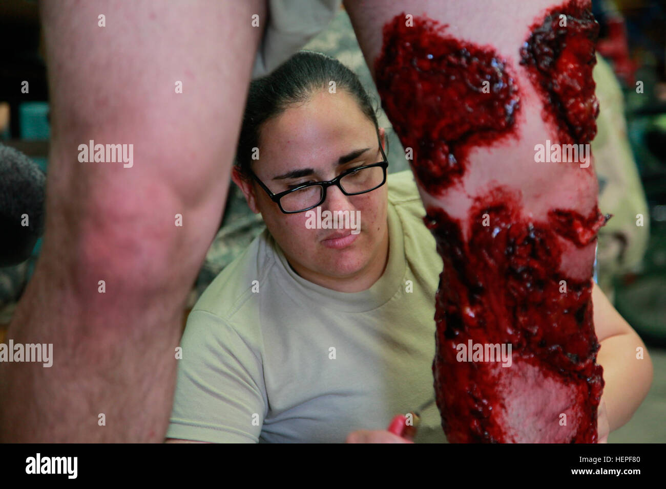 U.S. Army Reserve Spc. Brenda Pruitt, of 228th Combat Support Hospital, applies moulage to a soldier's leg to portray muscle tendon abrasions, during Global Medic at Fort McCoy, Wis., June 17, 2015. Specialist Pruitt training during Global medic has allowed her to create various trauma effects to mannequins and real simulated patients. Global Medic is the premier medical field training event in the Department of Defense and is the only joint accredited exercise conceived, planned and executed by Army Reserve Soldiers, who are part of the Medical Readiness and Training Command, Army Reserve Me Stock Photohttps://www.alamy.com/image-license-details/?v=1https://www.alamy.com/stock-photo-us-army-reserve-spc-brenda-pruitt-of-228th-combat-support-hospital-129572688.html
RMHEPF80–U.S. Army Reserve Spc. Brenda Pruitt, of 228th Combat Support Hospital, applies moulage to a soldier's leg to portray muscle tendon abrasions, during Global Medic at Fort McCoy, Wis., June 17, 2015. Specialist Pruitt training during Global medic has allowed her to create various trauma effects to mannequins and real simulated patients. Global Medic is the premier medical field training event in the Department of Defense and is the only joint accredited exercise conceived, planned and executed by Army Reserve Soldiers, who are part of the Medical Readiness and Training Command, Army Reserve Me
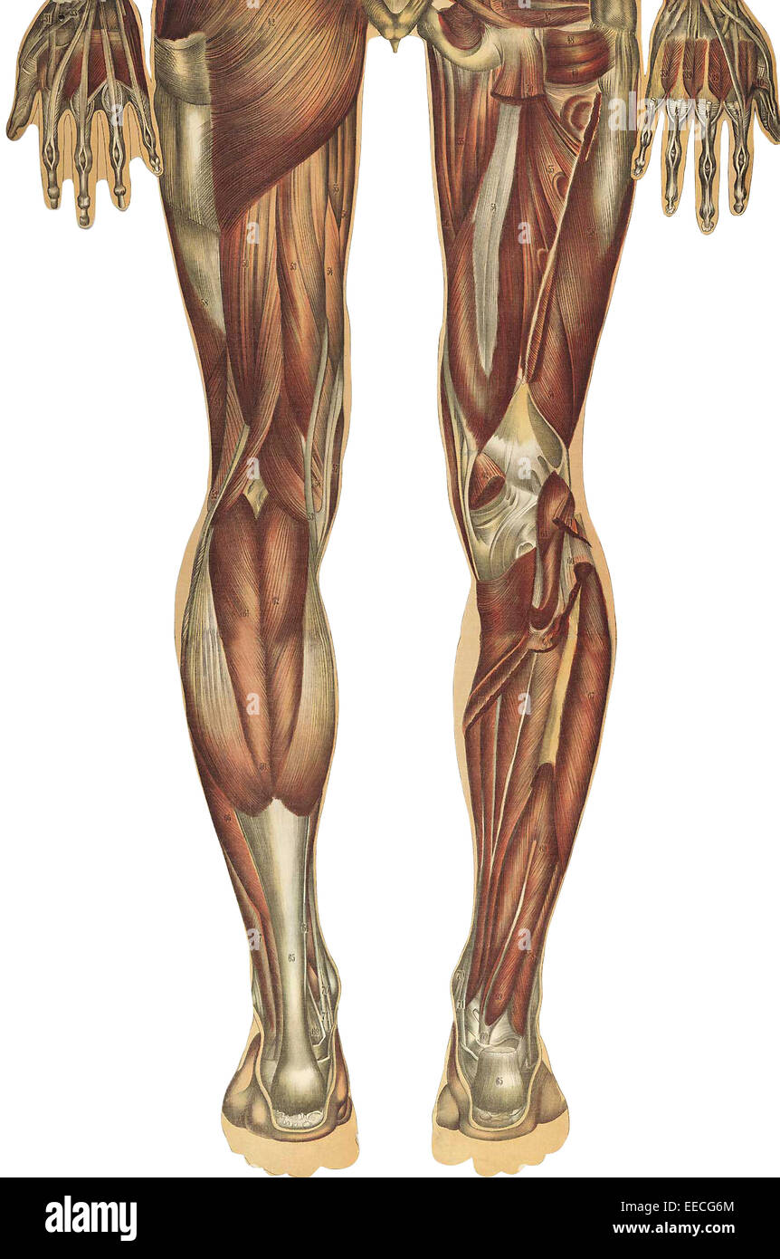 The human body with superimposed colored plates, by Julien Bougle, circa 1899. Stock Photohttps://www.alamy.com/image-license-details/?v=1https://www.alamy.com/stock-photo-the-human-body-with-superimposed-colored-plates-by-julien-bougle-circa-77722812.html
RFEECG6M–The human body with superimposed colored plates, by Julien Bougle, circa 1899.
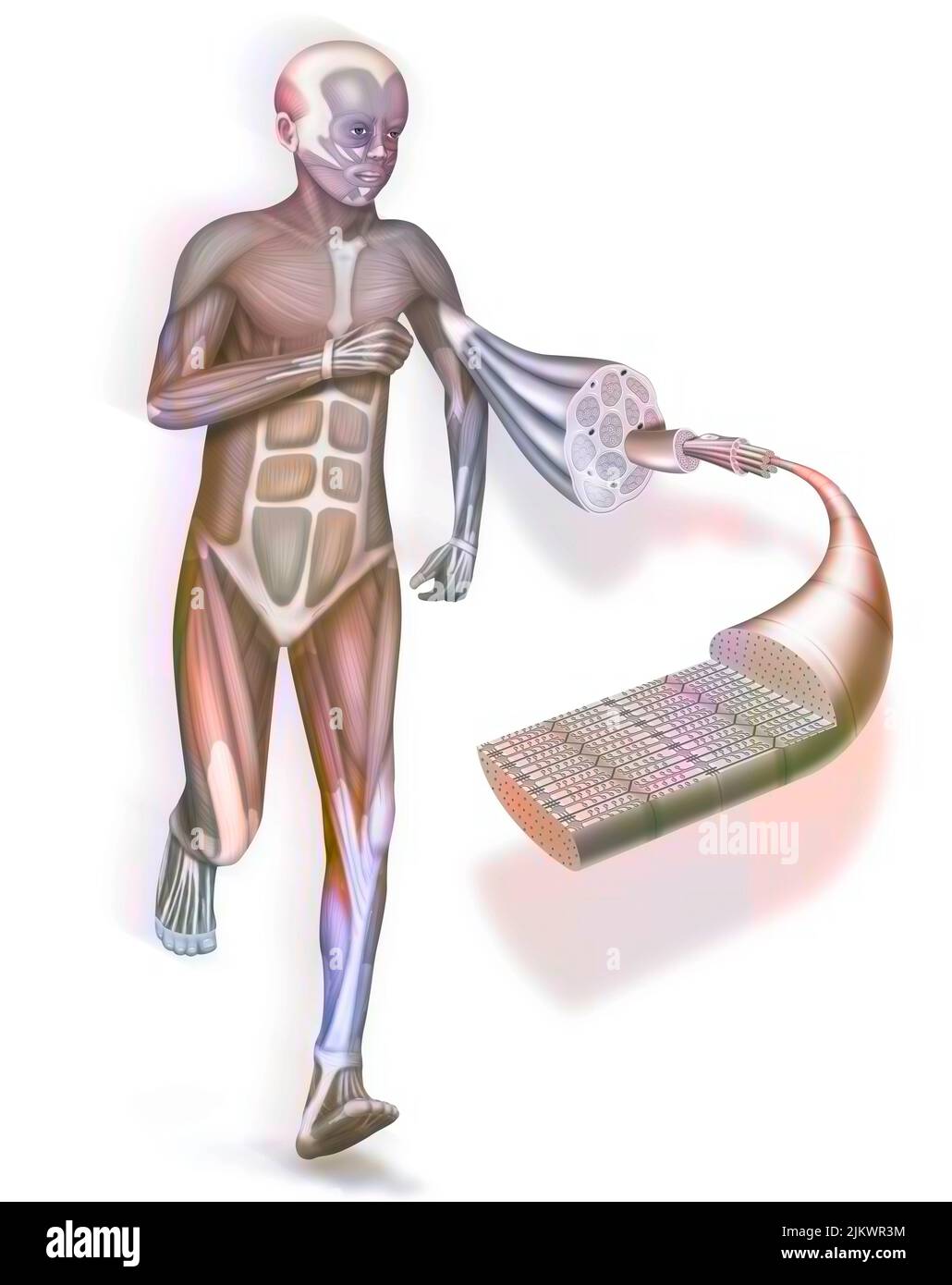 A cut away and zoom on a muscle and its structure: tendon, muscle. Stock Photohttps://www.alamy.com/image-license-details/?v=1https://www.alamy.com/a-cut-away-and-zoom-on-a-muscle-and-its-structure-tendon-muscle-image476925336.html
RF2JKWR3M–A cut away and zoom on a muscle and its structure: tendon, muscle.
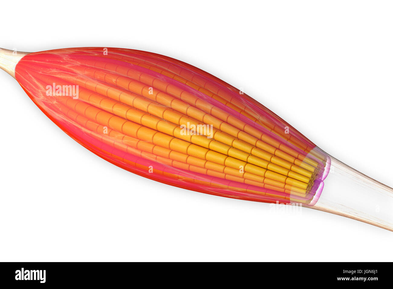 Skeletal muscle, illustration. The muscle is surrounded by epimysium connective tissue (transparent), which forms the tendon that attaches the muscle to bone. Bundles of muscle fibres (yellow) are surrounded by perimysium connective tissue (pink). Stock Photohttps://www.alamy.com/image-license-details/?v=1https://www.alamy.com/stock-photo-skeletal-muscle-illustration-the-muscle-is-surrounded-by-epimysium-147983641.html
RFJGN6J1–Skeletal muscle, illustration. The muscle is surrounded by epimysium connective tissue (transparent), which forms the tendon that attaches the muscle to bone. Bundles of muscle fibres (yellow) are surrounded by perimysium connective tissue (pink).
 The biceps muscles relaxed, '1 and 3', representing, two heads of the muscle; '2 and 3', representing, muscular portion and tendon fastening to the fo Stock Vectorhttps://www.alamy.com/image-license-details/?v=1https://www.alamy.com/the-biceps-muscles-relaxed-1-and-3-representing-two-heads-of-the-muscle-2-and-3-representing-muscular-portion-and-tendon-fastening-to-the-fo-image359344943.html
RF2BTHG3Y–The biceps muscles relaxed, '1 and 3', representing, two heads of the muscle; '2 and 3', representing, muscular portion and tendon fastening to the fo
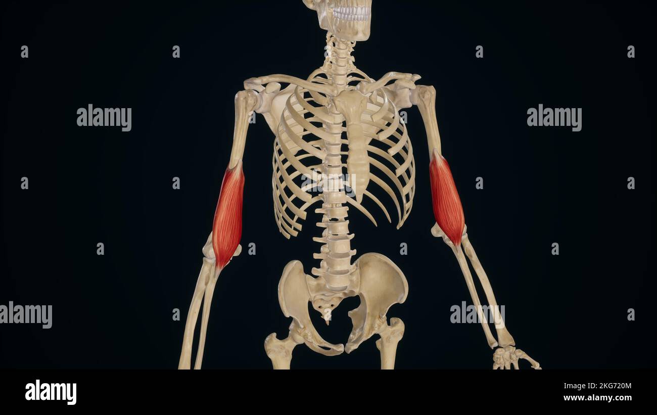 Brachialis Muscle Stock Photohttps://www.alamy.com/image-license-details/?v=1https://www.alamy.com/brachialis-muscle-image491880052.html
RF2KG720M–Brachialis Muscle
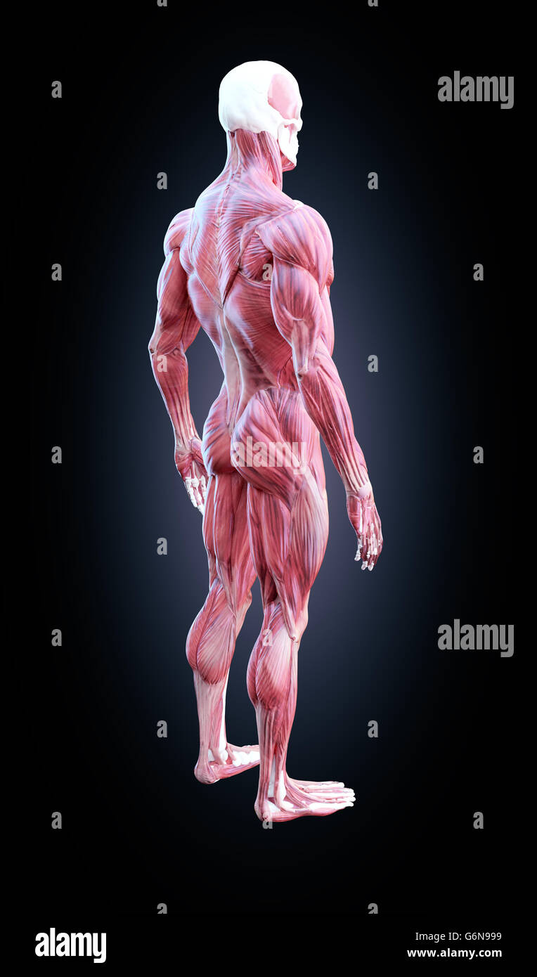 Detailed muscle human anatomy illustration Stock Photohttps://www.alamy.com/image-license-details/?v=1https://www.alamy.com/stock-photo-detailed-muscle-human-anatomy-illustration-107418453.html
RFG6N999–Detailed muscle human anatomy illustration
 This image shows the structure of a skeletal muscle, revealing the muscle fibers bundle, the motor neuron, the muscle fiber and the myofibril. Stock Photohttps://www.alamy.com/image-license-details/?v=1https://www.alamy.com/this-image-shows-the-structure-of-a-skeletal-muscle-revealing-the-image156174431.html
RMK22A27–This image shows the structure of a skeletal muscle, revealing the muscle fibers bundle, the motor neuron, the muscle fiber and the myofibril.
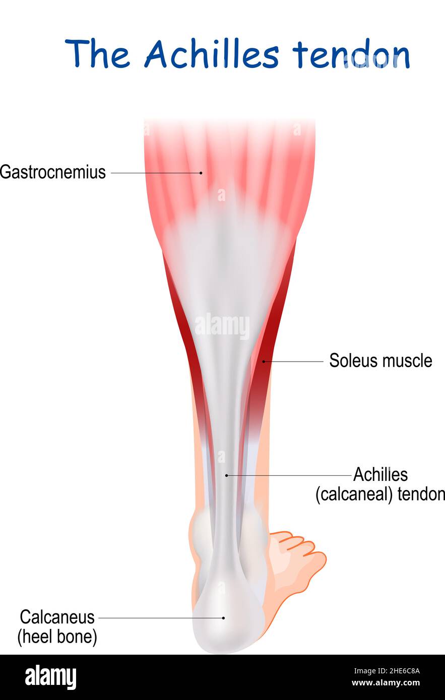 The Achilles tendon serves to attach the plantaris, gastrocnemius (calf) soleus muscles to the calcaneus (heel) bone. heel cord or calcaneal tendon Stock Vectorhttps://www.alamy.com/image-license-details/?v=1https://www.alamy.com/the-achilles-tendon-serves-to-attach-the-plantaris-gastrocnemius-calf-soleus-muscles-to-the-calcaneus-heel-bone-heel-cord-or-calcaneal-tendon-image456216106.html
RF2HE6C8A–The Achilles tendon serves to attach the plantaris, gastrocnemius (calf) soleus muscles to the calcaneus (heel) bone. heel cord or calcaneal tendon
 Hispanic indian business man muscle join neck pain from office syndrome after hard working day. Stock Photohttps://www.alamy.com/image-license-details/?v=1https://www.alamy.com/hispanic-indian-business-man-muscle-join-neck-pain-from-office-syndrome-after-hard-working-day-image627648342.html
RF2YD3RT6–Hispanic indian business man muscle join neck pain from office syndrome after hard working day.
 Anatomy of Highlighted Abdominal Muscle Sections Stock Photohttps://www.alamy.com/image-license-details/?v=1https://www.alamy.com/anatomy-of-highlighted-abdominal-muscle-sections-image635142514.html
RF2YW96N6–Anatomy of Highlighted Abdominal Muscle Sections
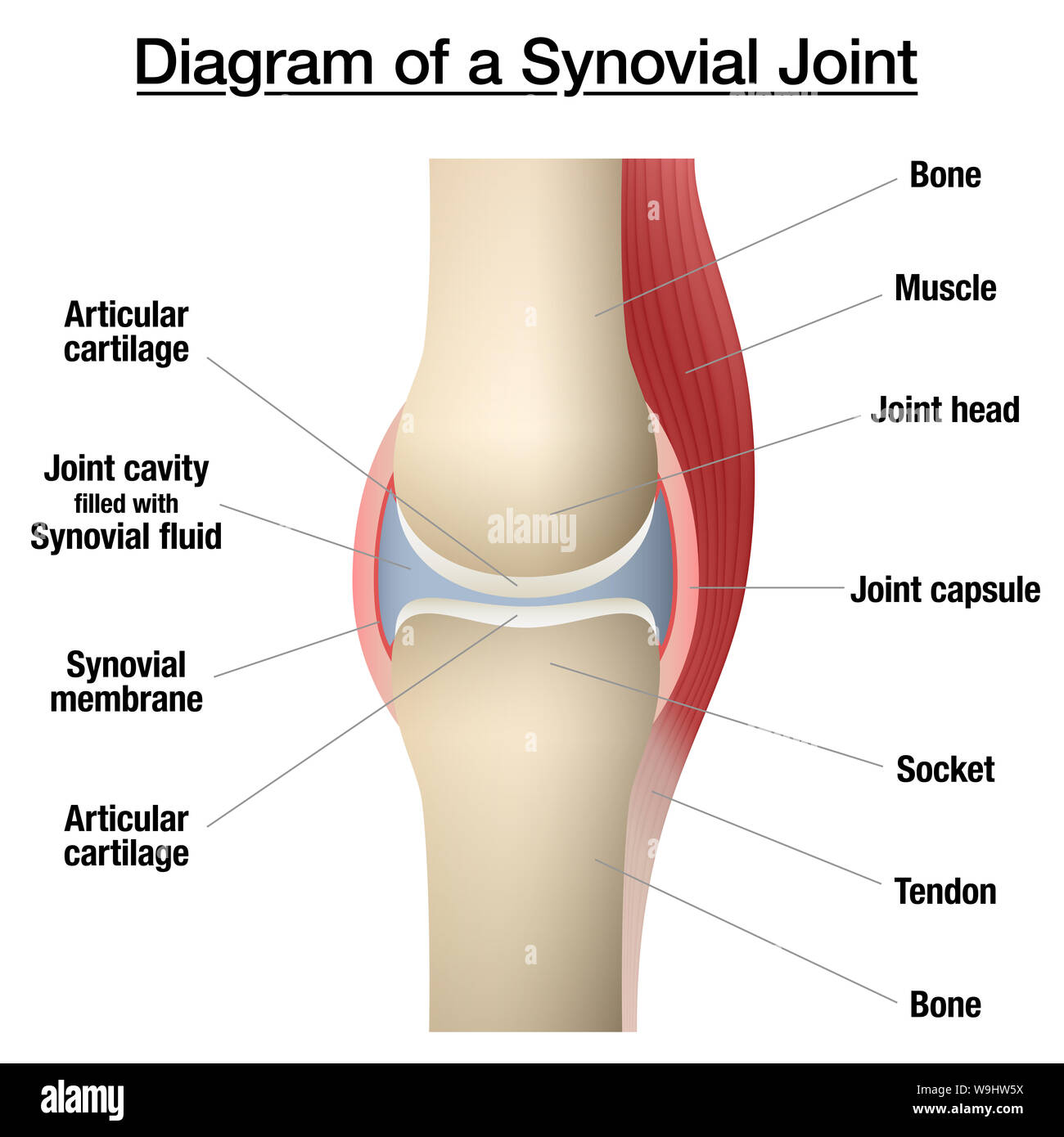 Synovial joint chart. Labeled anatomy infographic with two bones, articular cartilage, joint cavity, synovial fluid, muscle and tendon. Stock Photohttps://www.alamy.com/image-license-details/?v=1https://www.alamy.com/synovial-joint-chart-labeled-anatomy-infographic-with-two-bones-articular-cartilage-joint-cavity-synovial-fluid-muscle-and-tendon-image264080374.html
RFW9HW5X–Synovial joint chart. Labeled anatomy infographic with two bones, articular cartilage, joint cavity, synovial fluid, muscle and tendon.
 Achilles tendon Stock Photohttps://www.alamy.com/image-license-details/?v=1https://www.alamy.com/stock-photo-achilles-tendon-54596190.html
RFD4R1YX–Achilles tendon
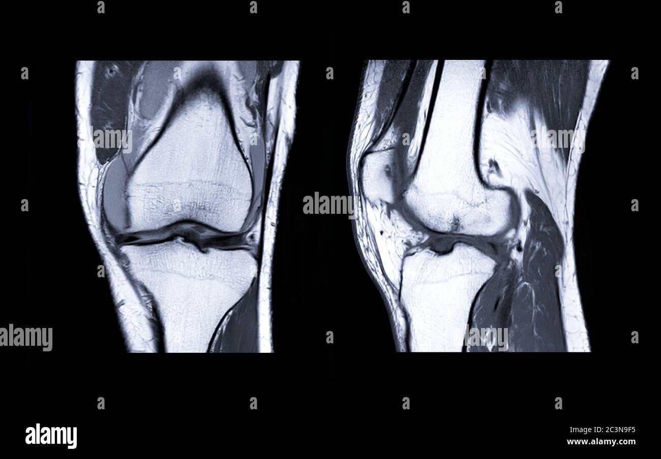 MRI Knee joint or Magnetic resonance imaging compare coronal and sagittal view for detect tear or sprain of the anterior cruciate ligament (ACL). Stock Photohttps://www.alamy.com/image-license-details/?v=1https://www.alamy.com/mri-knee-joint-or-magnetic-resonance-imaging-compare-coronal-and-sagittal-view-for-detect-tear-or-sprain-of-the-anterior-cruciate-ligament-acl-image363730169.html
RF2C3N9F5–MRI Knee joint or Magnetic resonance imaging compare coronal and sagittal view for detect tear or sprain of the anterior cruciate ligament (ACL).
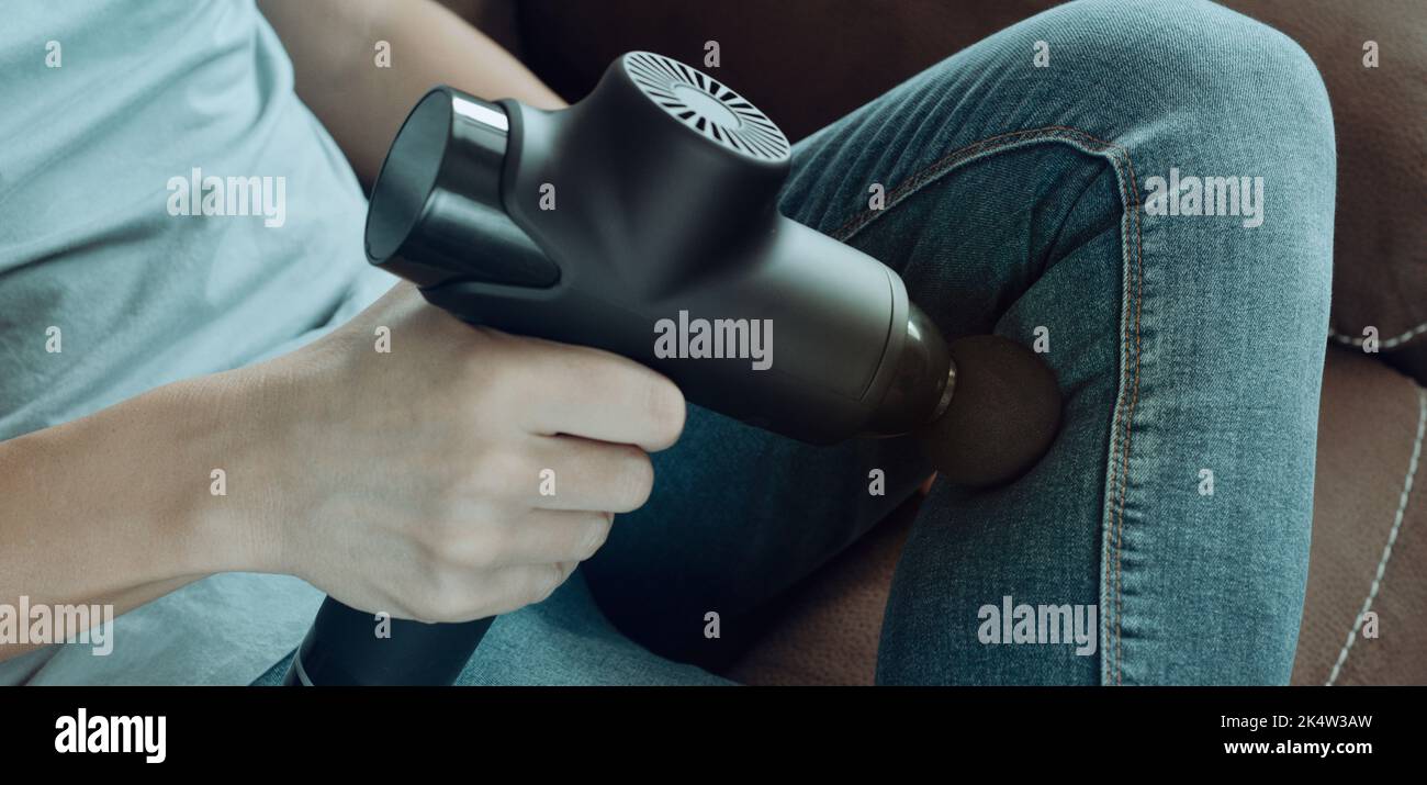 a young caucasian man, in jeans, uses a massage gun to massage the muscles of his leg, sitting on a couch, in a panoramic format to use as web banner Stock Photohttps://www.alamy.com/image-license-details/?v=1https://www.alamy.com/a-young-caucasian-man-in-jeans-uses-a-massage-gun-to-massage-the-muscles-of-his-leg-sitting-on-a-couch-in-a-panoramic-format-to-use-as-web-banner-image484900385.html
RF2K4W3AW–a young caucasian man, in jeans, uses a massage gun to massage the muscles of his leg, sitting on a couch, in a panoramic format to use as web banner
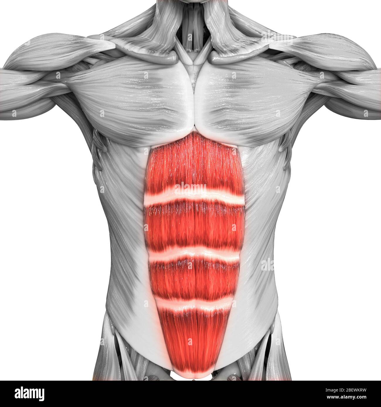 Human Muscular System Parts Rectus Abdominis Muscle Anatomy Stock Photohttps://www.alamy.com/image-license-details/?v=1https://www.alamy.com/human-muscular-system-parts-rectus-abdominis-muscle-anatomy-image353376909.html
RF2BEWKRW–Human Muscular System Parts Rectus Abdominis Muscle Anatomy
 X-ray normal human's foot lateral Stock Photohttps://www.alamy.com/image-license-details/?v=1https://www.alamy.com/x-ray-normal-humans-foot-lateral-image552049361.html
RF2R240GH–X-ray normal human's foot lateral
 ILLUSTRATION DISSECTION TENDONS BACK OF HAND Stock Photohttps://www.alamy.com/image-license-details/?v=1https://www.alamy.com/stock-photo-illustration-dissection-tendons-back-of-hand-18452509.html
RFB20GA5–ILLUSTRATION DISSECTION TENDONS BACK OF HAND
 Biceps reflex examination with a reflex hammer. Neurological assessment of biceps brachii. Stock Photohttps://www.alamy.com/image-license-details/?v=1https://www.alamy.com/biceps-reflex-examination-with-a-reflex-hammer-neurological-assessment-of-biceps-brachii-image455241079.html
RF2HCJ0HY–Biceps reflex examination with a reflex hammer. Neurological assessment of biceps brachii.
 U.S. Army Reserve Spc. Brenda Pruitt, of 228th Combat Support Hospital, applies moulage to a soldier's leg to portray muscle tendon abrasions, during Global Medic at Fort McCoy, Wis., June 17, 2015. Specialist Pruitt training during Global medic has allowed her to create various trauma effects to mannequins and real simulated patients. Global Medic is the premier medical field training event in the Department of Defense and is the only joint accredited exercise conceived, planned and executed by Army Reserve Soldiers, who are part of the Medical Readiness and Training Command, Army Reserve Me Stock Photohttps://www.alamy.com/image-license-details/?v=1https://www.alamy.com/stock-photo-us-army-reserve-spc-brenda-pruitt-of-228th-combat-support-hospital-129572691.html
RMHEPF83–U.S. Army Reserve Spc. Brenda Pruitt, of 228th Combat Support Hospital, applies moulage to a soldier's leg to portray muscle tendon abrasions, during Global Medic at Fort McCoy, Wis., June 17, 2015. Specialist Pruitt training during Global medic has allowed her to create various trauma effects to mannequins and real simulated patients. Global Medic is the premier medical field training event in the Department of Defense and is the only joint accredited exercise conceived, planned and executed by Army Reserve Soldiers, who are part of the Medical Readiness and Training Command, Army Reserve Me
 Male muscle anatomy of the human legs, side view. Stock Photohttps://www.alamy.com/image-license-details/?v=1https://www.alamy.com/stock-photo-male-muscle-anatomy-of-the-human-legs-side-view-59361111.html
RFDCG3KK–Male muscle anatomy of the human legs, side view.
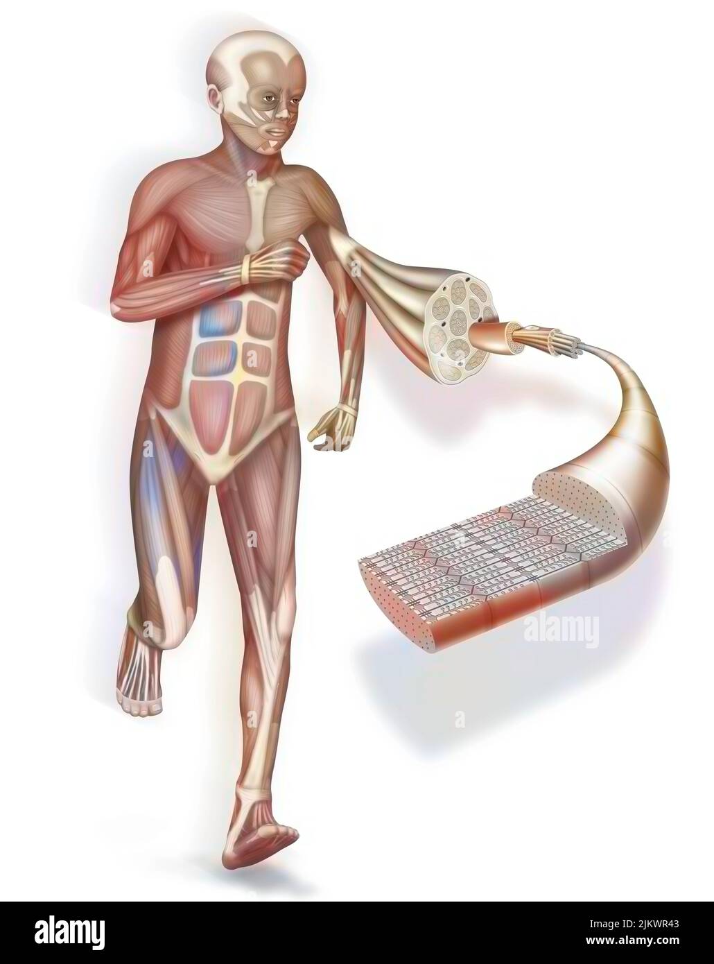 A cut away and zoom on a muscle and its structure: tendon, muscle. Stock Photohttps://www.alamy.com/image-license-details/?v=1https://www.alamy.com/a-cut-away-and-zoom-on-a-muscle-and-its-structure-tendon-muscle-image476925347.html
RF2JKWR43–A cut away and zoom on a muscle and its structure: tendon, muscle.
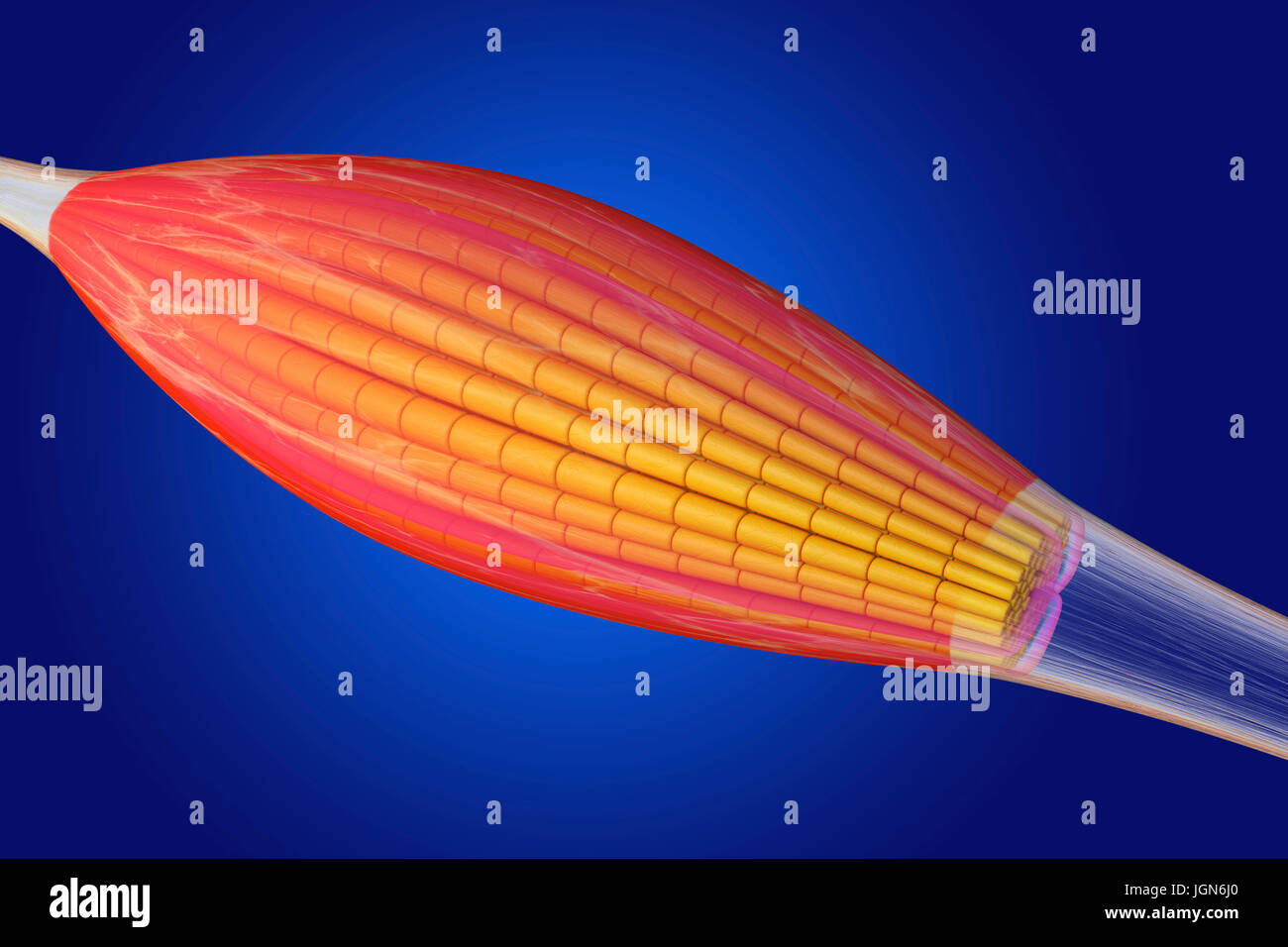 Skeletal muscle, illustration. The muscle is surrounded by epimysium connective tissue (transparent), which forms the tendon that attaches the muscle to bone. Bundles of muscle fibres (yellow) are surrounded by perimysium connective tissue (pink). Stock Photohttps://www.alamy.com/image-license-details/?v=1https://www.alamy.com/stock-photo-skeletal-muscle-illustration-the-muscle-is-surrounded-by-epimysium-147983640.html
RFJGN6J0–Skeletal muscle, illustration. The muscle is surrounded by epimysium connective tissue (transparent), which forms the tendon that attaches the muscle to bone. Bundles of muscle fibres (yellow) are surrounded by perimysium connective tissue (pink).
 A typical representation of the extensor muscles on the back of the forearm. The tendons at the wrist are noted, vintage line drawing or engraving ill Stock Vectorhttps://www.alamy.com/image-license-details/?v=1https://www.alamy.com/a-typical-representation-of-the-extensor-muscles-on-the-back-of-the-forearm-the-tendons-at-the-wrist-are-noted-vintage-line-drawing-or-engraving-ill-image348660127.html
RF2B76RFB–A typical representation of the extensor muscles on the back of the forearm. The tendons at the wrist are noted, vintage line drawing or engraving ill