Quick filters:
Microscopic organism Stock Photos and Images
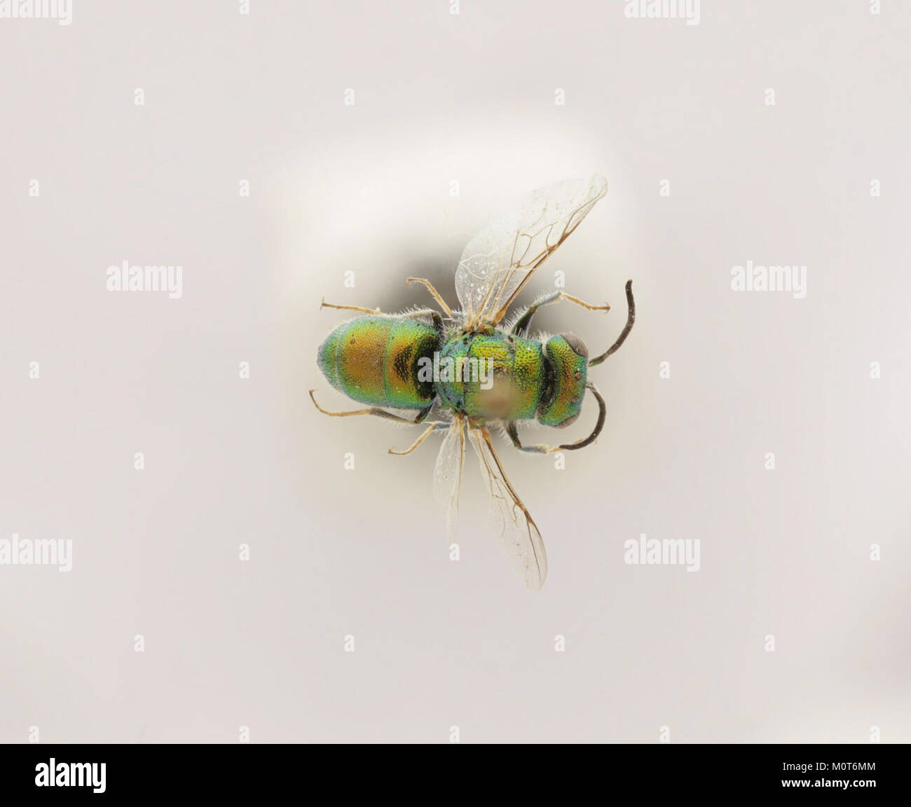 Cephalochrysis ehrenbergi is a species of ciliate protozoa, identified in the Zoosphere collection. It is a microscopic organism studied in biology and zoology for its unique characteristics. Stock Photohttps://www.alamy.com/image-license-details/?v=1https://www.alamy.com/stock-photo-cephalochrysis-ehrenbergi-is-a-species-of-ciliate-protozoa-identified-172635812.html
RMM0T6MM–Cephalochrysis ehrenbergi is a species of ciliate protozoa, identified in the Zoosphere collection. It is a microscopic organism studied in biology and zoology for its unique characteristics.
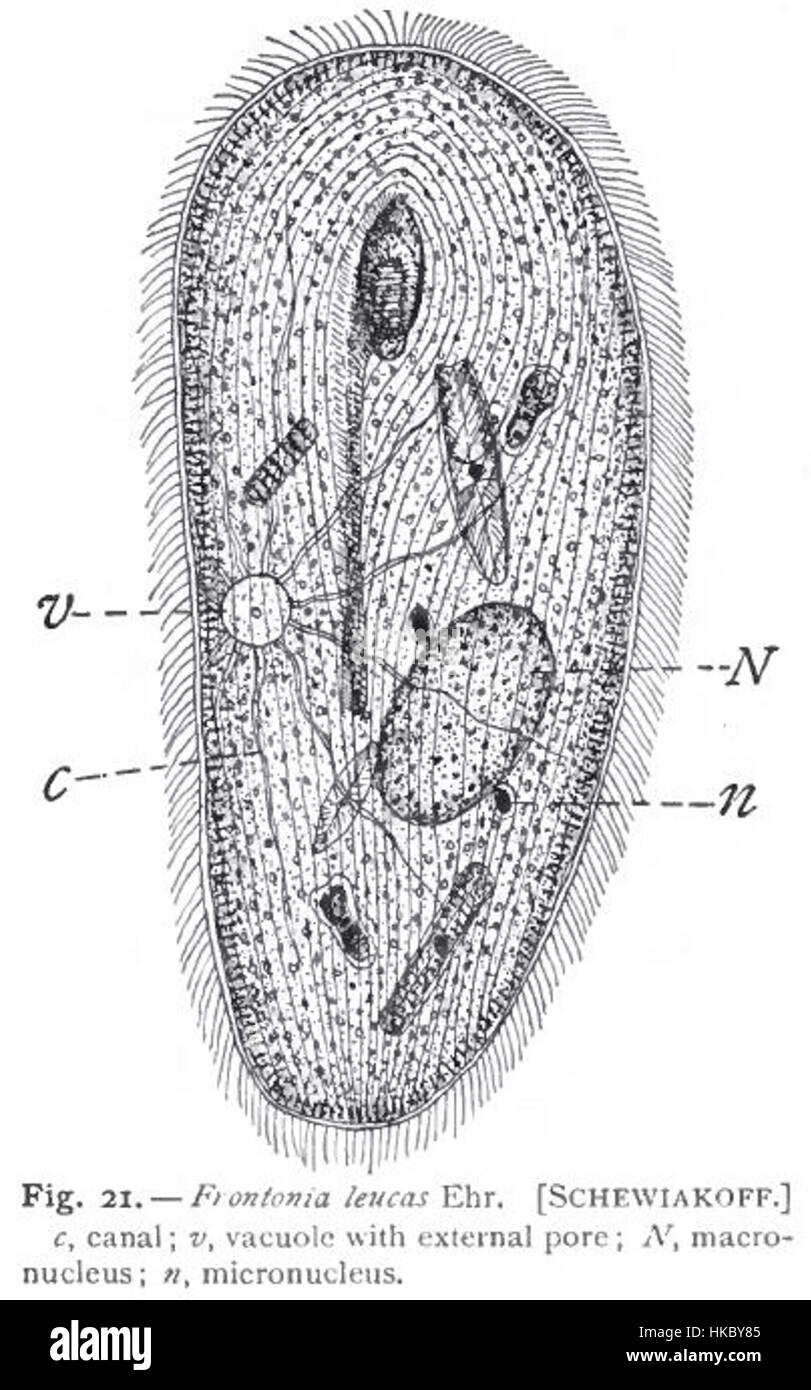 This detailed illustration of Frontonia leucas, created by Gary Calkins and drawn by Schewiakoff, showcases the microscopic organism, highlighting its unique structure. The work is a scientific representation of this species in the field of biology. Stock Photohttps://www.alamy.com/image-license-details/?v=1https://www.alamy.com/stock-photo-this-detailed-illustration-of-frontonia-leucas-created-by-gary-calkins-132413909.html
RMHKBY85–This detailed illustration of Frontonia leucas, created by Gary Calkins and drawn by Schewiakoff, showcases the microscopic organism, highlighting its unique structure. The work is a scientific representation of this species in the field of biology.
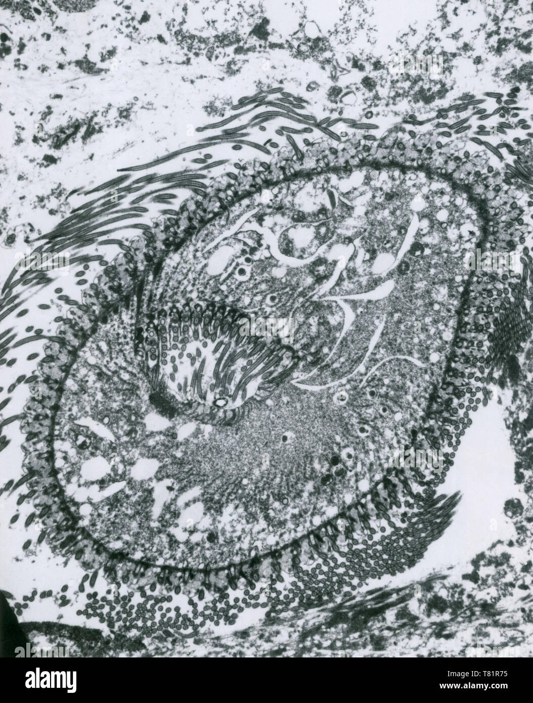 Balantidium Coli (TEM) Stock Photohttps://www.alamy.com/image-license-details/?v=1https://www.alamy.com/balantidium-coli-tem-image245902585.html
RMT81R75–Balantidium Coli (TEM)
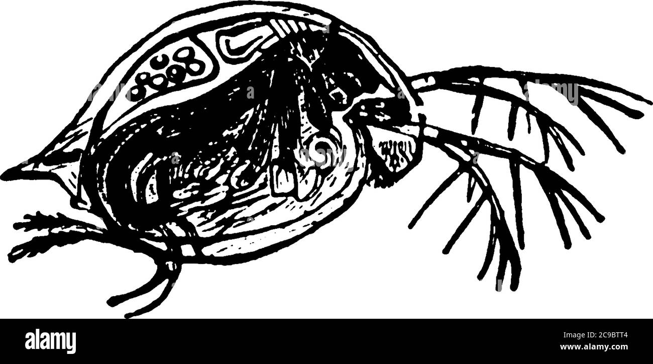 Water Flea is a microscopic organism of order Cladocera., vintage line drawing or engraving illustration. Stock Vectorhttps://www.alamy.com/image-license-details/?v=1https://www.alamy.com/water-flea-is-a-microscopic-organism-of-order-cladocera-vintage-line-drawing-or-engraving-illustration-image367210596.html
RF2C9BTT4–Water Flea is a microscopic organism of order Cladocera., vintage line drawing or engraving illustration.
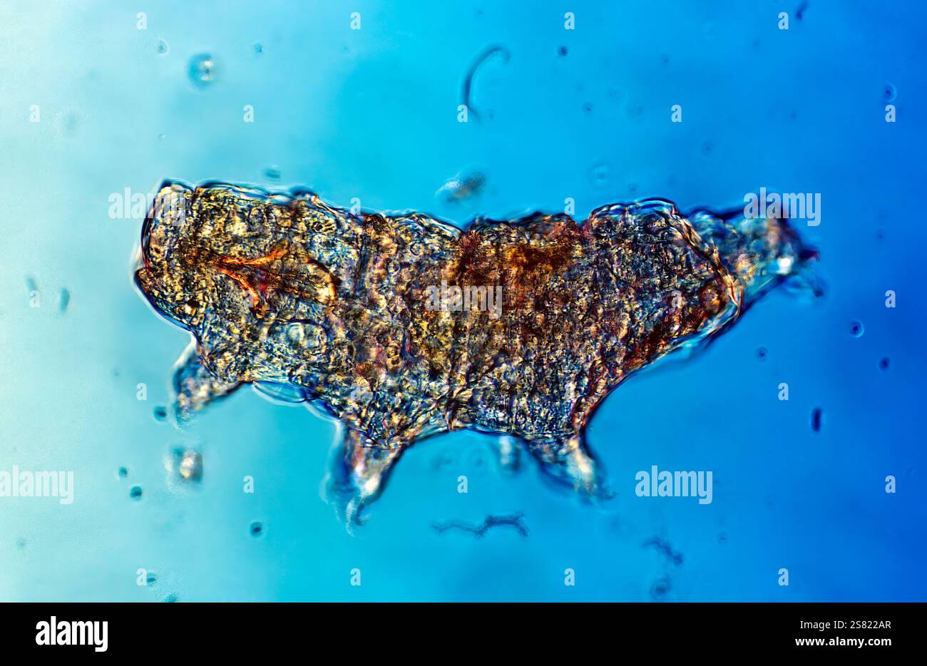 Tardigrade (commonly known as a water bear), a microscopic organism renowned for its resilience, imaged at 200x magnification. Stock Photohttps://www.alamy.com/image-license-details/?v=1https://www.alamy.com/tardigrade-commonly-known-as-a-water-bear-a-microscopic-organism-renowned-for-its-resilience-imaged-at-200x-magnification-image641746639.html
RM2S822AR–Tardigrade (commonly known as a water bear), a microscopic organism renowned for its resilience, imaged at 200x magnification.
RMP250FF–. Gonium rectangulum 141 Gonium rectangulum - - Print - Iconographia Zoologica - Special Collections University of Amsterdam - UBAINV0274 113 23 0017
 a large background image of cells or bacteria under the microscope Stock Photohttps://www.alamy.com/image-license-details/?v=1https://www.alamy.com/stock-photo-a-large-background-image-of-cells-or-bacteria-under-the-microscope-14558756.html
RFAHX48N–a large background image of cells or bacteria under the microscope
 3D illustration. Background image, abstract concept of microscopic life, virus and bacteria. Stock Photohttps://www.alamy.com/image-license-details/?v=1https://www.alamy.com/3d-illustration-background-image-abstract-concept-of-microscopic-life-virus-and-bacteria-image228149170.html
RFR752GJ–3D illustration. Background image, abstract concept of microscopic life, virus and bacteria.
 a large rendered image of bacteria or cells under a microscope Stock Photohttps://www.alamy.com/image-license-details/?v=1https://www.alamy.com/stock-photo-a-large-rendered-image-of-bacteria-or-cells-under-a-microscope-15109747.html
RFAM0M4M–a large rendered image of bacteria or cells under a microscope
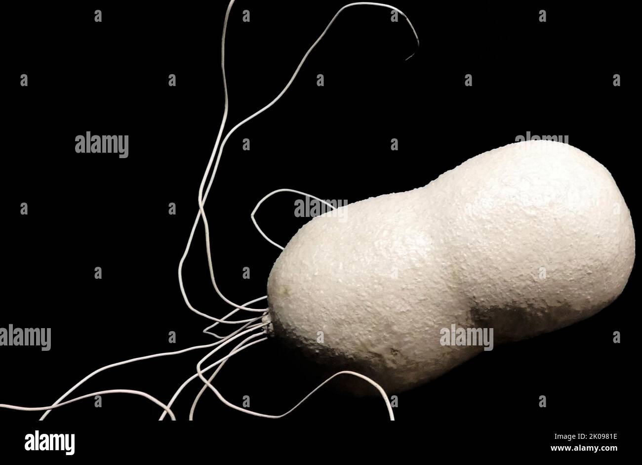 Model of a microbe from a laboratory. A microorganism, or microbe, is an organism of microscopic size, which may exist in its single-celled form or as a colony of cells. Stock Photohttps://www.alamy.com/image-license-details/?v=1https://www.alamy.com/model-of-a-microbe-from-a-laboratory-a-microorganism-or-microbe-is-an-organism-of-microscopic-size-which-may-exist-in-its-single-celled-form-or-as-a-colony-of-cells-image482094186.html
RM2K0981E–Model of a microbe from a laboratory. A microorganism, or microbe, is an organism of microscopic size, which may exist in its single-celled form or as a colony of cells.
 brightly backlit red cells on white background under the microscope Stock Photohttps://www.alamy.com/image-license-details/?v=1https://www.alamy.com/stock-photo-brightly-backlit-red-cells-on-white-background-under-the-microscope-15075485.html
RFAKW25J–brightly backlit red cells on white background under the microscope
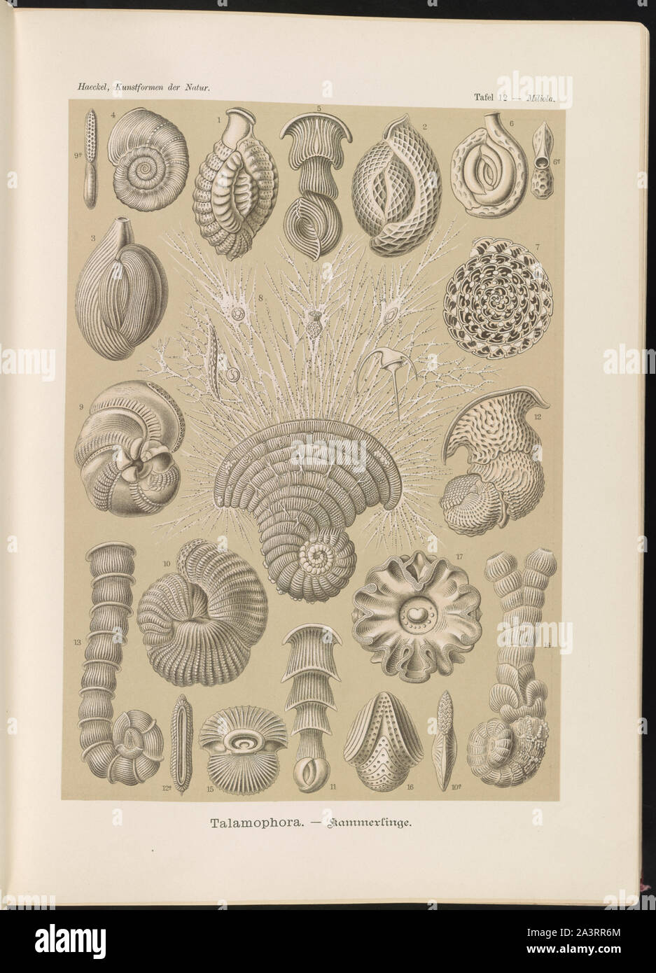 Talamophora. - Kammerlinge Stock Photohttps://www.alamy.com/image-license-details/?v=1https://www.alamy.com/talamophora-kammerlinge-image329364076.html
RM2A3RR6M–Talamophora. - Kammerlinge
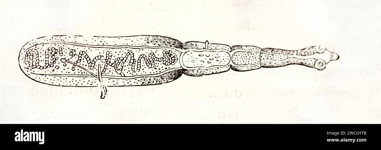 Echinococcus granulosus illustrated in anatomical engraving, showing parasitic structure in detailed view. Naturalist plate published in 1878. Stock Photohttps://www.alamy.com/image-license-details/?v=1https://www.alamy.com/echinococcus-granulosus-illustrated-in-anatomical-engraving-showing-parasitic-structure-in-detailed-view-naturalist-plate-published-in-1878-image592901467.html
RF2WCGYTB–Echinococcus granulosus illustrated in anatomical engraving, showing parasitic structure in detailed view. Naturalist plate published in 1878.
 Illustration of a virus with tentacles spreading infection through the body, a representation of illness and disease Stock Vectorhttps://www.alamy.com/image-license-details/?v=1https://www.alamy.com/illustration-of-a-virus-with-tentacles-spreading-infection-through-the-body-a-representation-of-illness-and-disease-image628386192.html
RF2YE9D00–Illustration of a virus with tentacles spreading infection through the body, a representation of illness and disease
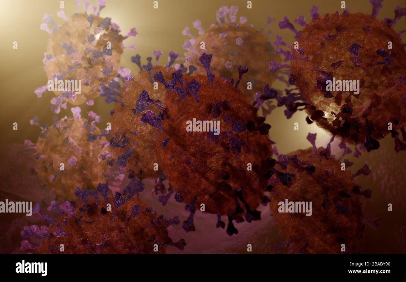 coronavirus covid19 inside the body microscopic illustration, 3D render based on virus microscopy photos Stock Photohttps://www.alamy.com/image-license-details/?v=1https://www.alamy.com/coronavirus-covid19-inside-the-body-microscopic-illustration-3d-render-based-on-virus-microscopy-photos-image350616812.html
RM2BABY90–coronavirus covid19 inside the body microscopic illustration, 3D render based on virus microscopy photos
 Set of bright colorful biology cells, bacteria and virus arranged in circle shape on white Stock Vectorhttps://www.alamy.com/image-license-details/?v=1https://www.alamy.com/set-of-bright-colorful-biology-cells-bacteria-and-virus-arranged-in-circle-shape-on-white-image228225646.html
RFR78G3X–Set of bright colorful biology cells, bacteria and virus arranged in circle shape on white
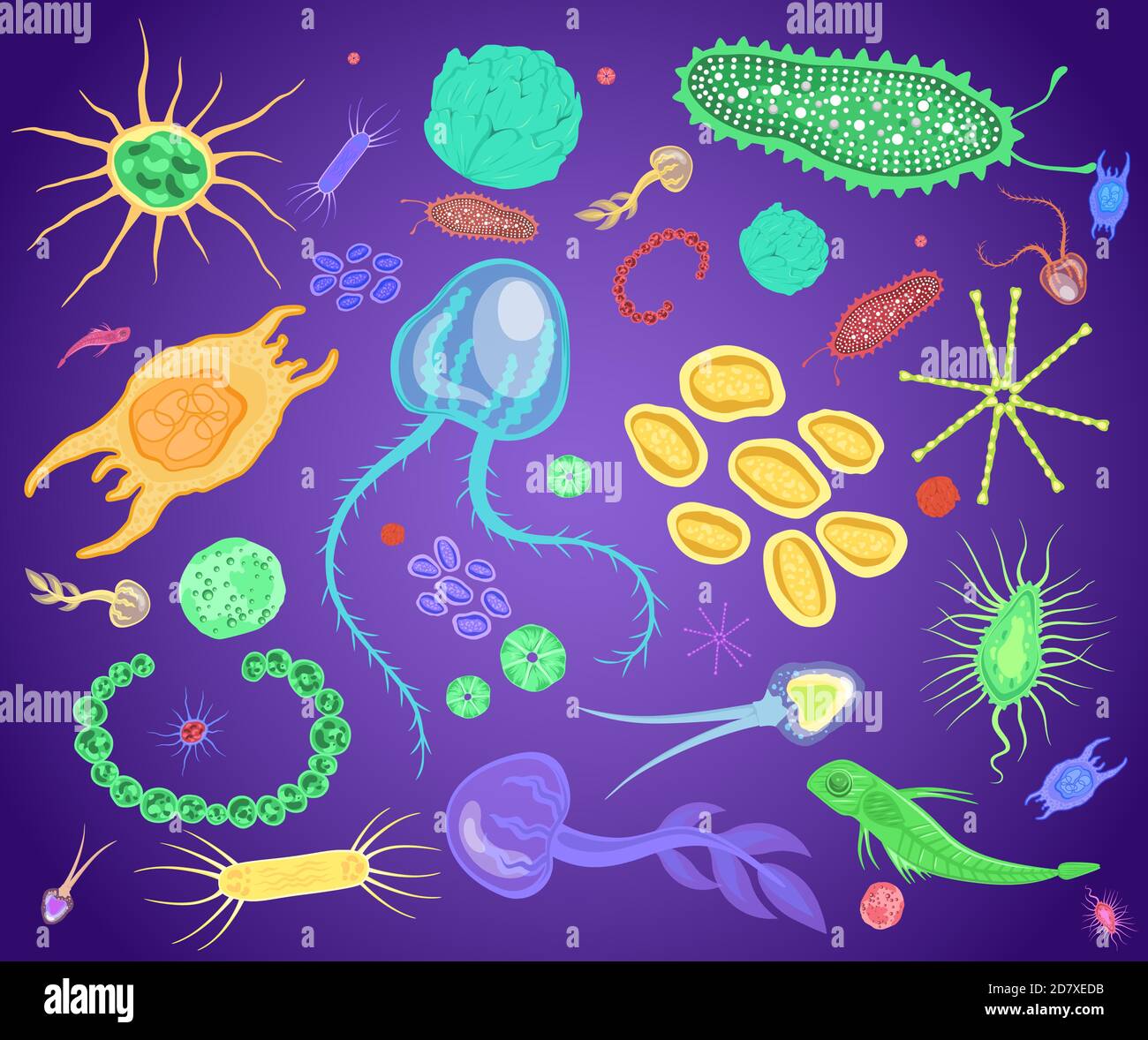 Plankton, marine and freshwater microscopic organism set, flat vector isolated illustration. Stock Vectorhttps://www.alamy.com/image-license-details/?v=1https://www.alamy.com/plankton-marine-and-freshwater-microscopic-organism-set-flat-vector-isolated-illustration-image383512791.html
RF2D7XEDB–Plankton, marine and freshwater microscopic organism set, flat vector isolated illustration.
 Proceedings of the Zoological Society of London, London, Academic Press periodicals, zoology, turtle, An intricate illustration depicting the dorsal view of an aspidochete, a type of microscopic organism. The detailed rendering showcases the organism’s distinctive shape, characterized by a symmetrical body structure and clearly defined appendages or regions. The texture is meticulously highlighted, emphasizing the fine details of its surface features, such as the small, granular formations that cover its body. The composition presents an educational perspective on the morphology of this organi Stock Photohttps://www.alamy.com/image-license-details/?v=1https://www.alamy.com/proceedings-of-the-zoological-society-of-london-london-academic-press-periodicals-zoology-turtle-an-intricate-illustration-depicting-the-dorsal-view-of-an-aspidochete-a-type-of-microscopic-organism-the-detailed-rendering-showcases-the-organisms-distinctive-shape-characterized-by-a-symmetrical-body-structure-and-clearly-defined-appendages-or-regions-the-texture-is-meticulously-highlighted-emphasizing-the-fine-details-of-its-surface-features-such-as-the-small-granular-formations-that-cover-its-body-the-composition-presents-an-educational-perspective-on-the-morphology-of-this-organi-image643623446.html
RM2SB3G7J–Proceedings of the Zoological Society of London, London, Academic Press periodicals, zoology, turtle, An intricate illustration depicting the dorsal view of an aspidochete, a type of microscopic organism. The detailed rendering showcases the organism’s distinctive shape, characterized by a symmetrical body structure and clearly defined appendages or regions. The texture is meticulously highlighted, emphasizing the fine details of its surface features, such as the small, granular formations that cover its body. The composition presents an educational perspective on the morphology of this organi
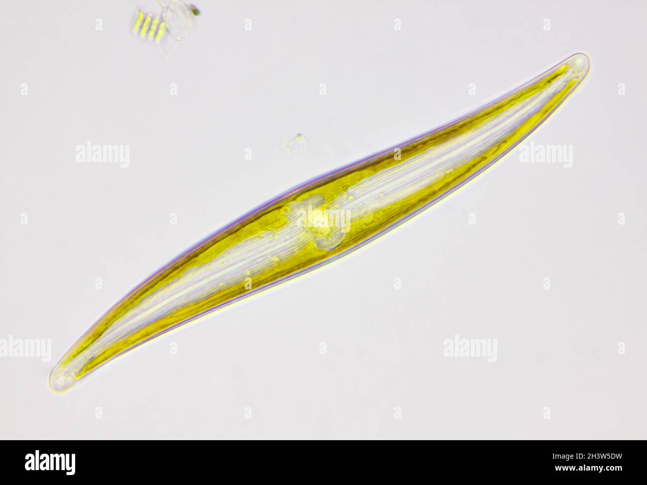 Microscopic view of a diatom (Gyrosigma). Brightfield illumination. Stock Photohttps://www.alamy.com/image-license-details/?v=1https://www.alamy.com/microscopic-view-of-a-diatom-gyrosigma-brightfield-illumination-image449866645.html
RF2H3W5DW–Microscopic view of a diatom (Gyrosigma). Brightfield illumination.
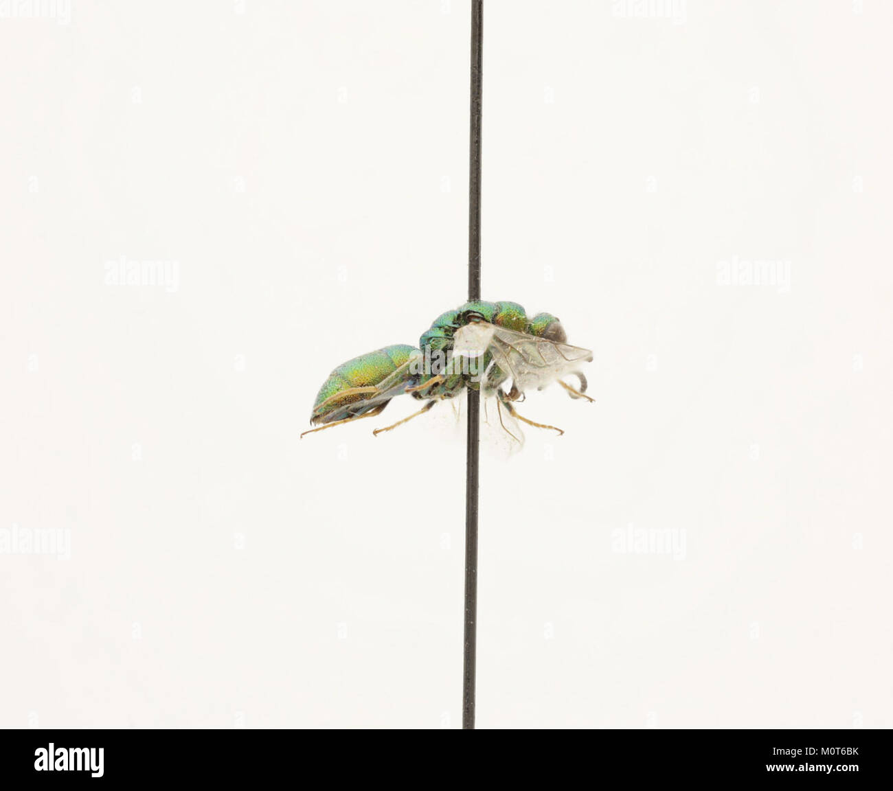 Cephalochrysis ehrenbergi is a species of parasitic copepod, part of the Zoosphere collection. This microscopic organism is significant in marine biology and its ecological role in the ocean’s ecosystem. Stock Photohttps://www.alamy.com/image-license-details/?v=1https://www.alamy.com/stock-photo-cephalochrysis-ehrenbergi-is-a-species-of-parasitic-copepod-part-of-172635559.html
RMM0T6BK–Cephalochrysis ehrenbergi is a species of parasitic copepod, part of the Zoosphere collection. This microscopic organism is significant in marine biology and its ecological role in the ocean’s ecosystem.
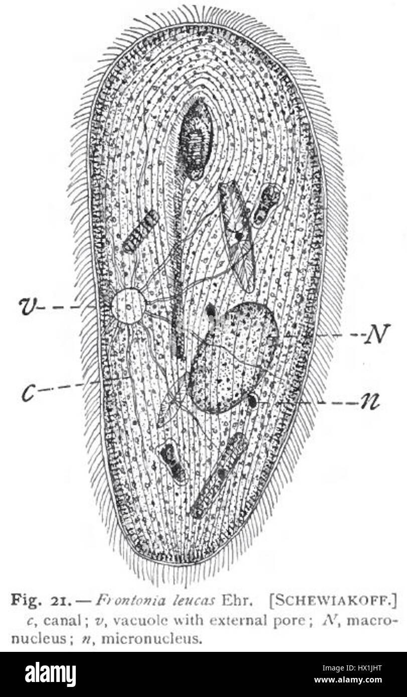 This illustration by Schewiakoff features Frontonia leucas, a species of ciliate protozoan. It highlights the detailed structure and features of this microscopic organism, as observed by Gary Calkins. Stock Photohttps://www.alamy.com/image-license-details/?v=1https://www.alamy.com/stock-photo-this-illustration-by-schewiakoff-features-frontonia-leucas-a-species-136490196.html
RMHX1JHT–This illustration by Schewiakoff features Frontonia leucas, a species of ciliate protozoan. It highlights the detailed structure and features of this microscopic organism, as observed by Gary Calkins.
 Abstract microscopic particles background, fluid inner space, medical illustration Stock Photohttps://www.alamy.com/image-license-details/?v=1https://www.alamy.com/abstract-microscopic-particles-background-fluid-inner-space-medical-illustration-image470349083.html
RF2J9671F–Abstract microscopic particles background, fluid inner space, medical illustration
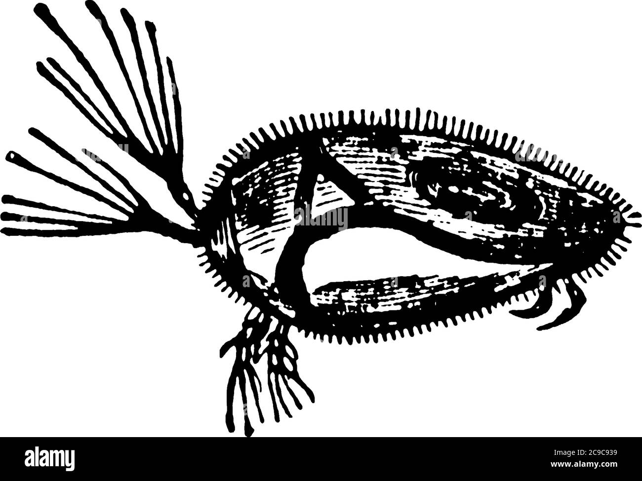 Water Flea is a microscopic organism of order Cladocera., vintage line drawing or engraving illustration. Stock Vectorhttps://www.alamy.com/image-license-details/?v=1https://www.alamy.com/water-flea-is-a-microscopic-organism-of-order-cladocera-vintage-line-drawing-or-engraving-illustration-image367220205.html
RF2C9C939–Water Flea is a microscopic organism of order Cladocera., vintage line drawing or engraving illustration.
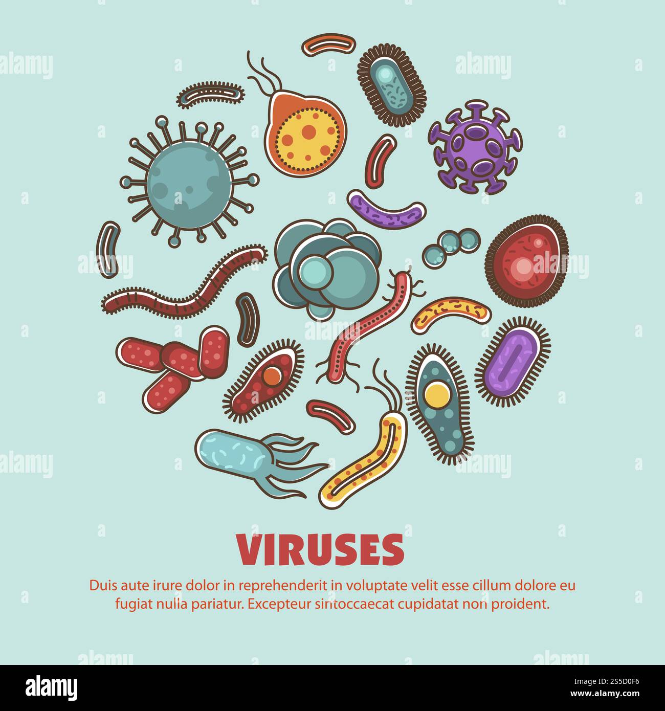 Viruses bacteria small healthy and harmful organism vector. Microscopic creatures of different shapes and form, molecular cells and micro bacillus germs. Biological study and research of probiotics. Viruses bacteria harmful and healthy harmful organism vector Stock Vectorhttps://www.alamy.com/image-license-details/?v=1https://www.alamy.com/viruses-bacteria-small-healthy-and-harmful-organism-vector-microscopic-creatures-of-different-shapes-and-form-molecular-cells-and-micro-bacillus-germs-biological-study-and-research-of-probiotics-viruses-bacteria-harmful-and-healthy-harmful-organism-vector-image640142698.html
RF2S5D0F6–Viruses bacteria small healthy and harmful organism vector. Microscopic creatures of different shapes and form, molecular cells and micro bacillus germs. Biological study and research of probiotics. Viruses bacteria harmful and healthy harmful organism vector
RMP2502R–. Enchelis punctifera 117 Enchelis punctifera - - Print - Iconographia Zoologica - Special Collections University of Amsterdam - UBAINV0274 113 13 0024
 Microscopic organism, germ, virus Stock Photohttps://www.alamy.com/image-license-details/?v=1https://www.alamy.com/microscopic-organism-germ-virus-image443553669.html
RF2GNHH6D–Microscopic organism, germ, virus
 3D illustration. Background image, abstract concept of microscopic life, virus and bacteria. Stock Photohttps://www.alamy.com/image-license-details/?v=1https://www.alamy.com/3d-illustration-background-image-abstract-concept-of-microscopic-life-virus-and-bacteria-image228122941.html
RFR73W3W–3D illustration. Background image, abstract concept of microscopic life, virus and bacteria.
 Abstract cluster of orange organic spheres Stock Photohttps://www.alamy.com/image-license-details/?v=1https://www.alamy.com/stock-photo-abstract-cluster-of-orange-organic-spheres-133497717.html
RFHN59KH–Abstract cluster of orange organic spheres
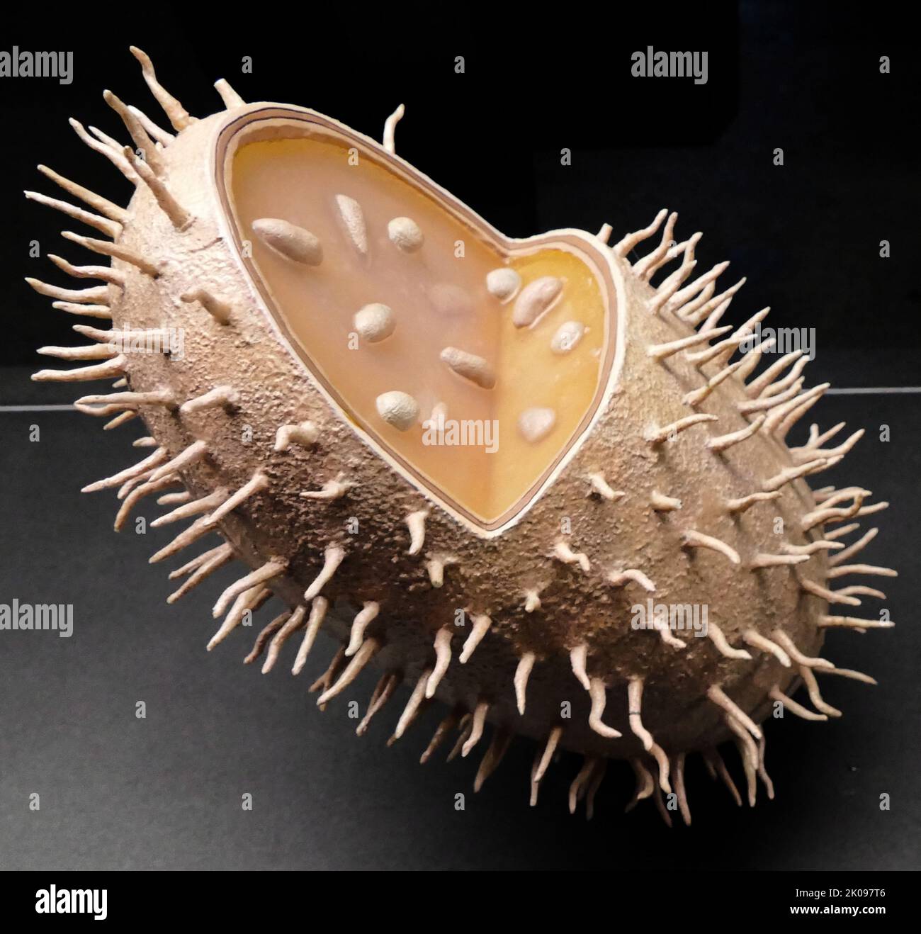 A photograph of a microbe from a laboratory. A microorganism, or microbe, is an organism of microscopic size, which may exist in its single-celled form or as a colony of cells. Stock Photohttps://www.alamy.com/image-license-details/?v=1https://www.alamy.com/a-photograph-of-a-microbe-from-a-laboratory-a-microorganism-or-microbe-is-an-organism-of-microscopic-size-which-may-exist-in-its-single-celled-form-or-as-a-colony-of-cells-image482094038.html
RM2K097T6–A photograph of a microbe from a laboratory. A microorganism, or microbe, is an organism of microscopic size, which may exist in its single-celled form or as a colony of cells.
 An image of a microscopic organism set. Stock Photohttps://www.alamy.com/image-license-details/?v=1https://www.alamy.com/stock-photo-an-image-of-a-microscopic-organism-set-78839663.html
RFEG7CP7–An image of a microscopic organism set.
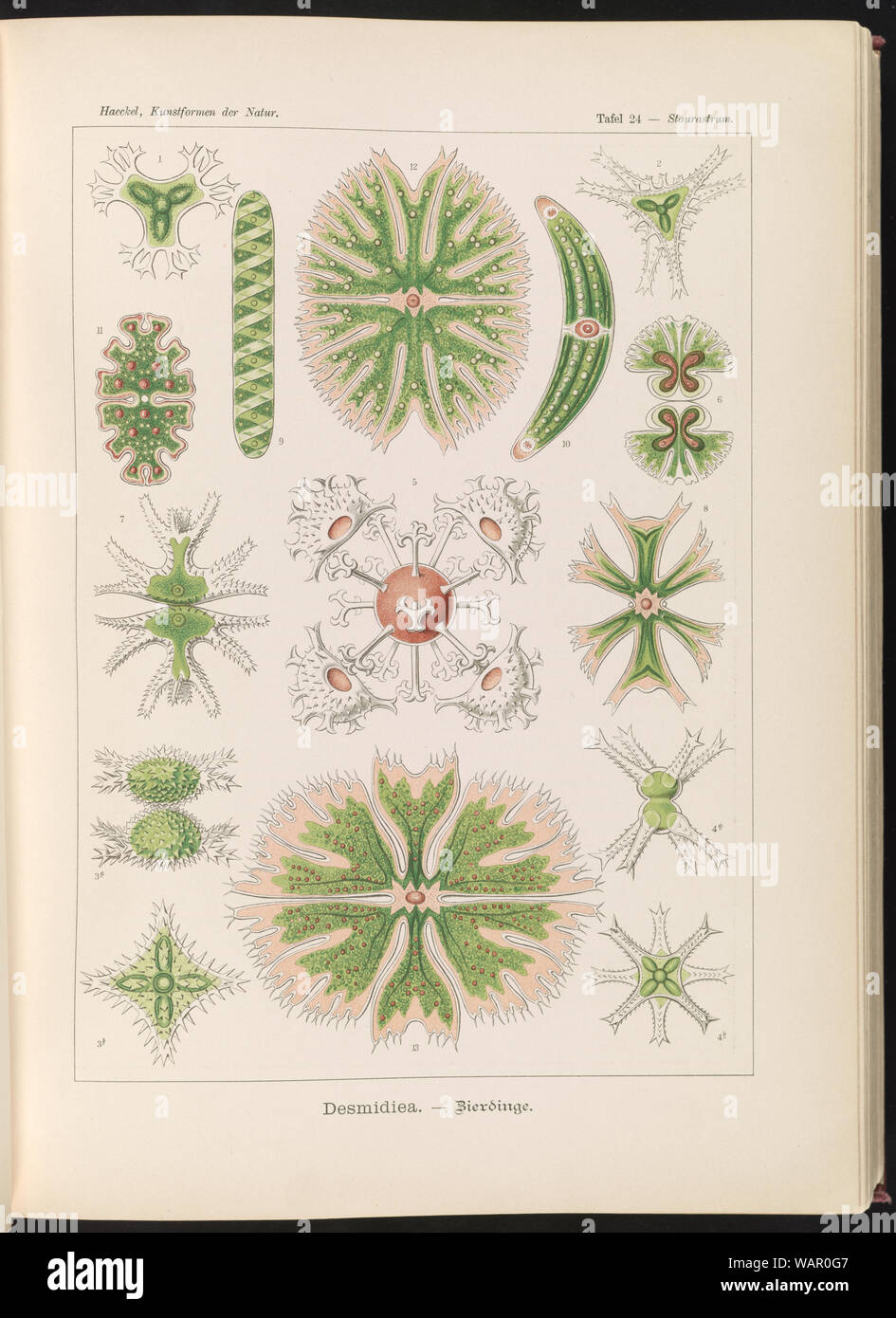 Desmidiea. - Bierdinge Stock Photohttps://www.alamy.com/image-license-details/?v=1https://www.alamy.com/desmidiea-bierdinge-image264807431.html
RMWAR0G7–Desmidiea. - Bierdinge
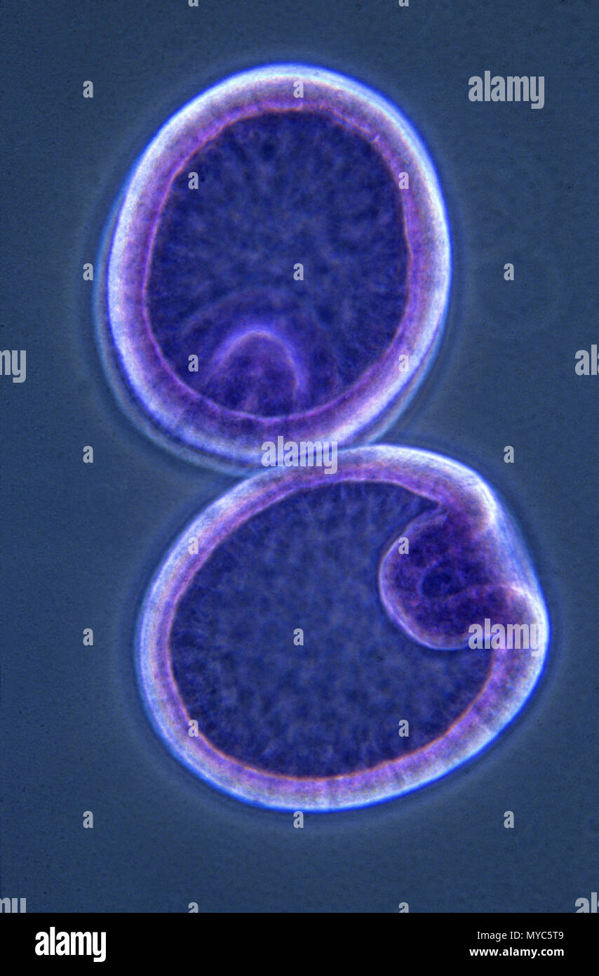 Gastrula.Starfish Stock Photohttps://www.alamy.com/image-license-details/?v=1https://www.alamy.com/gastrulastarfish-image188967417.html
RFMYC5T9–Gastrula.Starfish
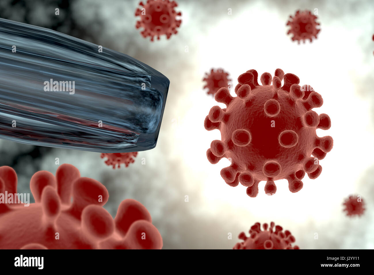 Rendering of a virus isolated in an organism Stock Photohttps://www.alamy.com/image-license-details/?v=1https://www.alamy.com/stock-photo-rendering-of-a-virus-isolated-in-an-organism-139526157.html
RFJ2YY11–Rendering of a virus isolated in an organism
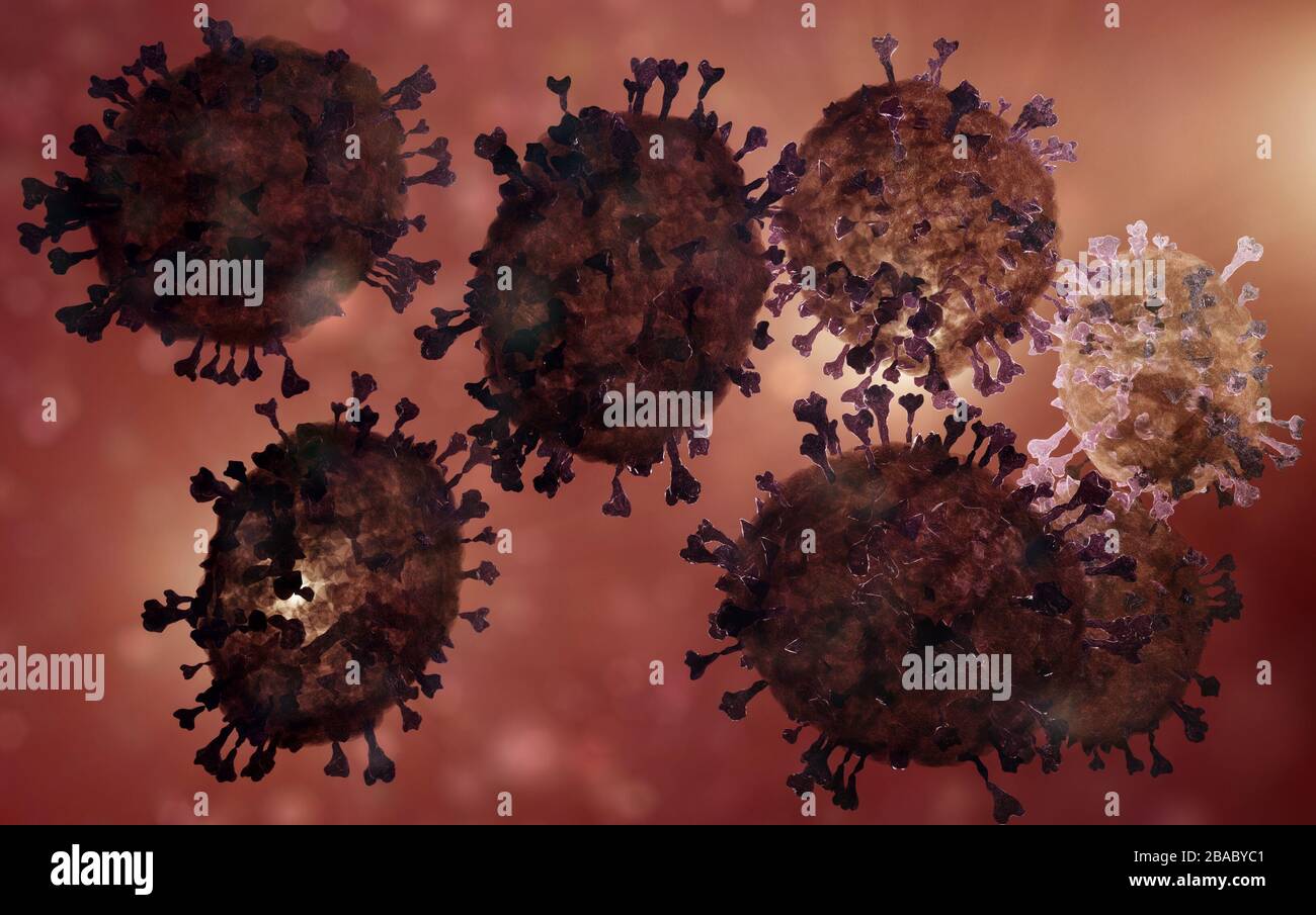 coronavirus covid19 inside the body microscopic illustration, 3D render based on virus microscopy photos Stock Photohttps://www.alamy.com/image-license-details/?v=1https://www.alamy.com/coronavirus-covid19-inside-the-body-microscopic-illustration-3d-render-based-on-virus-microscopy-photos-image350616897.html
RM2BABYC1–coronavirus covid19 inside the body microscopic illustration, 3D render based on virus microscopy photos
RFR648T2–Large set of bright colorful biology cells, bacteria and virus icons on white
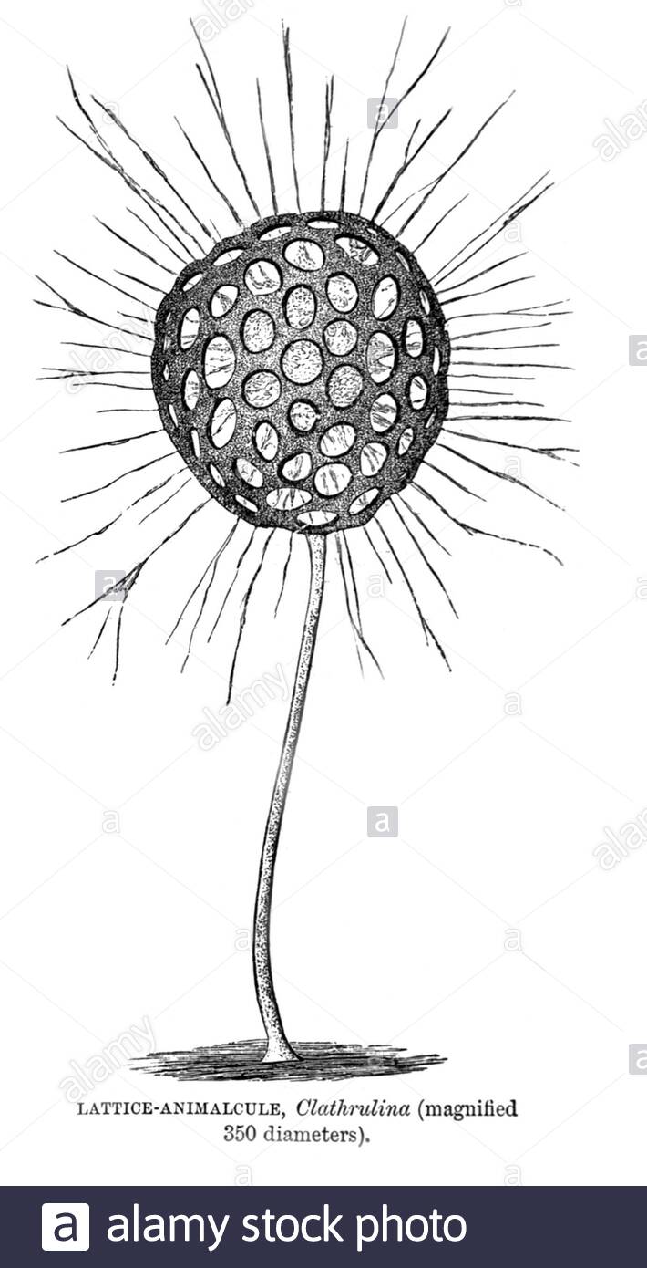 Lattice Animalcule (microorganism), vintage illustration from 1896 Stock Photohttps://www.alamy.com/image-license-details/?v=1https://www.alamy.com/lattice-animalcule-microorganism-vintage-illustration-from-1896-image387699888.html
RM2DEN74G–Lattice Animalcule (microorganism), vintage illustration from 1896
 Genesi, organizzazione e metamorfosi degli infusori, Firenze, Tipo. di S. Landi, 1895, infusoria, protista, Museum of Comparative Zoology, microscopy, The illustration showcases several detailed representations of a microscopic organism from the genus Floscularia. Each specimen is depicted in varying orientations, highlighting distinct anatomical features such as the structure of the body, feeding apparatus, and reproductive systems. The labels indicating different parts of the organisms (not shown here) suggest a scientific focus on anatomy and function. The contrasting colors enhance the vis Stock Photohttps://www.alamy.com/image-license-details/?v=1https://www.alamy.com/genesi-organizzazione-e-metamorfosi-degli-infusori-firenze-tipo-di-s-landi-1895-infusoria-protista-museum-of-comparative-zoology-microscopy-the-illustration-showcases-several-detailed-representations-of-a-microscopic-organism-from-the-genus-floscularia-each-specimen-is-depicted-in-varying-orientations-highlighting-distinct-anatomical-features-such-as-the-structure-of-the-body-feeding-apparatus-and-reproductive-systems-the-labels-indicating-different-parts-of-the-organisms-not-shown-here-suggest-a-scientific-focus-on-anatomy-and-function-the-contrasting-colors-enhance-the-vis-image643603425.html
RM2SB2JMH–Genesi, organizzazione e metamorfosi degli infusori, Firenze, Tipo. di S. Landi, 1895, infusoria, protista, Museum of Comparative Zoology, microscopy, The illustration showcases several detailed representations of a microscopic organism from the genus Floscularia. Each specimen is depicted in varying orientations, highlighting distinct anatomical features such as the structure of the body, feeding apparatus, and reproductive systems. The labels indicating different parts of the organisms (not shown here) suggest a scientific focus on anatomy and function. The contrasting colors enhance the vis
 Natural Microscopic Organism Material for Research 4k uhd 3d illustration Background Stock Photohttps://www.alamy.com/image-license-details/?v=1https://www.alamy.com/natural-microscopic-organism-material-for-research-4k-uhd-3d-illustration-background-image371276365.html
RF2CG12P5–Natural Microscopic Organism Material for Research 4k uhd 3d illustration Background
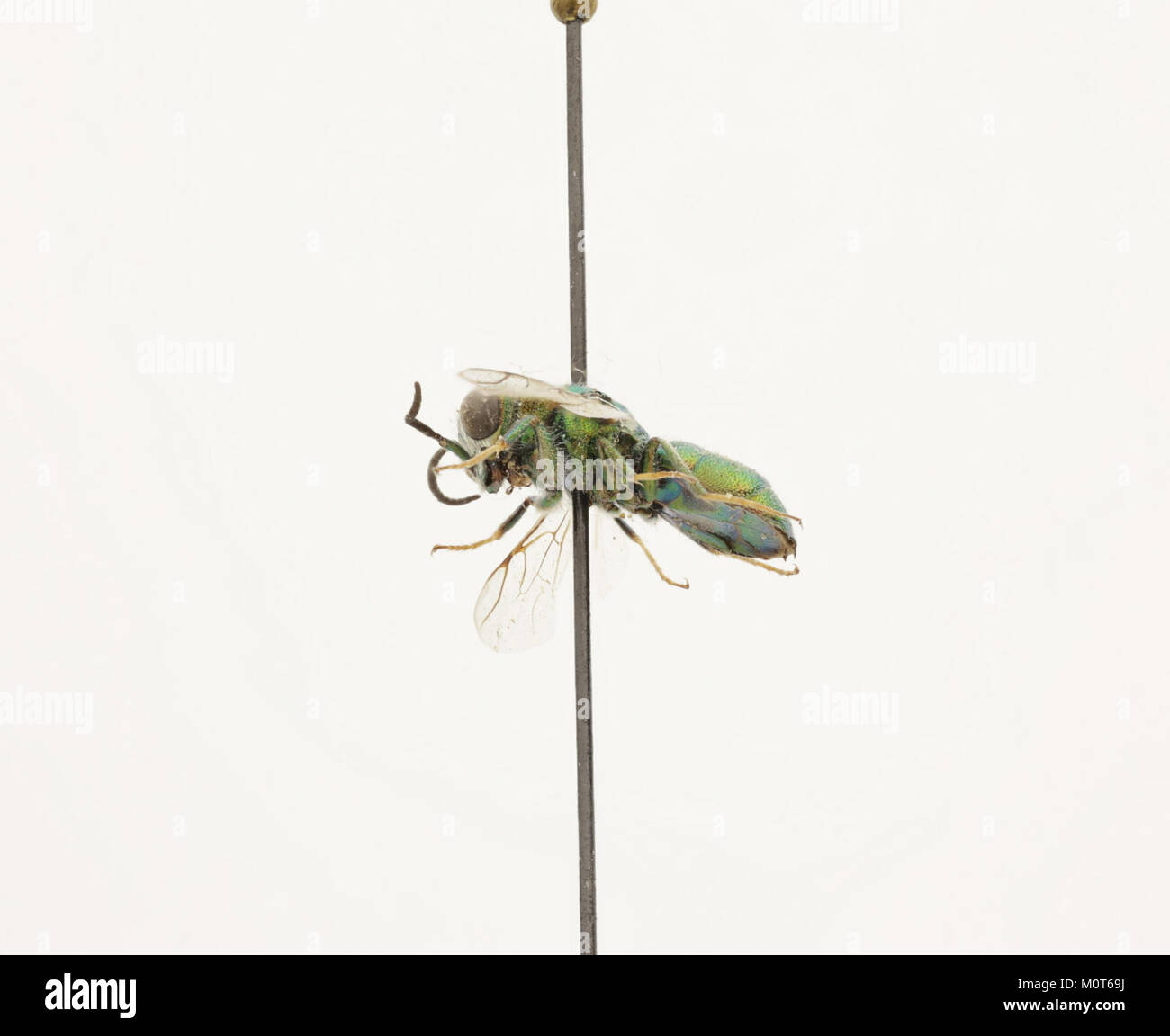 Cephalochrysis ehrenbergi is a species of golden algae in the class Chrysophyceae, known for its role in aquatic ecosystems. This microscopic organism contributes to the base of the food web in freshwater environments. Stock Photohttps://www.alamy.com/image-license-details/?v=1https://www.alamy.com/stock-photo-cephalochrysis-ehrenbergi-is-a-species-of-golden-algae-in-the-class-172635502.html
RMM0T69J–Cephalochrysis ehrenbergi is a species of golden algae in the class Chrysophyceae, known for its role in aquatic ecosystems. This microscopic organism contributes to the base of the food web in freshwater environments.
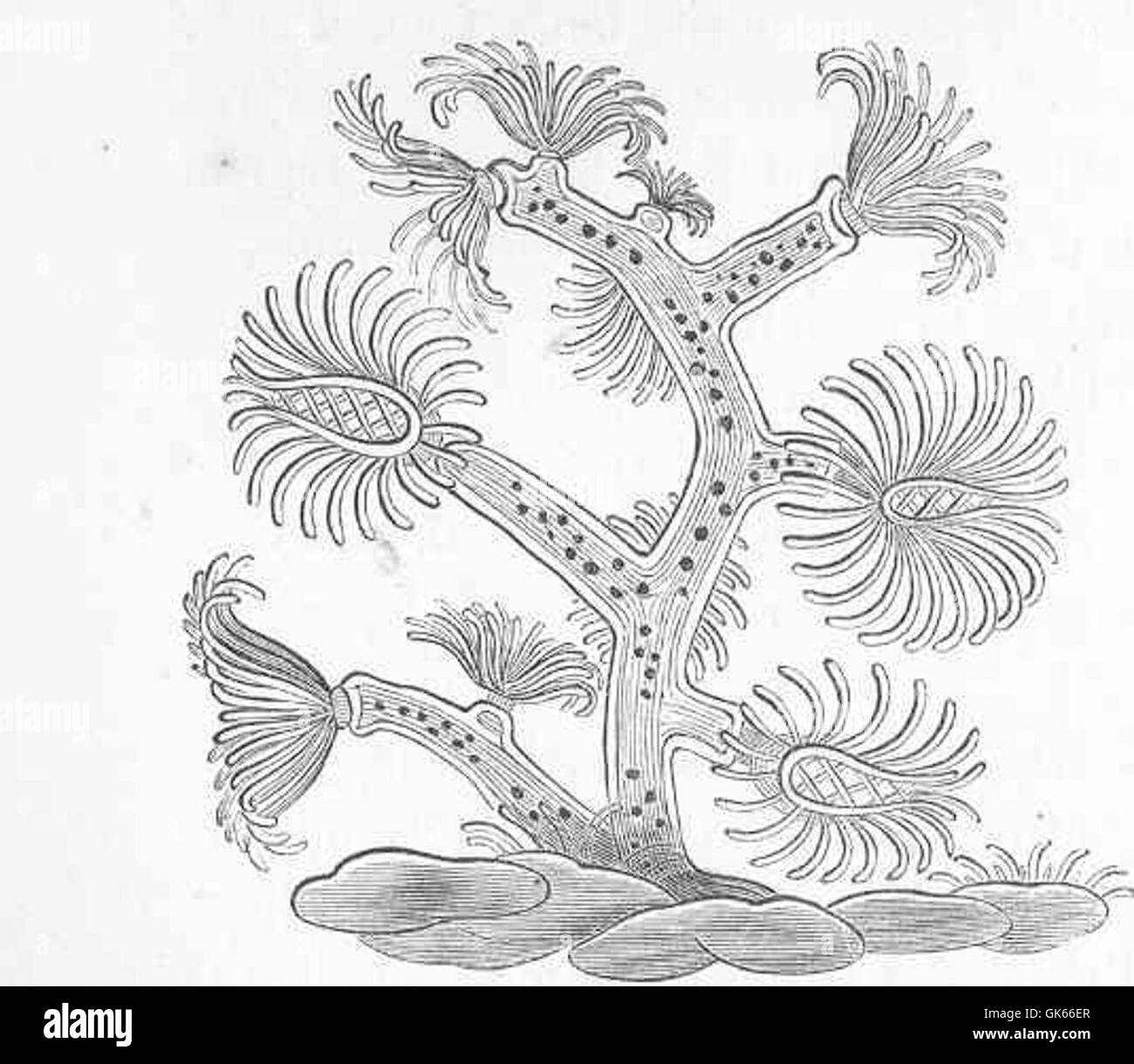 Plumatella cristalina, a freshwater bryozoan species, depicted as drawn by Roesel. This microscopic organism is part of the phylum Bryozoa and is known for its colonial structure. Stock Photohttps://www.alamy.com/image-license-details/?v=1https://www.alamy.com/stock-photo-plumatella-cristalina-a-freshwater-bryozoan-species-depicted-as-drawn-115077503.html
RMGK66ER–Plumatella cristalina, a freshwater bryozoan species, depicted as drawn by Roesel. This microscopic organism is part of the phylum Bryozoa and is known for its colonial structure.
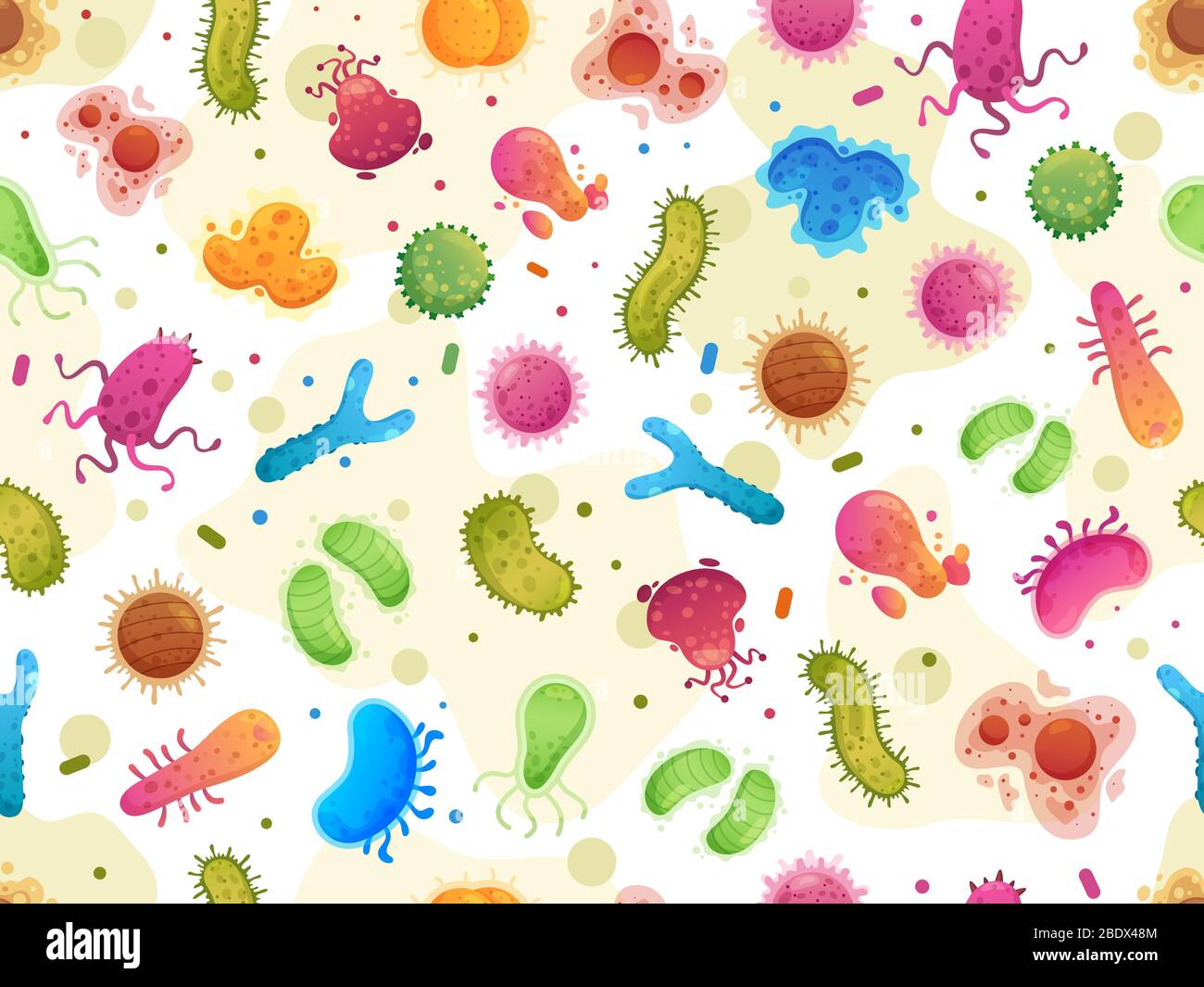 Seamless bacteria pattern. Color germs, microorganism cells microscopic organisms and viruses cartoon vector illustration Stock Vectorhttps://www.alamy.com/image-license-details/?v=1https://www.alamy.com/seamless-bacteria-pattern-color-germs-microorganism-cells-microscopic-organisms-and-viruses-cartoon-vector-illustration-image352772020.html
RF2BDX48M–Seamless bacteria pattern. Color germs, microorganism cells microscopic organisms and viruses cartoon vector illustration
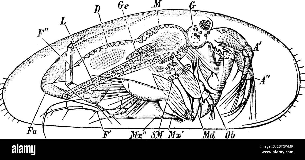 Figure showing Cypris, which are ostracods and related to mussels and shrimp, vintage line drawing or engraving illustration. Stock Vectorhttps://www.alamy.com/image-license-details/?v=1https://www.alamy.com/figure-showing-cypris-which-are-ostracods-and-related-to-mussels-and-shrimp-vintage-line-drawing-or-engraving-illustration-image359330519.html
RF2BTGWMR–Figure showing Cypris, which are ostracods and related to mussels and shrimp, vintage line drawing or engraving illustration.
 Microbes, viruses, bacteria, microorganism cells and primitive organism line vector concept. Virus cell and microbe, bacteria organism, medical microscopic illustration Stock Vectorhttps://www.alamy.com/image-license-details/?v=1https://www.alamy.com/microbes-viruses-bacteria-microorganism-cells-and-primitive-organism-line-vector-concept-virus-cell-and-microbe-bacteria-organism-medical-microscopic-illustration-image354210326.html
RF2BG7JTP–Microbes, viruses, bacteria, microorganism cells and primitive organism line vector concept. Virus cell and microbe, bacteria organism, medical microscopic illustration
RMP250GH–. Monas atomus 195 Monas atomus - - Print - Iconographia Zoologica - Special Collections University of Amsterdam - UBAINV0274 113 25 0007
 Cell with spikes. Illustration of a round cell with multiple spikes and blue spots, representing a microscopic organism. Stock Vectorhttps://www.alamy.com/image-license-details/?v=1https://www.alamy.com/cell-with-spikes-illustration-of-a-round-cell-with-multiple-spikes-and-blue-spots-representing-a-microscopic-organism-image620274358.html
RF2Y13X7J–Cell with spikes. Illustration of a round cell with multiple spikes and blue spots, representing a microscopic organism.
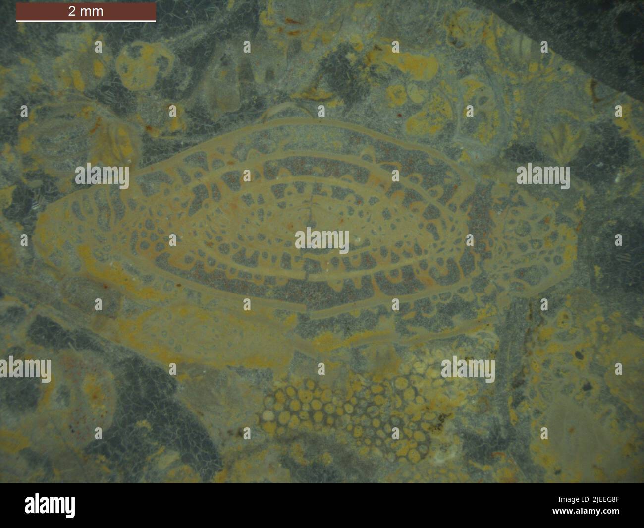 Schwagerina tersa Ross. Stock Photohttps://www.alamy.com/image-license-details/?v=1https://www.alamy.com/schwagerina-tersa-ross-image473605231.html
RM2JEEG8F–Schwagerina tersa Ross.
RM3BEFHYE–Print of *Vibrio olor* (now Lacrymaria olor), a microscopic organism, by Theodoor Gerard van Lidth de Jeude, Abraham Oltmans, and R. T. Maitland, 1923. Published in *Iconographia Zoologica*, Special Collections of the University of Amsterdam.
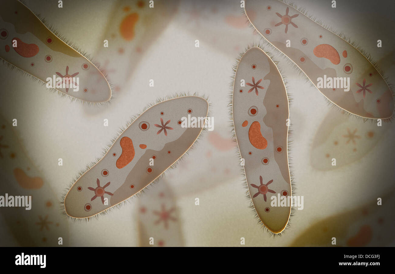 Microscopic view of paramecium. Stock Photohttps://www.alamy.com/image-license-details/?v=1https://www.alamy.com/stock-photo-microscopic-view-of-paramecium-59360998.html
RFDCG3FJ–Microscopic view of paramecium.
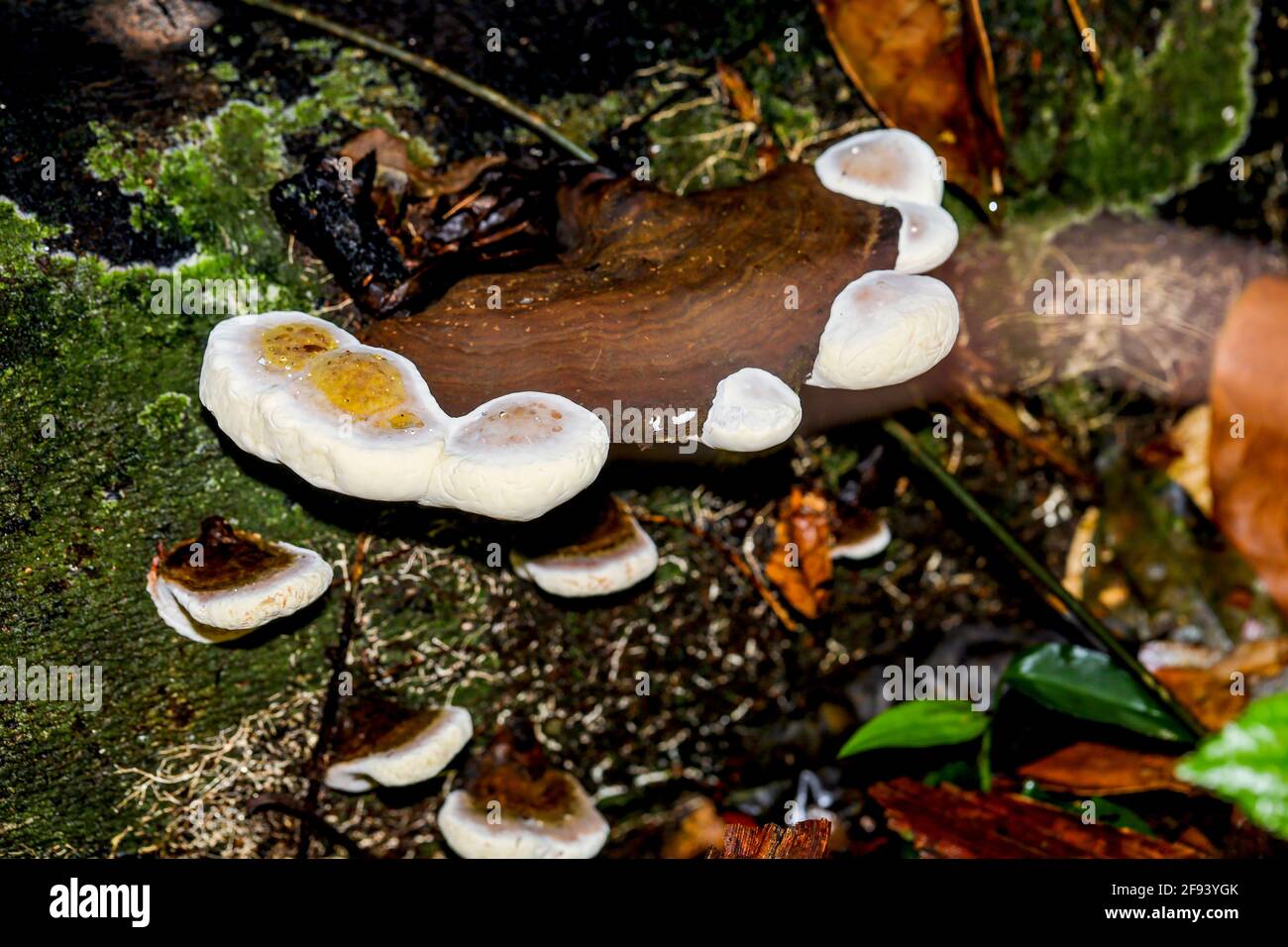 An interesting wild fungi growing on a rotting tree trunk in a tropical jungle. Stock Photohttps://www.alamy.com/image-license-details/?v=1https://www.alamy.com/an-interesting-wild-fungi-growing-on-a-rotting-tree-trunk-in-a-tropical-jungle-image418668227.html
RF2F93YGK–An interesting wild fungi growing on a rotting tree trunk in a tropical jungle.
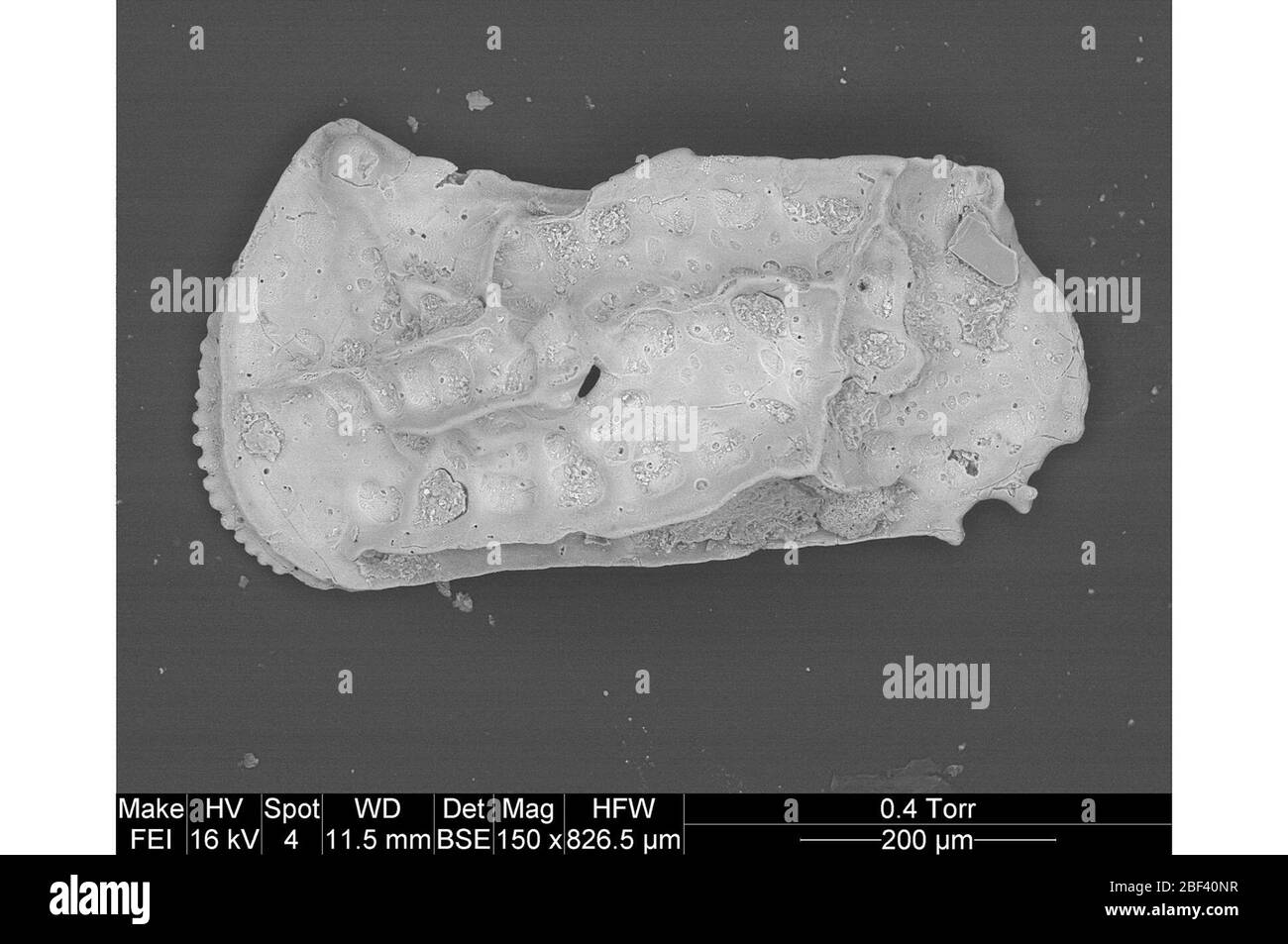 Ostracod. This object is part of the Education and Outreach collection, some of which are in the Q?rius science education center and available to see.Cenozoic - Paleogene - PaleoceneLeft side of the outer carapace, or shell, of a fossil adult male ostracod, approximately 0.69 mm long. Stock Photohttps://www.alamy.com/image-license-details/?v=1https://www.alamy.com/ostracod-this-object-is-part-of-the-education-and-outreach-collection-some-of-which-are-in-the-qrius-science-education-center-and-available-to-seecenozoic-paleogene-paleoceneleft-side-of-the-outer-carapace-or-shell-of-a-fossil-adult-male-ostracod-approximately-069-mm-long-image353515619.html
RM2BF40NR–Ostracod. This object is part of the Education and Outreach collection, some of which are in the Q?rius science education center and available to see.Cenozoic - Paleogene - PaleoceneLeft side of the outer carapace, or shell, of a fossil adult male ostracod, approximately 0.69 mm long.
 Covid-19 virus vector illustration on the black Stock Vectorhttps://www.alamy.com/image-license-details/?v=1https://www.alamy.com/covid-19-virus-vector-illustration-on-the-black-image354014517.html
RF2BFXN3H–Covid-19 virus vector illustration on the black
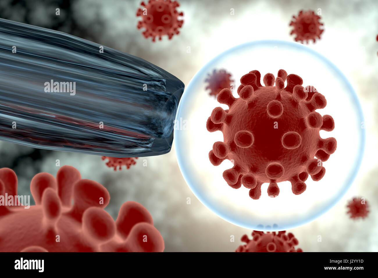 Rendering of a virus isolated in an organism Stock Photohttps://www.alamy.com/image-license-details/?v=1https://www.alamy.com/stock-photo-rendering-of-a-virus-isolated-in-an-organism-139526169.html
RFJ2YY1D–Rendering of a virus isolated in an organism
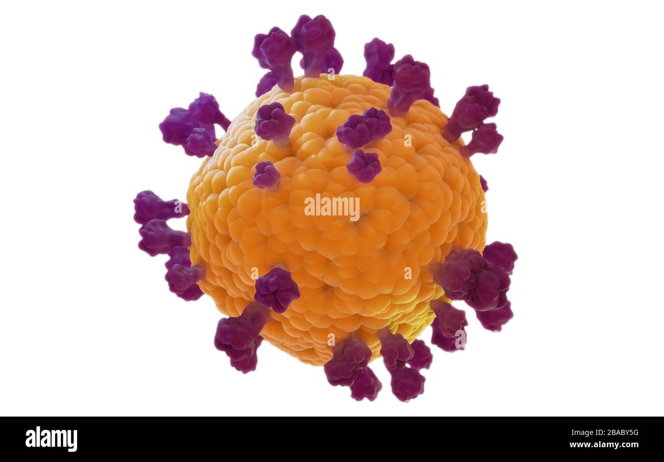 Coronavirus covid19 isolated model, 3D render on white background based on the virus microscopic images Stock Photohttps://www.alamy.com/image-license-details/?v=1https://www.alamy.com/coronavirus-covid19-isolated-model-3d-render-on-white-background-based-on-the-virus-microscopic-images-image350616716.html
RF2BABY5G–Coronavirus covid19 isolated model, 3D render on white background based on the virus microscopic images
 Background for medicine, science or technology. Vector illustration with dynamic effect. Stock Vectorhttps://www.alamy.com/image-license-details/?v=1https://www.alamy.com/background-for-medicine-science-or-technology-vector-illustration-with-dynamic-effect-image683467018.html
RF3BKXH4A–Background for medicine, science or technology. Vector illustration with dynamic effect.
RF2ATTMDC–Virus vector illustration icon template design
 Report on the scientific results of the voyage of H.M.S. Challenger during the years 1873-76 under the command of Captain George S. Nares, Edinburgh, Neill, 1880-1895., This visual representation showcases various forms and structures of the genus Lychnosphaera, a type of microscopic organism. The central figure, marked with a prominent nucleus, is surrounded by intricate, branching appendages that extend outward, illustrating the complex morphology typical of these protists. Additional labeled diagrams highlight specific features, such as the radiating spines and filamentous extensions that c Stock Photohttps://www.alamy.com/image-license-details/?v=1https://www.alamy.com/report-on-the-scientific-results-of-the-voyage-of-hms-challenger-during-the-years-1873-76-under-the-command-of-captain-george-s-nares-edinburgh-neill-1880-1895-this-visual-representation-showcases-various-forms-and-structures-of-the-genus-lychnosphaera-a-type-of-microscopic-organism-the-central-figure-marked-with-a-prominent-nucleus-is-surrounded-by-intricate-branching-appendages-that-extend-outward-illustrating-the-complex-morphology-typical-of-these-protists-additional-labeled-diagrams-highlight-specific-features-such-as-the-radiating-spines-and-filamentous-extensions-that-c-image643619316.html
RM2SB3B04–Report on the scientific results of the voyage of H.M.S. Challenger during the years 1873-76 under the command of Captain George S. Nares, Edinburgh, Neill, 1880-1895., This visual representation showcases various forms and structures of the genus Lychnosphaera, a type of microscopic organism. The central figure, marked with a prominent nucleus, is surrounded by intricate, branching appendages that extend outward, illustrating the complex morphology typical of these protists. Additional labeled diagrams highlight specific features, such as the radiating spines and filamentous extensions that c
RF2C703R7–Virus bacteria icons set. Cartoon flat color vector illustration
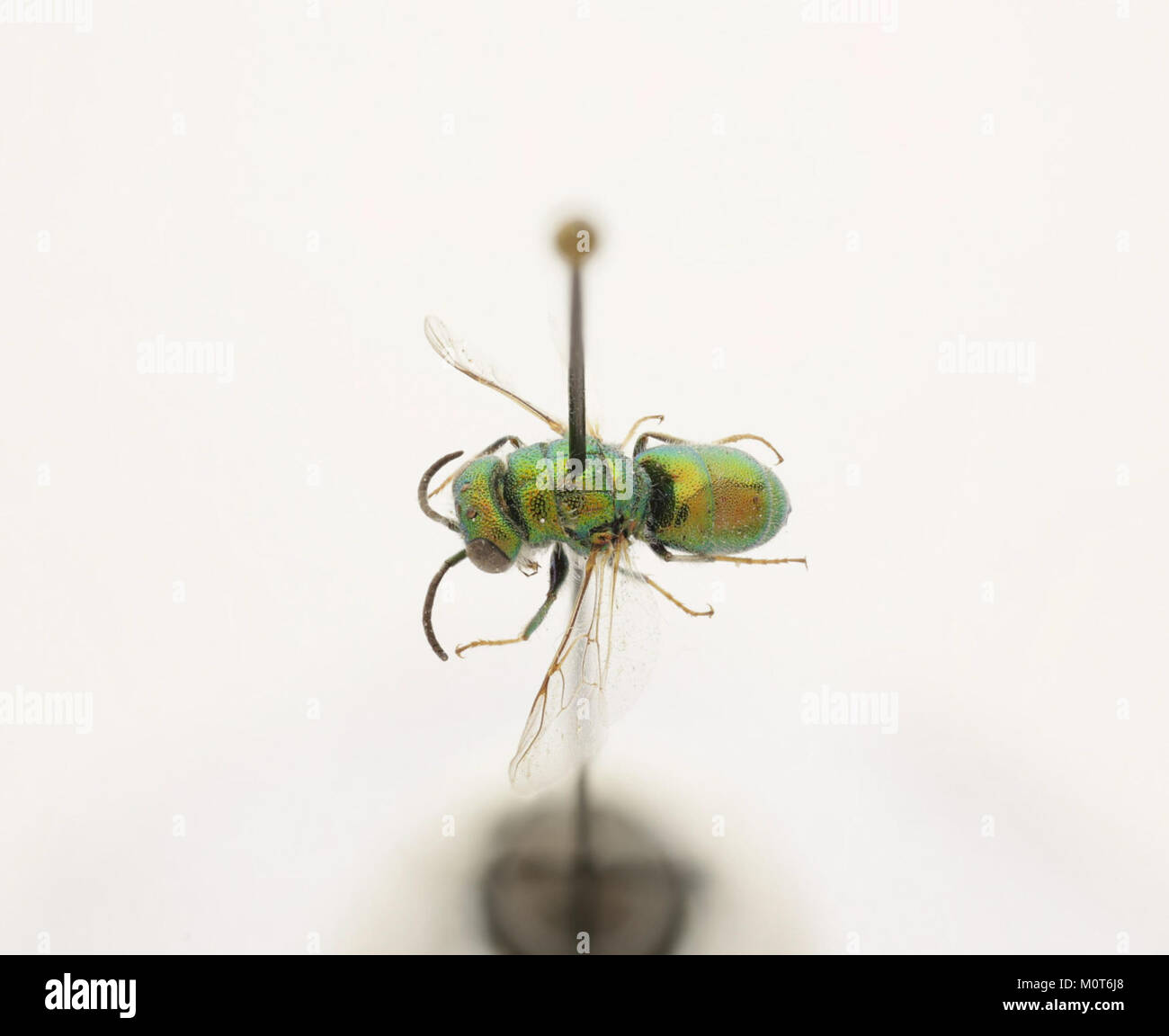 Cephalochrysis ehrenbergi is a species of microscopic organism, noted for its role in the zoosphere and its importance in biological research. It appears in Zoosphere 128 271 as part of a study on marine life. Stock Photohttps://www.alamy.com/image-license-details/?v=1https://www.alamy.com/stock-photo-cephalochrysis-ehrenbergi-is-a-species-of-microscopic-organism-noted-172635744.html
RMM0T6J8–Cephalochrysis ehrenbergi is a species of microscopic organism, noted for its role in the zoosphere and its importance in biological research. It appears in Zoosphere 128 271 as part of a study on marine life.
 Mastigocerca bicupses, observed from a dorsal view, is a species of parasitic copepod. This microscopic organism inhabits the gills of fish and plays a role in marine ecosystems. Stock Photohttps://www.alamy.com/image-license-details/?v=1https://www.alamy.com/stock-photo-mastigocerca-bicupses-observed-from-a-dorsal-view-is-a-species-of-115088848.html
RMGK6N00–Mastigocerca bicupses, observed from a dorsal view, is a species of parasitic copepod. This microscopic organism inhabits the gills of fish and plays a role in marine ecosystems.
 Microscopic bacteria. Bacterium microorganism, viruses and microbes backdrop. Microbiology virus science 3d vector background Stock Vectorhttps://www.alamy.com/image-license-details/?v=1https://www.alamy.com/microscopic-bacteria-bacterium-microorganism-viruses-and-microbes-backdrop-microbiology-virus-science-3d-vector-background-image344826738.html
RF2B1060J–Microscopic bacteria. Bacterium microorganism, viruses and microbes backdrop. Microbiology virus science 3d vector background
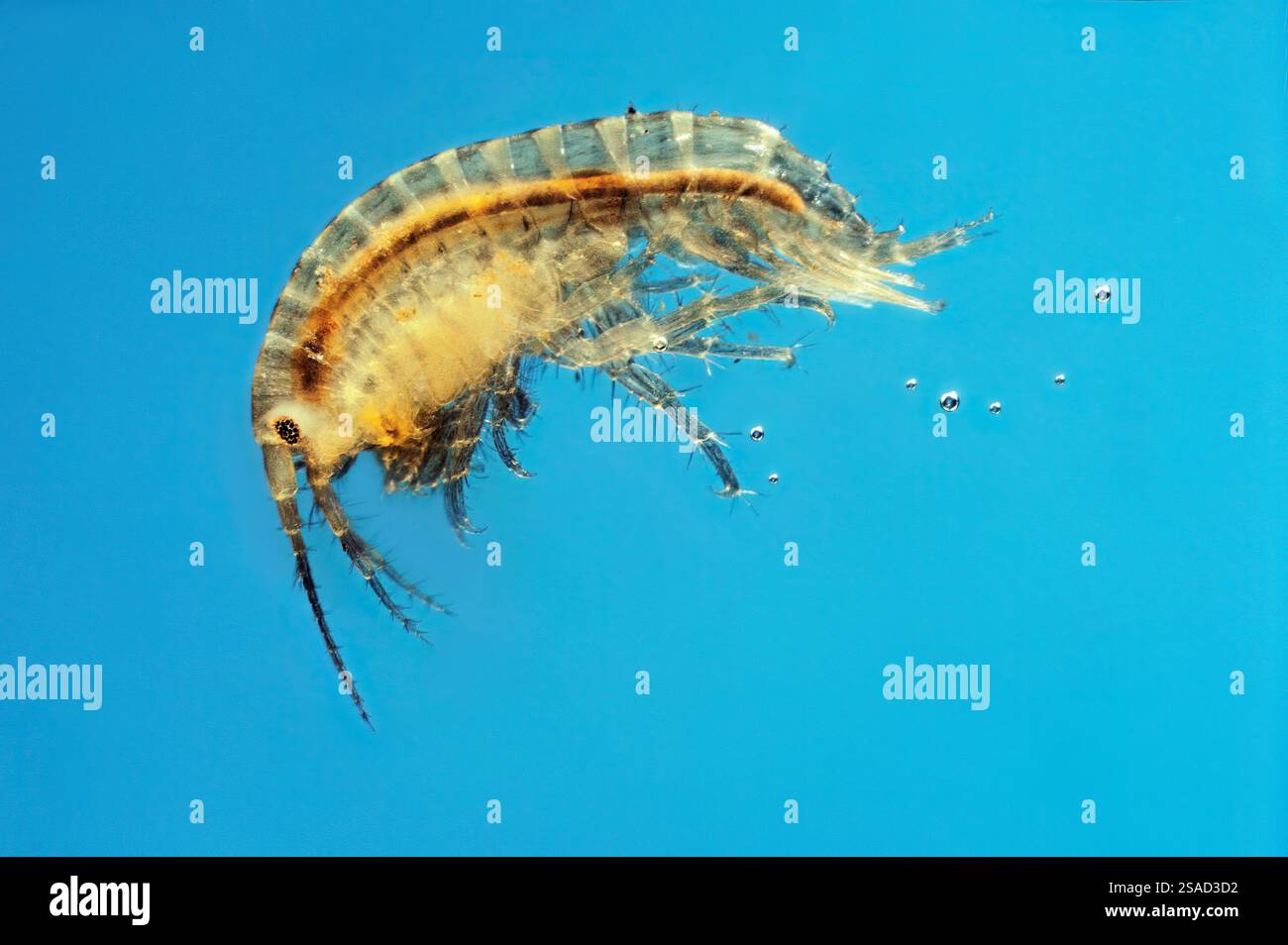 Krill (Euphausiacea), a small crustacean vital to marine ecosystems, imaged at 10x magnification. Stock Photohttps://www.alamy.com/image-license-details/?v=1https://www.alamy.com/krill-euphausiacea-a-small-crustacean-vital-to-marine-ecosystems-imaged-at-10x-magnification-image643218270.html
RM2SAD3D2–Krill (Euphausiacea), a small crustacean vital to marine ecosystems, imaged at 10x magnification.
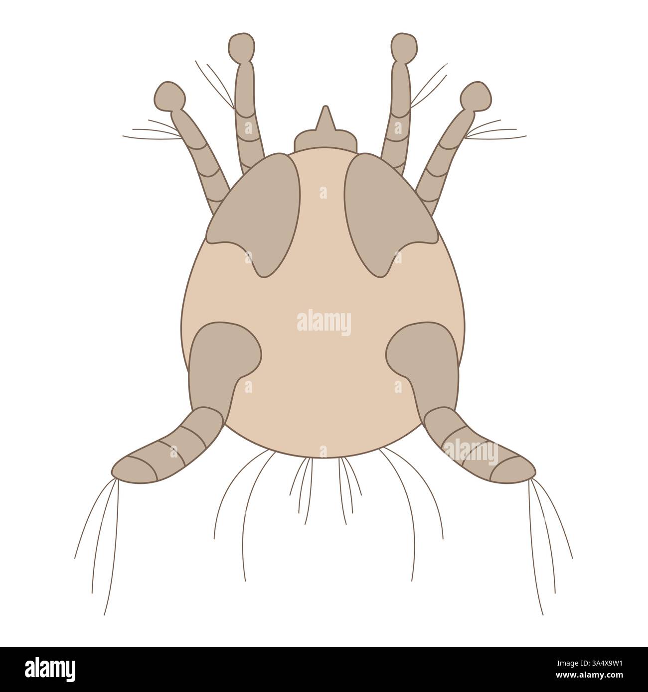
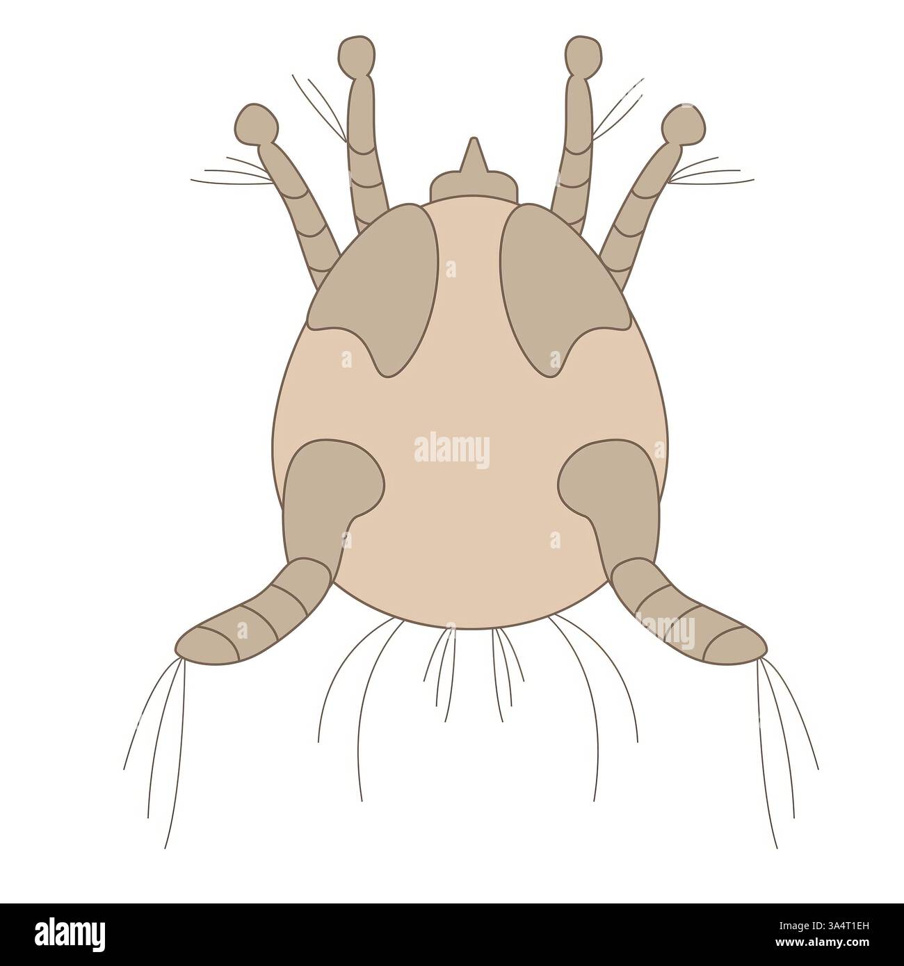 Detailed illustration of a dust mite on a white background Stock Photohttps://www.alamy.com/image-license-details/?v=1https://www.alamy.com/detailed-illustration-of-a-dust-mite-on-a-white-background-image656980649.html
RF3A4T1EH–Detailed illustration of a dust mite on a white background
RMP24YYP–. Frondicularia complanata 129 Frondicularia complanata - - Print - Iconographia Zoologica - Special Collections University of Amsterdam - UBAINV0274 113 07 0008
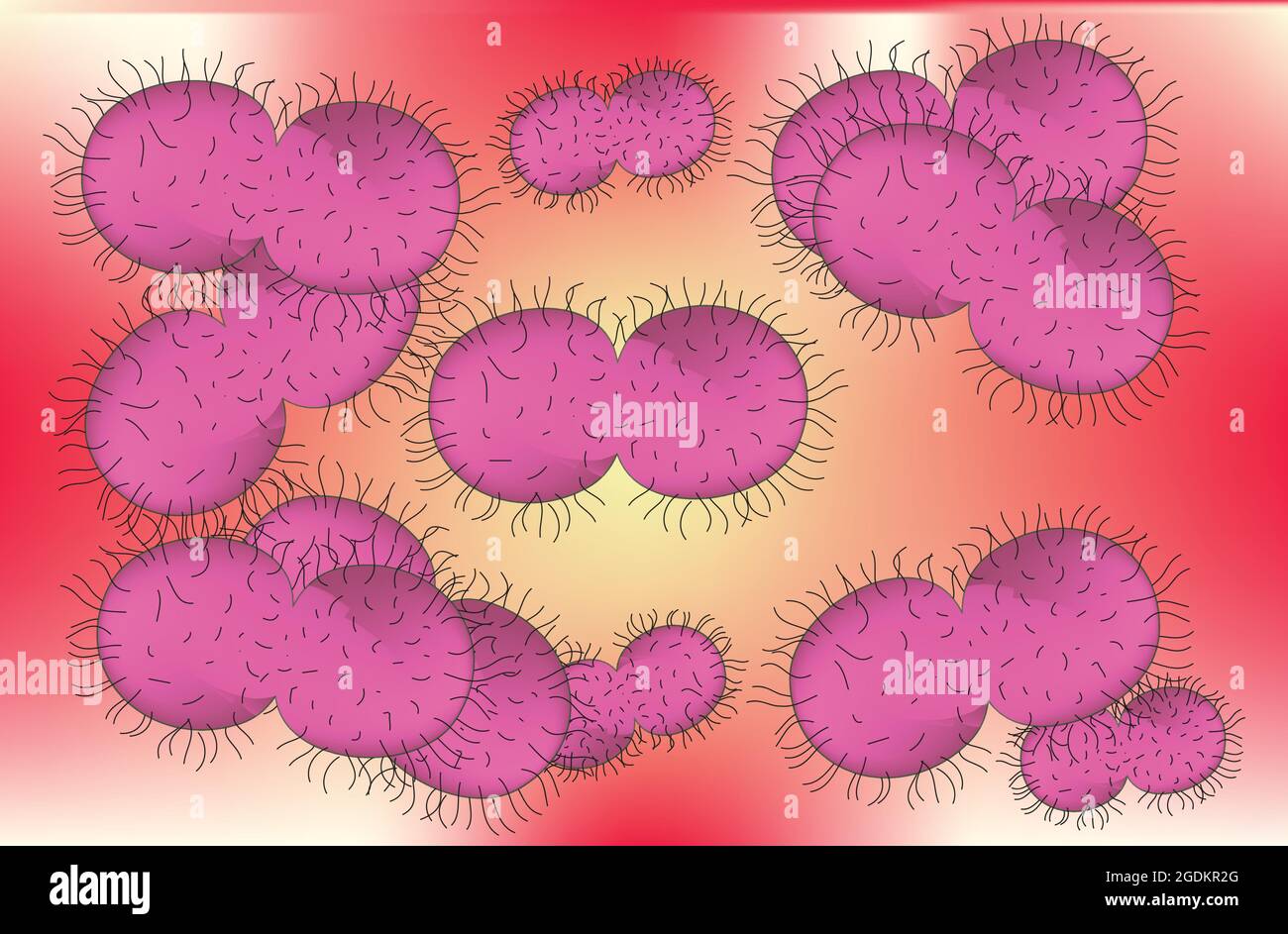 Neisseria gonorrhoeae, gonorrhea, gonorrhea bacteria, gonococcus, anatomy of gonococcus, gonococcus structure Stock Vectorhttps://www.alamy.com/image-license-details/?v=1https://www.alamy.com/neisseria-gonorrhoeae-gonorrhea-gonorrhea-bacteria-gonococcus-anatomy-of-gonococcus-gonococcus-structure-image438684920.html
RF2GDKR2G–Neisseria gonorrhoeae, gonorrhea, gonorrhea bacteria, gonococcus, anatomy of gonococcus, gonococcus structure
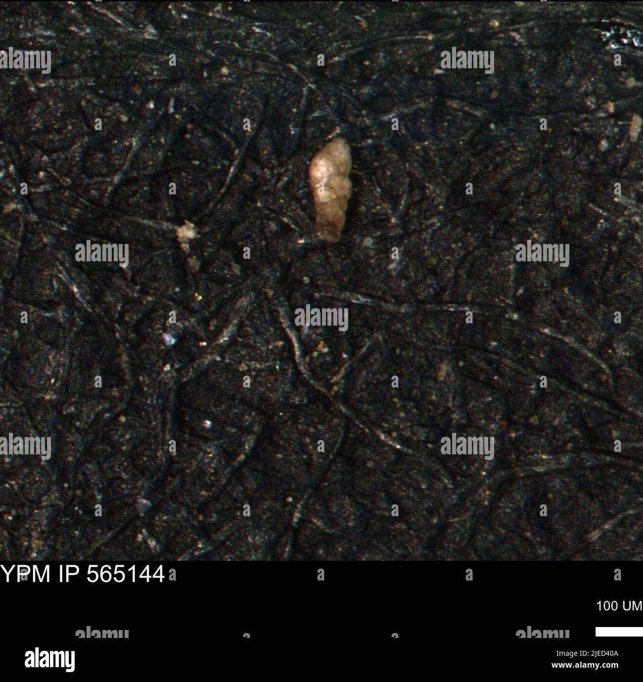 Verneuilinoides kansasensis Loeblich & Tappan. Stock Photohttps://www.alamy.com/image-license-details/?v=1https://www.alamy.com/verneuilinoides-kansasensis-loeblich-tappan-image473573642.html
RM2JED40A–Verneuilinoides kansasensis Loeblich & Tappan.
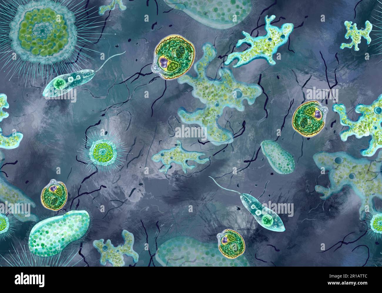 Bacteria, unicellular protozoa, algae. Seamless pattern for printing in textbooks, medical brochures, packaging, fabrics and other polygraphy. Color illustration of microbiology. High quality illustration Stock Photohttps://www.alamy.com/image-license-details/?v=1https://www.alamy.com/bacteria-unicellular-protozoa-algae-seamless-pattern-for-printing-in-textbooks-medical-brochures-packaging-fabrics-and-other-polygraphy-color-illustration-of-microbiology-high-quality-illustration-image551585452.html
RF2R1ATTC–Bacteria, unicellular protozoa, algae. Seamless pattern for printing in textbooks, medical brochures, packaging, fabrics and other polygraphy. Color illustration of microbiology. High quality illustration
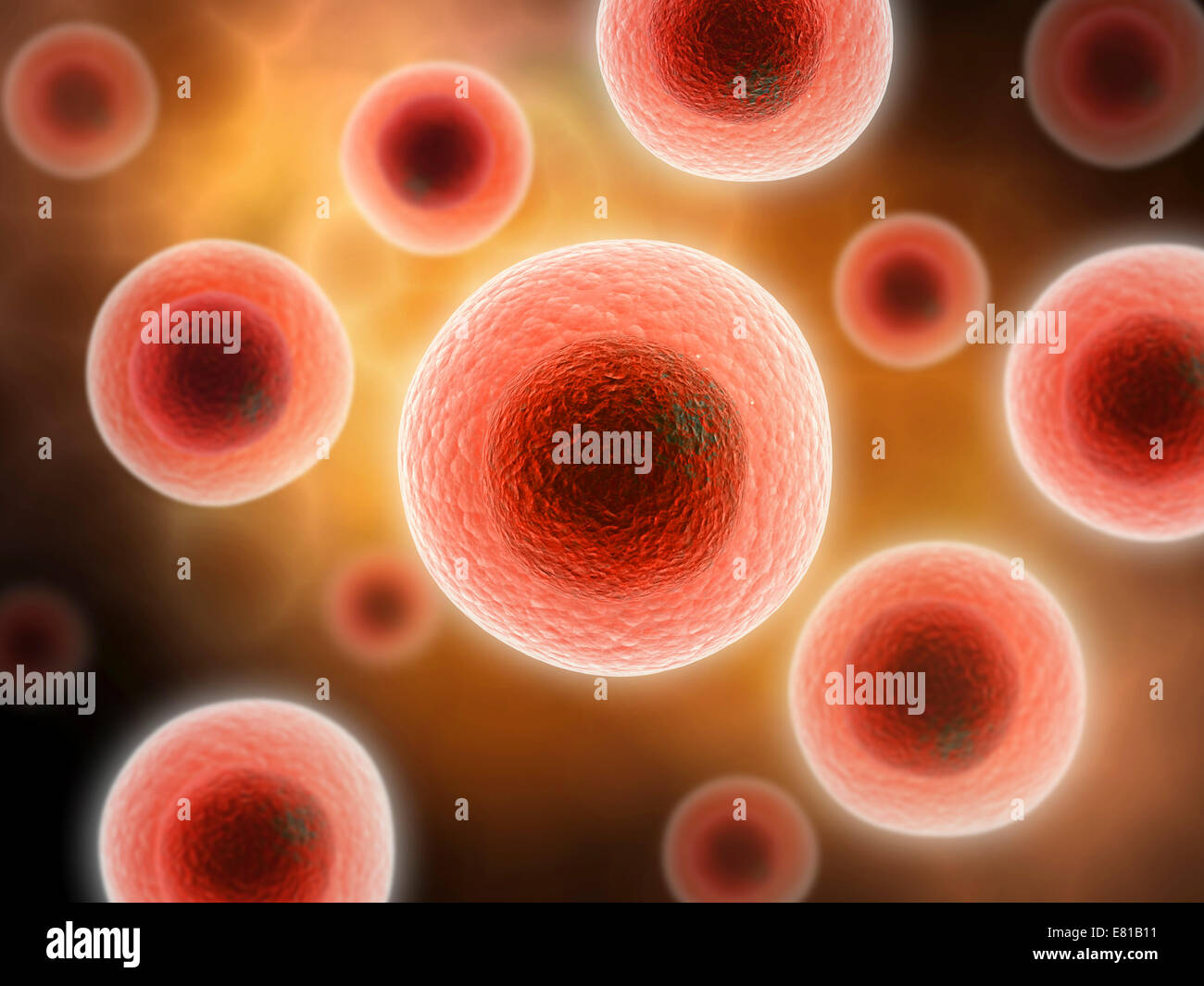 Microscopic view of cell. Stock Photohttps://www.alamy.com/image-license-details/?v=1https://www.alamy.com/stock-photo-microscopic-view-of-cell-73789325.html
RFE81B11–Microscopic view of cell.
RF2E4D361–Virus solid icon. Biology microbe bacterium and germ glyph style pictogram on white background. Science and microbiology signs for mobile concept and
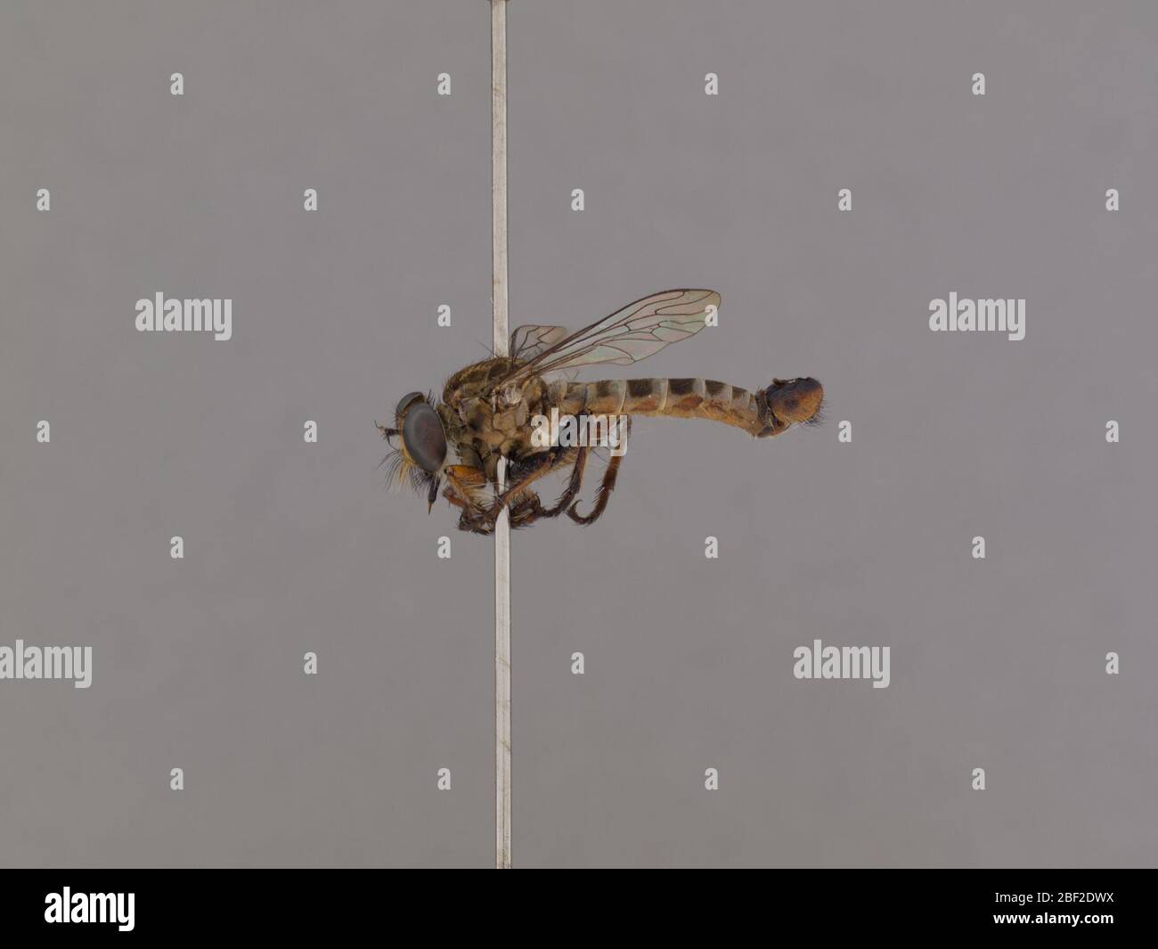 Wilcoxius tumidus. 17 Mar 20201 Stock Photohttps://www.alamy.com/image-license-details/?v=1https://www.alamy.com/wilcoxius-tumidus-17-mar-20201-image353482022.html
RM2BF2DWX–Wilcoxius tumidus. 17 Mar 20201
 Covid-19 virus vector sign on the black and white Stock Vectorhttps://www.alamy.com/image-license-details/?v=1https://www.alamy.com/covid-19-virus-vector-sign-on-the-black-and-white-image354913802.html
RF2BHBM4X–Covid-19 virus vector sign on the black and white
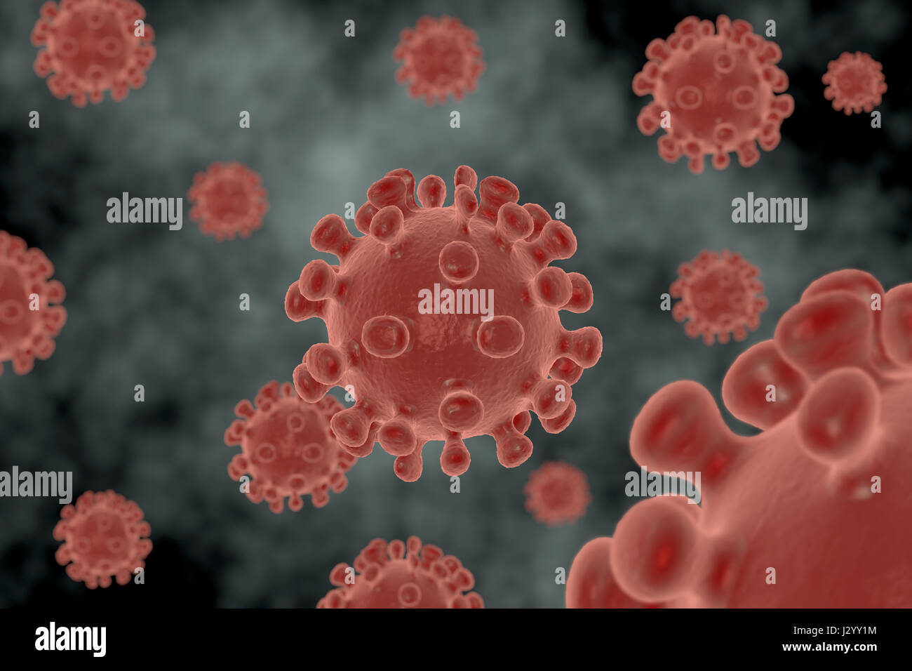 Rendering of a virus isolated in an organism Stock Photohttps://www.alamy.com/image-license-details/?v=1https://www.alamy.com/stock-photo-rendering-of-a-virus-isolated-in-an-organism-139526176.html
RFJ2YY1M–Rendering of a virus isolated in an organism
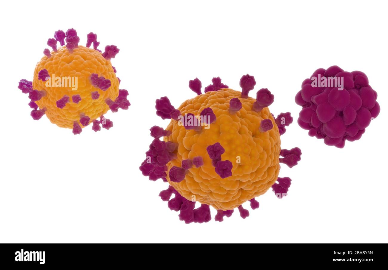 Coronavirus covid19 isolated model, 3D render on white background based on the virus microscopic images Stock Photohttps://www.alamy.com/image-license-details/?v=1https://www.alamy.com/coronavirus-covid19-isolated-model-3d-render-on-white-background-based-on-the-virus-microscopic-images-image350616721.html
RF2BABY5N–Coronavirus covid19 isolated model, 3D render on white background based on the virus microscopic images
RF2MJFYPH–Cell, collagen, regeneration vector icon on transparent background. Outline Cell, collagen, regeneration vector icon
RF2ATTM8R–Virus vector illustration icon template design
 Botanisk Tidsskrift København Dansk Botanisk Forening Illustration Denmark Drawing Snake Archives Botany Periodicals Fieldbook Jenny Hansen Eugen Warming, The illustration showcases a series of cellular structures, likely representing various stages of cell division or developmental forms in a microscopic organism. The figures are arranged in a grid pattern, with each labeled from 1 to 28, indicating different morphological characteristics or states. Some depict clusters of cells with distinct shapes, such as spherical or elongated forms, while others illustrate the gradual process of cell rep Stock Photohttps://www.alamy.com/image-license-details/?v=1https://www.alamy.com/botanisk-tidsskrift-kbenhavn-dansk-botanisk-forening-illustration-denmark-drawing-snake-archives-botany-periodicals-fieldbook-jenny-hansen-eugen-warming-the-illustration-showcases-a-series-of-cellular-structures-likely-representing-various-stages-of-cell-division-or-developmental-forms-in-a-microscopic-organism-the-figures-are-arranged-in-a-grid-pattern-with-each-labeled-from-1-to-28-indicating-different-morphological-characteristics-or-states-some-depict-clusters-of-cells-with-distinct-shapes-such-as-spherical-or-elongated-forms-while-others-illustrate-the-gradual-process-of-cell-rep-image643596109.html
RM2SB29B9–Botanisk Tidsskrift København Dansk Botanisk Forening Illustration Denmark Drawing Snake Archives Botany Periodicals Fieldbook Jenny Hansen Eugen Warming, The illustration showcases a series of cellular structures, likely representing various stages of cell division or developmental forms in a microscopic organism. The figures are arranged in a grid pattern, with each labeled from 1 to 28, indicating different morphological characteristics or states. Some depict clusters of cells with distinct shapes, such as spherical or elongated forms, while others illustrate the gradual process of cell rep
RF2C70793–Virus bacteria icons set. Cartoon flat color vector illustration
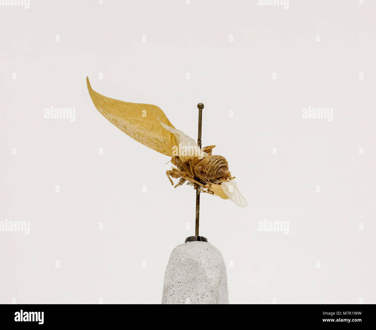 This is an image of Hemidictya frondosa, a species of freshwater diatom. It highlights the intricate structure of this microscopic organism found in aquatic ecosystems. Stock Photohttps://www.alamy.com/image-license-details/?v=1https://www.alamy.com/stock-photo-this-is-an-image-of-hemidictya-frondosa-a-species-of-freshwater-diatom-176824869.html
RMM7K1WW–This is an image of Hemidictya frondosa, a species of freshwater diatom. It highlights the intricate structure of this microscopic organism found in aquatic ecosystems.
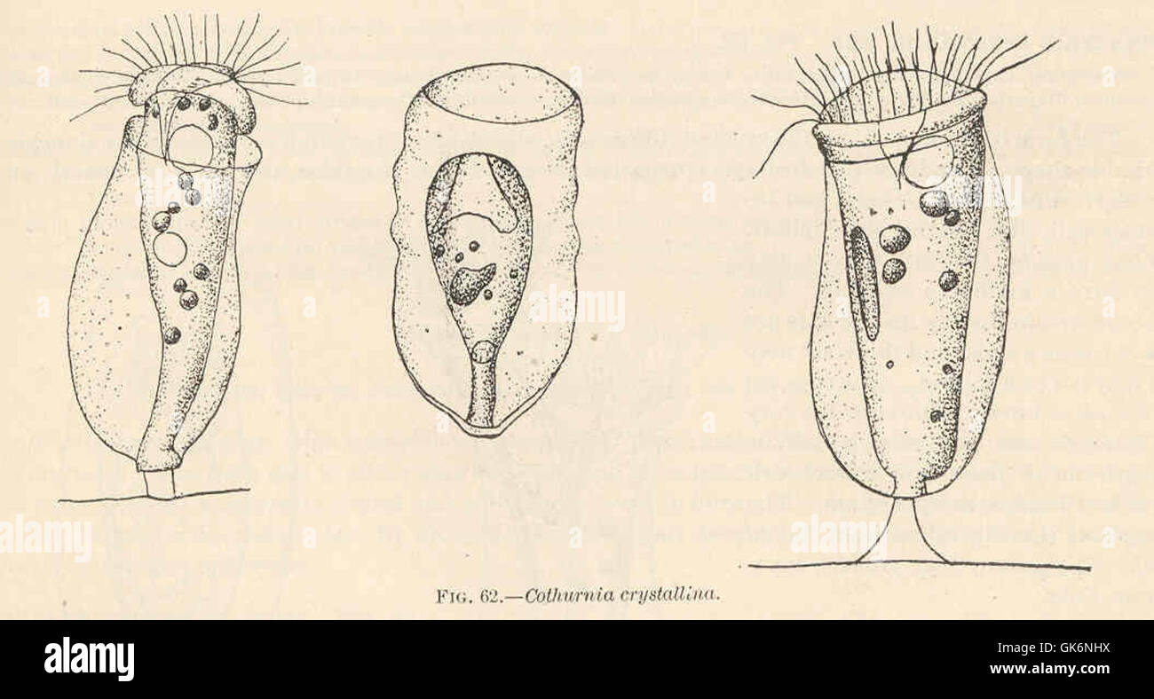 *Cothurnia crystallini*, a species of freshwater rotifer, a microscopic organism found in various aquatic environments. This species is studied for its role in aquatic food webs and ecosystem functioning. Stock Photohttps://www.alamy.com/image-license-details/?v=1https://www.alamy.com/stock-photo-cothurnia-crystallini-a-species-of-freshwater-rotifer-a-microscopic-115089350.html
RMGK6NHX–*Cothurnia crystallini*, a species of freshwater rotifer, a microscopic organism found in various aquatic environments. This species is studied for its role in aquatic food webs and ecosystem functioning.
RF2WW1AJ4–Microbes round icon. Biology symbol. Microscopic organism
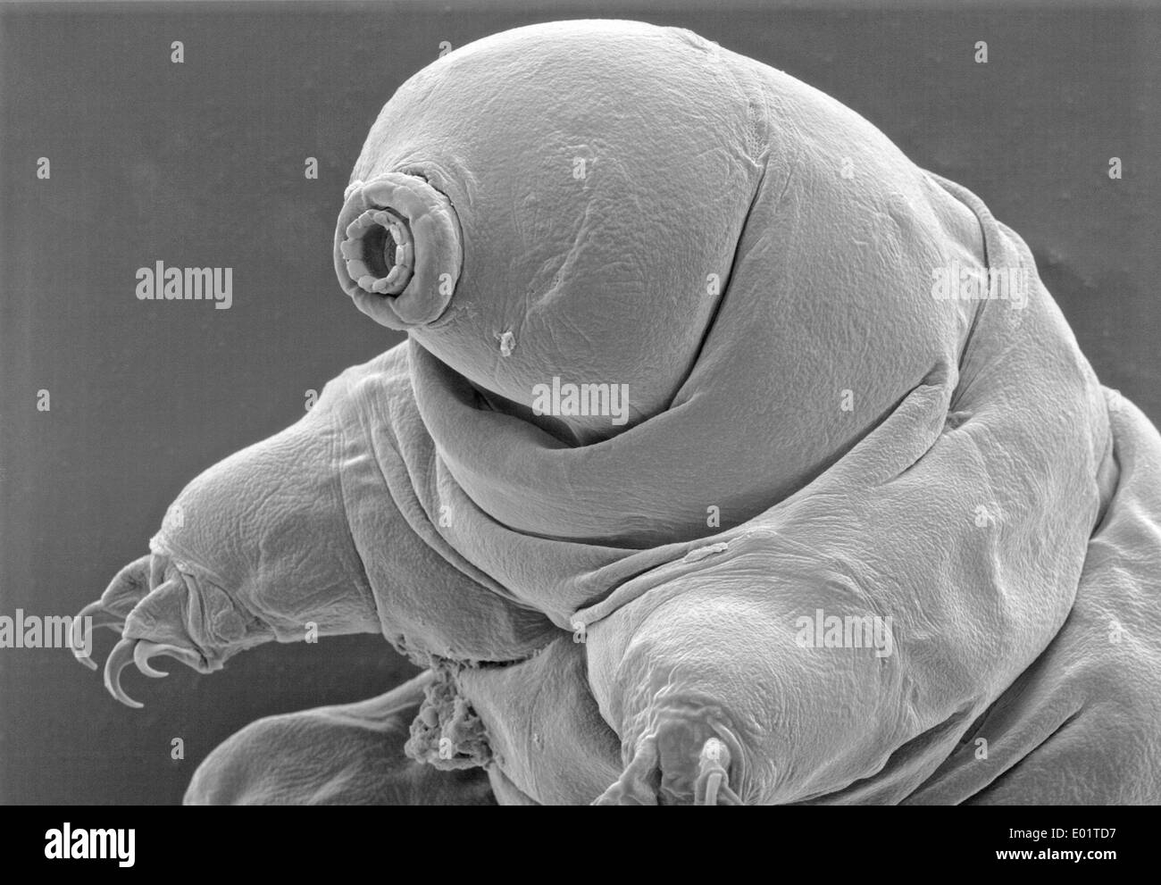 Microscopic view of a Tardigrades or water bear (Milnesium tardigradum) a extremophile organism that can thrive in extreme conditions captured October 21, 2011 in Saguaro National Park, Arizona. Stock Photohttps://www.alamy.com/image-license-details/?v=1https://www.alamy.com/microscopic-view-of-a-tardigrades-or-water-bear-milnesium-tardigradum-image68882611.html
RME01TD7–Microscopic view of a Tardigrades or water bear (Milnesium tardigradum) a extremophile organism that can thrive in extreme conditions captured October 21, 2011 in Saguaro National Park, Arizona.
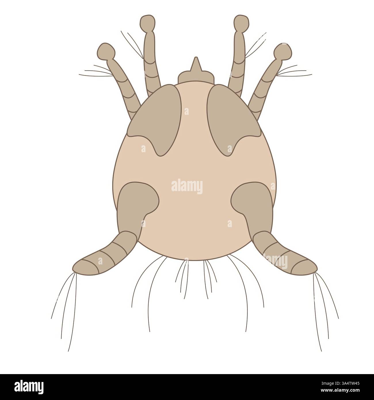 Detailed illustration of a dust mite on a white background Stock Photohttps://www.alamy.com/image-license-details/?v=1https://www.alamy.com/detailed-illustration-of-a-dust-mite-on-a-white-background-image656999173.html
RF3A4TW45–Detailed illustration of a dust mite on a white background
RMP250CA–. Stylonychia pustulata 298 Stylonychia pustulata - - Print - Iconographia Zoologica - Special Collections University of Amsterdam - UBAINV0274 113 18 0010
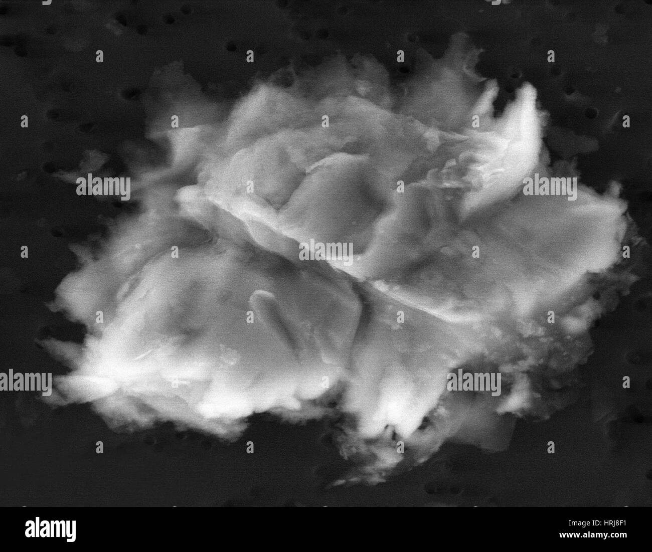 Lake Vostok Micro-Organism, SEM Stock Photohttps://www.alamy.com/image-license-details/?v=1https://www.alamy.com/stock-photo-lake-vostok-micro-organism-sem-135011493.html
RMHRJ8F1–Lake Vostok Micro-Organism, SEM
 Foraminiferida. Stock Photohttps://www.alamy.com/image-license-details/?v=1https://www.alamy.com/foraminiferida-image473935511.html
RM2JF1HG7–Foraminiferida.
 Bacteria, unicellular protozoa, algae. Seamless pattern for printing in textbooks, medical brochures, packaging, fabrics and other polygraphy. Color illustration of microbiology. High quality illustration Stock Photohttps://www.alamy.com/image-license-details/?v=1https://www.alamy.com/bacteria-unicellular-protozoa-algae-seamless-pattern-for-printing-in-textbooks-medical-brochures-packaging-fabrics-and-other-polygraphy-color-illustration-of-microbiology-high-quality-illustration-image551585151.html
RF2R1ATDK–Bacteria, unicellular protozoa, algae. Seamless pattern for printing in textbooks, medical brochures, packaging, fabrics and other polygraphy. Color illustration of microbiology. High quality illustration
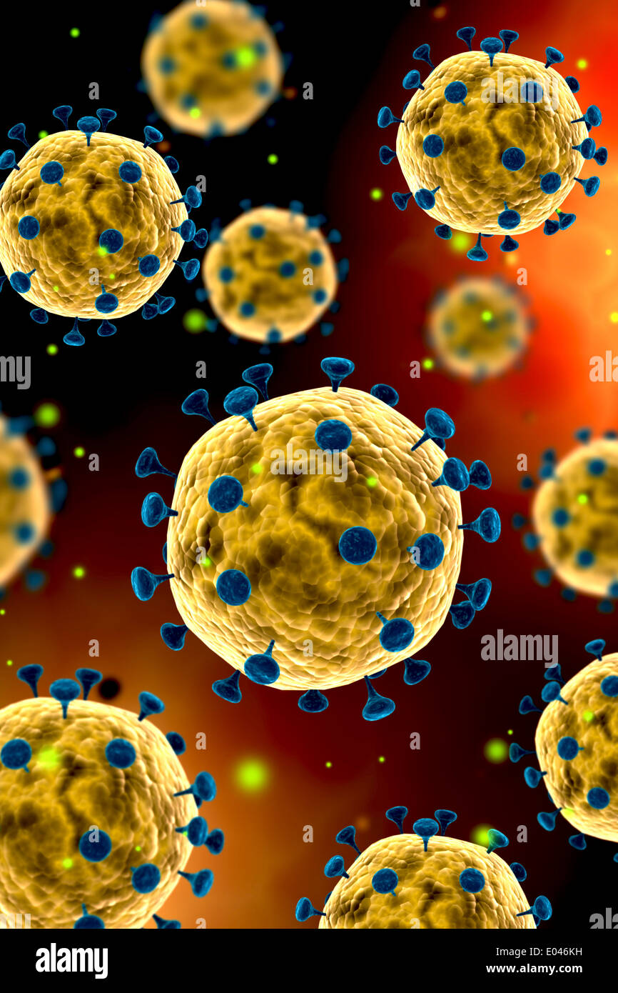 Microscopic view of coronavirus. Stock Photohttps://www.alamy.com/image-license-details/?v=1https://www.alamy.com/microscopic-view-of-coronavirus-image68934533.html
RFE046KH–Microscopic view of coronavirus.
RF2E4D3PP–Virus line and solid icon. Biology microbe bacterium and germ outline style pictogram on white background. Science and microbiology signs for mobile
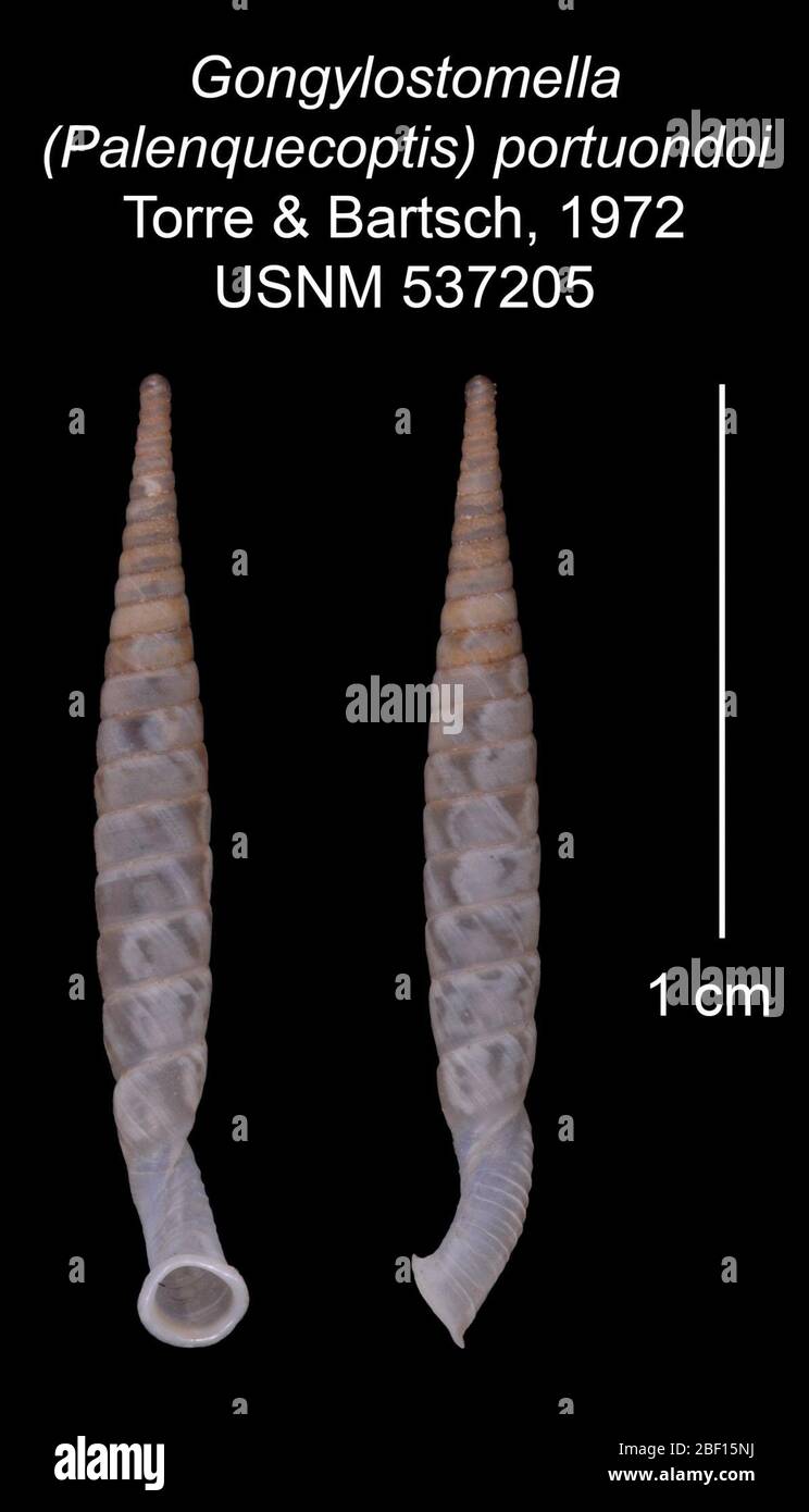 Gongylostomella Palenquecoptis portuondoi. 20 Jan 20161 Stock Photohttps://www.alamy.com/image-license-details/?v=1https://www.alamy.com/gongylostomella-palenquecoptis-portuondoi-20-jan-20161-image353453678.html
RM2BF15NJ–Gongylostomella Palenquecoptis portuondoi. 20 Jan 20161
 microscopic organism made from oil and water Stock Photohttps://www.alamy.com/image-license-details/?v=1https://www.alamy.com/stock-image-microscopic-organism-made-from-oil-and-water-161777561.html
RFKB5GX1–microscopic organism made from oil and water
 Black and white drawing of a paramecium swimming gracefully, displaying delicate cilia, perfect for educational purposes Stock Vectorhttps://www.alamy.com/image-license-details/?v=1https://www.alamy.com/black-and-white-drawing-of-a-paramecium-swimming-gracefully-displaying-delicate-cilia-perfect-for-educational-purposes-image628386111.html
RF2YE9CW3–Black and white drawing of a paramecium swimming gracefully, displaying delicate cilia, perfect for educational purposes
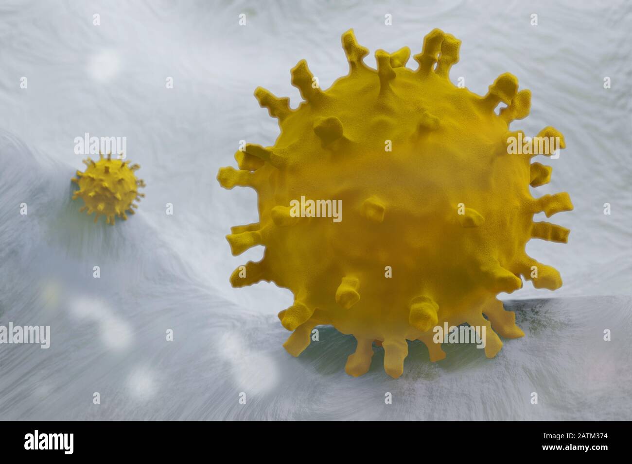 Scientific illustration of the Corona virus, 3D render based on microscopic images of the virus from th 2020 China outbreak Stock Photohttps://www.alamy.com/image-license-details/?v=1https://www.alamy.com/scientific-illustration-of-the-corona-virus-3d-render-based-on-microscopic-images-of-the-virus-from-th-2020-china-outbreak-image342190328.html
RF2ATM374–Scientific illustration of the Corona virus, 3D render based on microscopic images of the virus from th 2020 China outbreak
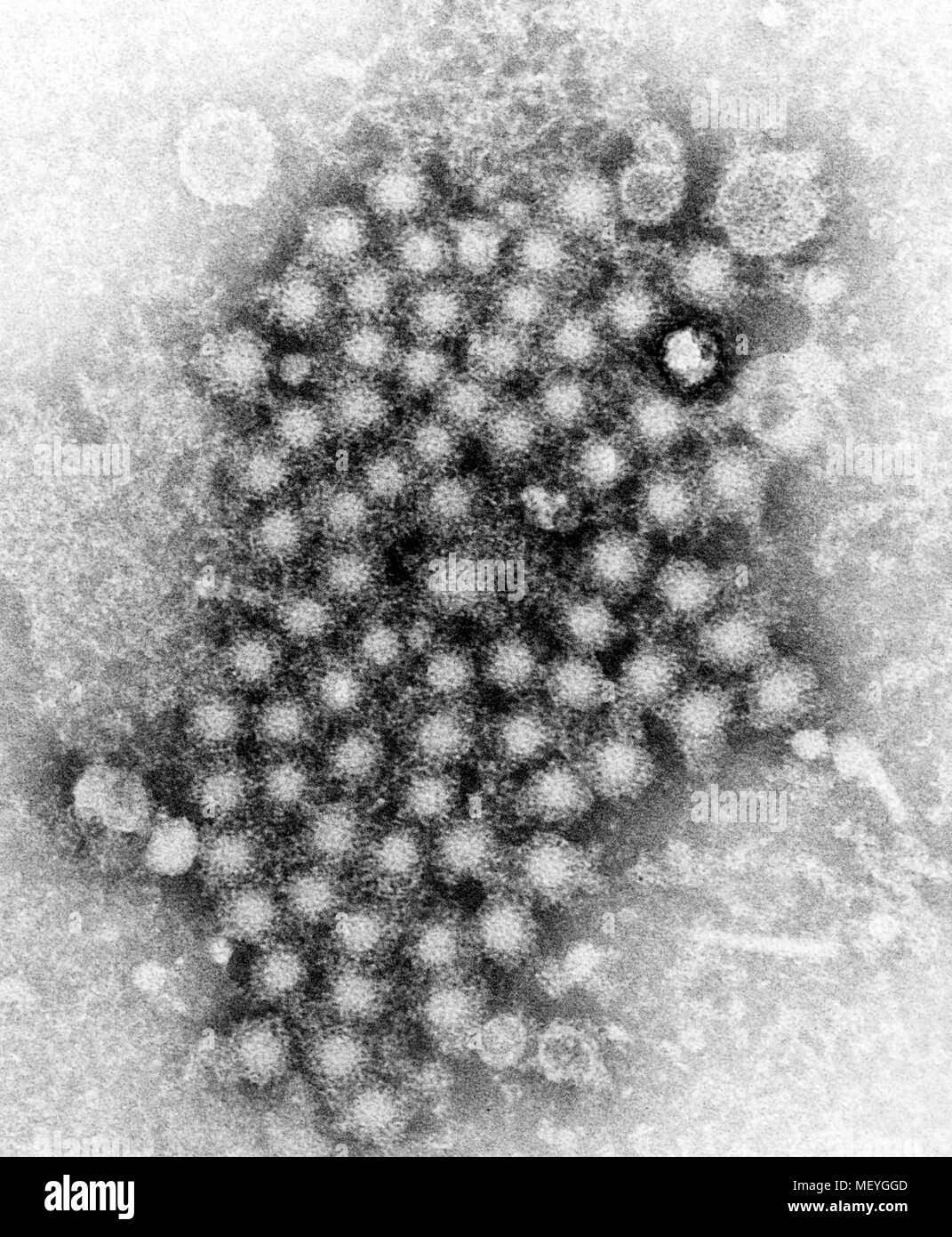 Hepatitis virions of an unknown strain of the organism revealed in the transmission electron microscopic (TEM) image, 1962. Image courtesy Centers for Disease Control (CDC) / E.H. Cook, Jr. () Stock Photohttps://www.alamy.com/image-license-details/?v=1https://www.alamy.com/hepatitis-virions-of-an-unknown-strain-of-the-organism-revealed-in-the-transmission-electron-microscopic-tem-image-1962-image-courtesy-centers-for-disease-control-cdc-eh-cook-jr-image181314573.html
RMMEYGGD–Hepatitis virions of an unknown strain of the organism revealed in the transmission electron microscopic (TEM) image, 1962. Image courtesy Centers for Disease Control (CDC) / E.H. Cook, Jr. ()
RF2ATTM8F–Virus vector illustration icon template design
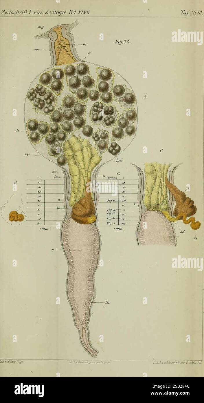 Zeitschrift für wissenschaftliche Zoologie, Leipzig, Wilhelm Engelmann, 1849-, anatomy, comparative, bibliography, periodicals, zoology, Museum of Comparative Zoology, A detailed scientific illustration depicting various stages of a microscopic organism. The main figure showcases a large, bulbous body, densely populated with smaller circular structures resembling cells or spores. Tentacle-like appendages extend from the lower part of the body, suggesting a means of movement or feeding. Accompanying labels and a scale bar indicate measurements, while additional illustrations on the side present Stock Photohttps://www.alamy.com/image-license-details/?v=1https://www.alamy.com/zeitschrift-fr-wissenschaftliche-zoologie-leipzig-wilhelm-engelmann-1849-anatomy-comparative-bibliography-periodicals-zoology-museum-of-comparative-zoology-a-detailed-scientific-illustration-depicting-various-stages-of-a-microscopic-organism-the-main-figure-showcases-a-large-bulbous-body-densely-populated-with-smaller-circular-structures-resembling-cells-or-spores-tentacle-like-appendages-extend-from-the-lower-part-of-the-body-suggesting-a-means-of-movement-or-feeding-accompanying-labels-and-a-scale-bar-indicate-measurements-while-additional-illustrations-on-the-side-present-image643595916.html
RM2SB294C–Zeitschrift für wissenschaftliche Zoologie, Leipzig, Wilhelm Engelmann, 1849-, anatomy, comparative, bibliography, periodicals, zoology, Museum of Comparative Zoology, A detailed scientific illustration depicting various stages of a microscopic organism. The main figure showcases a large, bulbous body, densely populated with smaller circular structures resembling cells or spores. Tentacle-like appendages extend from the lower part of the body, suggesting a means of movement or feeding. Accompanying labels and a scale bar indicate measurements, while additional illustrations on the side present
RF2B0TJ5R–Infection bacteria and pandemic virus vector biology icons. Illustration of bacteria and microbe organism allergen
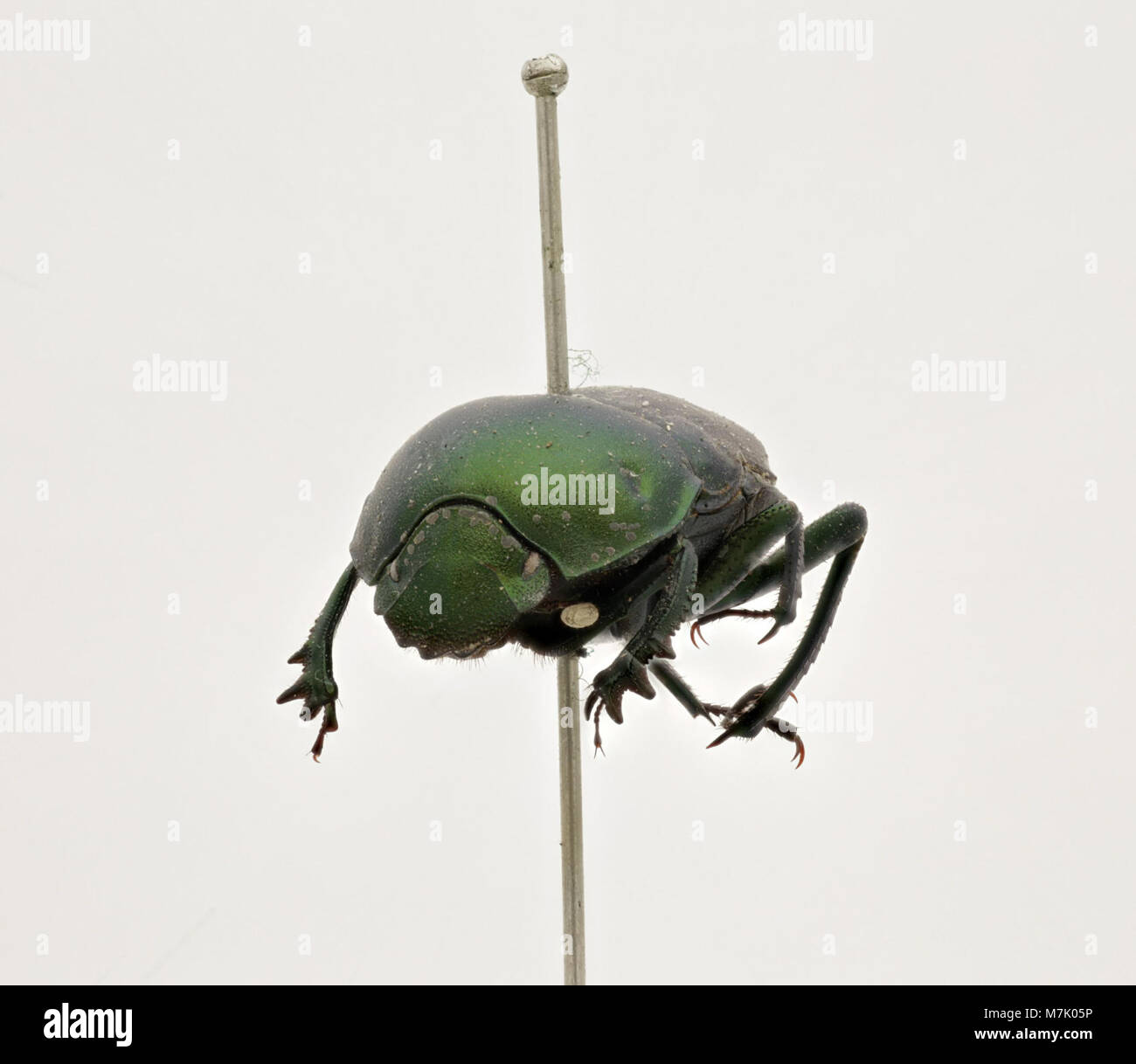 A close-up image of Garreta azureus, a species of zooplankton, showing the detailed features of this microscopic organism, important in aquatic ecosystems. Stock Photohttps://www.alamy.com/image-license-details/?v=1https://www.alamy.com/stock-photo-a-close-up-image-of-garreta-azureus-a-species-of-zooplankton-showing-176823522.html
RMM7K05P–A close-up image of Garreta azureus, a species of zooplankton, showing the detailed features of this microscopic organism, important in aquatic ecosystems.
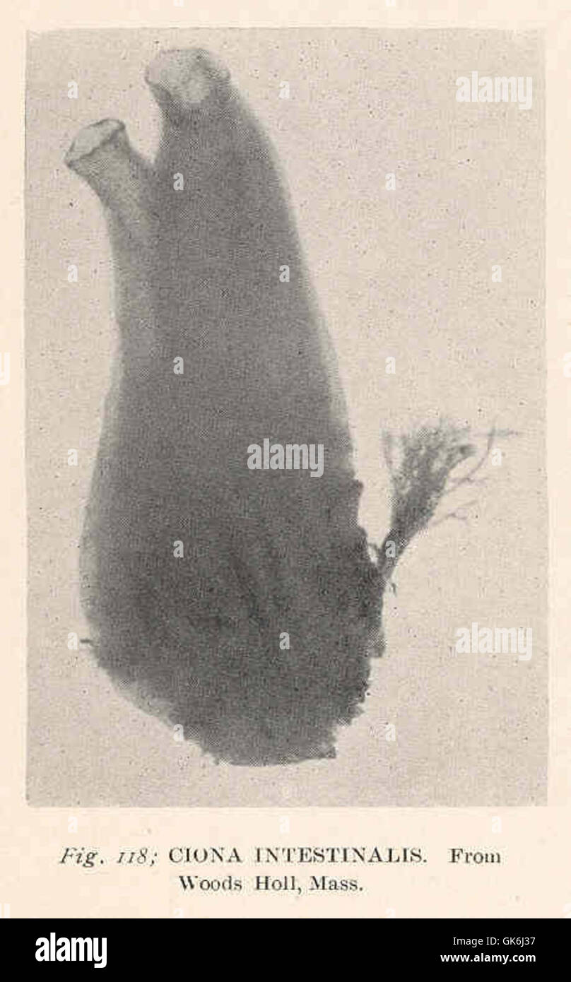 Cioona intestinalis is a species of marine ciliate found in the waters of Woods Hole, Massachusetts. This microscopic organism plays a role in the microbial food web of marine ecosystems. Stock Photohttps://www.alamy.com/image-license-details/?v=1https://www.alamy.com/stock-photo-cioona-intestinalis-is-a-species-of-marine-ciliate-found-in-the-waters-115086587.html
RMGK6J37–Cioona intestinalis is a species of marine ciliate found in the waters of Woods Hole, Massachusetts. This microscopic organism plays a role in the microbial food web of marine ecosystems.
 Microbes, viruses, bacteria, microorganism cells and primitive organism line vector concept. Virus cell and microbe, bacteria organism, medical microscopic illustration. Microbes, viruses, bacteria, microorganism cells and primitive organism line vector concept Stock Vectorhttps://www.alamy.com/image-license-details/?v=1https://www.alamy.com/microbes-viruses-bacteria-microorganism-cells-and-primitive-organism-line-vector-concept-virus-cell-and-microbe-bacteria-organism-medical-microscopic-illustration-microbes-viruses-bacteria-microorganism-cells-and-primitive-organism-line-vector-concept-image639969079.html
RF2S5532F–Microbes, viruses, bacteria, microorganism cells and primitive organism line vector concept. Virus cell and microbe, bacteria organism, medical microscopic illustration. Microbes, viruses, bacteria, microorganism cells and primitive organism line vector concept
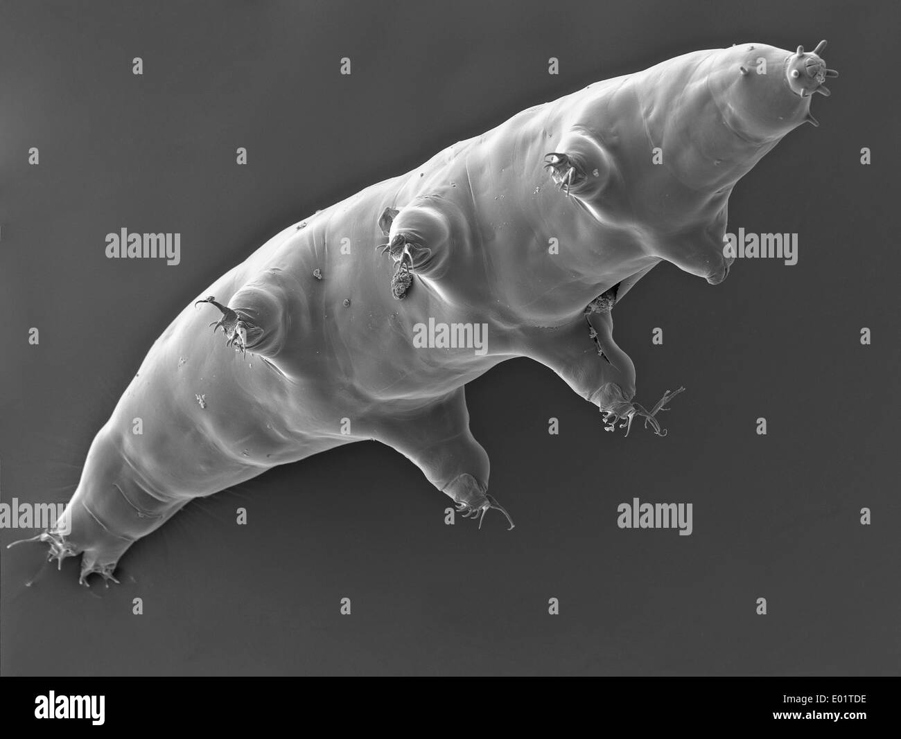 Microscopic view of a Tardigrades or water bear (Milnesium tardigradum) a extremophile organism that can thrive in extreme conditions captured October 21, 2011 in Saguaro National Park, Arizona. Stock Photohttps://www.alamy.com/image-license-details/?v=1https://www.alamy.com/microscopic-view-of-a-tardigrades-or-water-bear-milnesium-tardigradum-image68882618.html
RME01TDE–Microscopic view of a Tardigrades or water bear (Milnesium tardigradum) a extremophile organism that can thrive in extreme conditions captured October 21, 2011 in Saguaro National Park, Arizona.
