Quick filters:
Female pelvis Stock Photos and Images
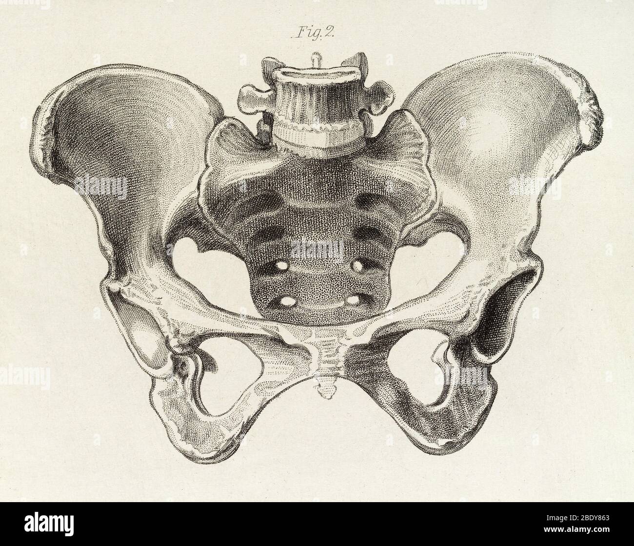 Female Pelvis, Illustration, c.1856 Stock Photohttps://www.alamy.com/image-license-details/?v=1https://www.alamy.com/female-pelvis-illustration-c1856-image352797035.html
RM2BDY863–Female Pelvis, Illustration, c.1856
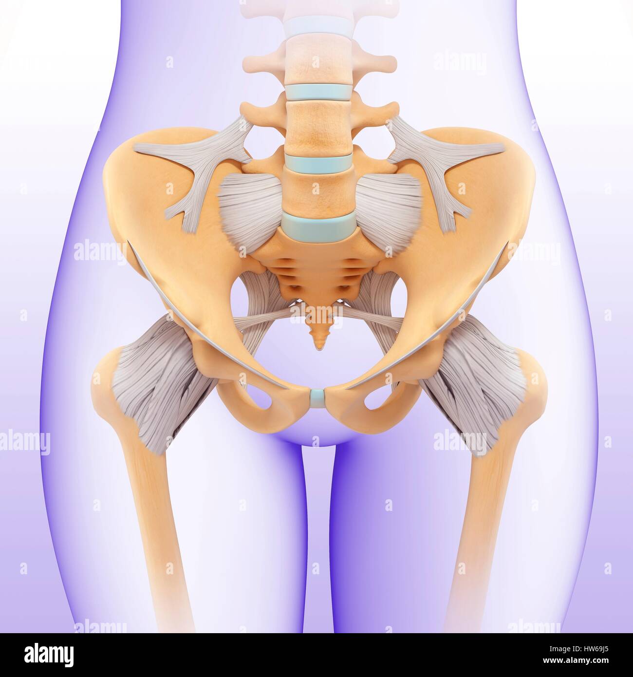 Illustration of a female pelvis. Stock Photohttps://www.alamy.com/image-license-details/?v=1https://www.alamy.com/stock-photo-illustration-of-a-female-pelvis-135978253.html
RFHW69J5–Illustration of a female pelvis.
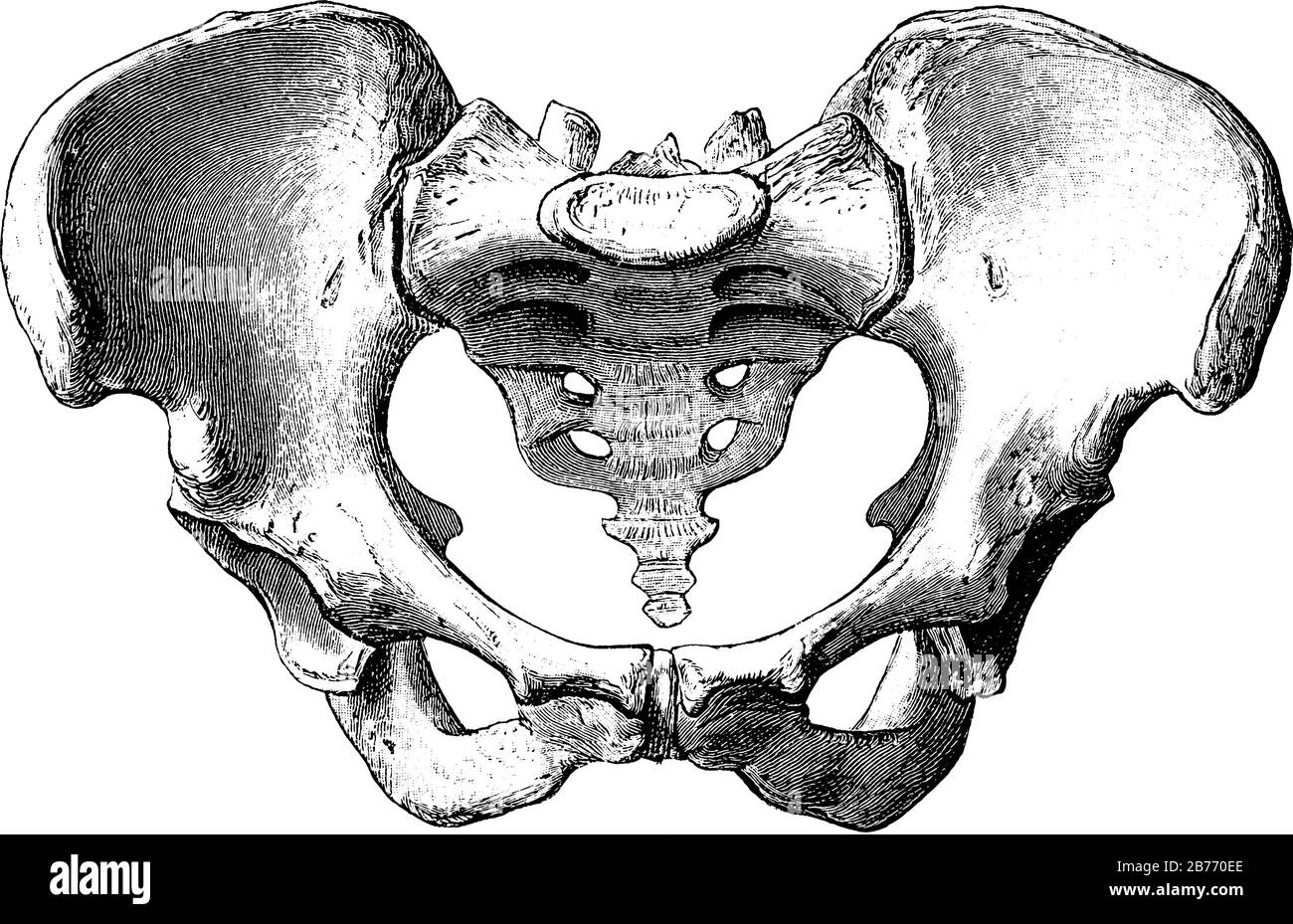 Female Pelvis, it is a basin-shaped complex of bones that connects the trunk and the legs, its primary role is to support the weight of the upper body Stock Vectorhttps://www.alamy.com/image-license-details/?v=1https://www.alamy.com/female-pelvis-it-is-a-basin-shaped-complex-of-bones-that-connects-the-trunk-and-the-legs-its-primary-role-is-to-support-the-weight-of-the-upper-body-image348664022.html
RF2B770EE–Female Pelvis, it is a basin-shaped complex of bones that connects the trunk and the legs, its primary role is to support the weight of the upper body
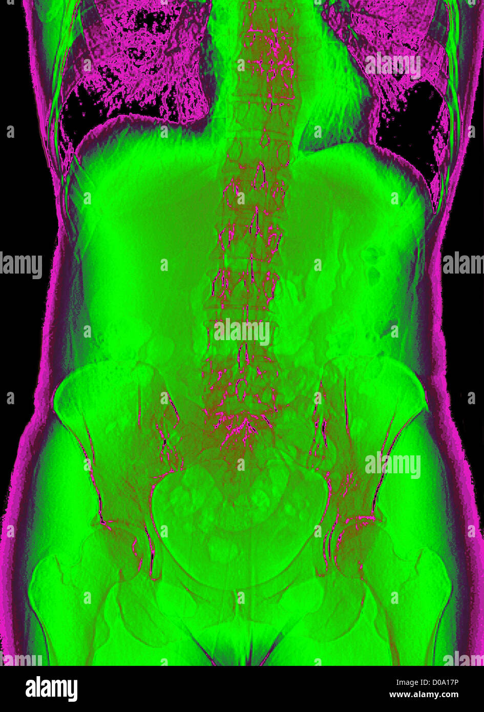 PELVIS, X-RAY Stock Photohttps://www.alamy.com/image-license-details/?v=1https://www.alamy.com/stock-photo-pelvis-x-ray-51851626.html
RMD0A17P–PELVIS, X-RAY
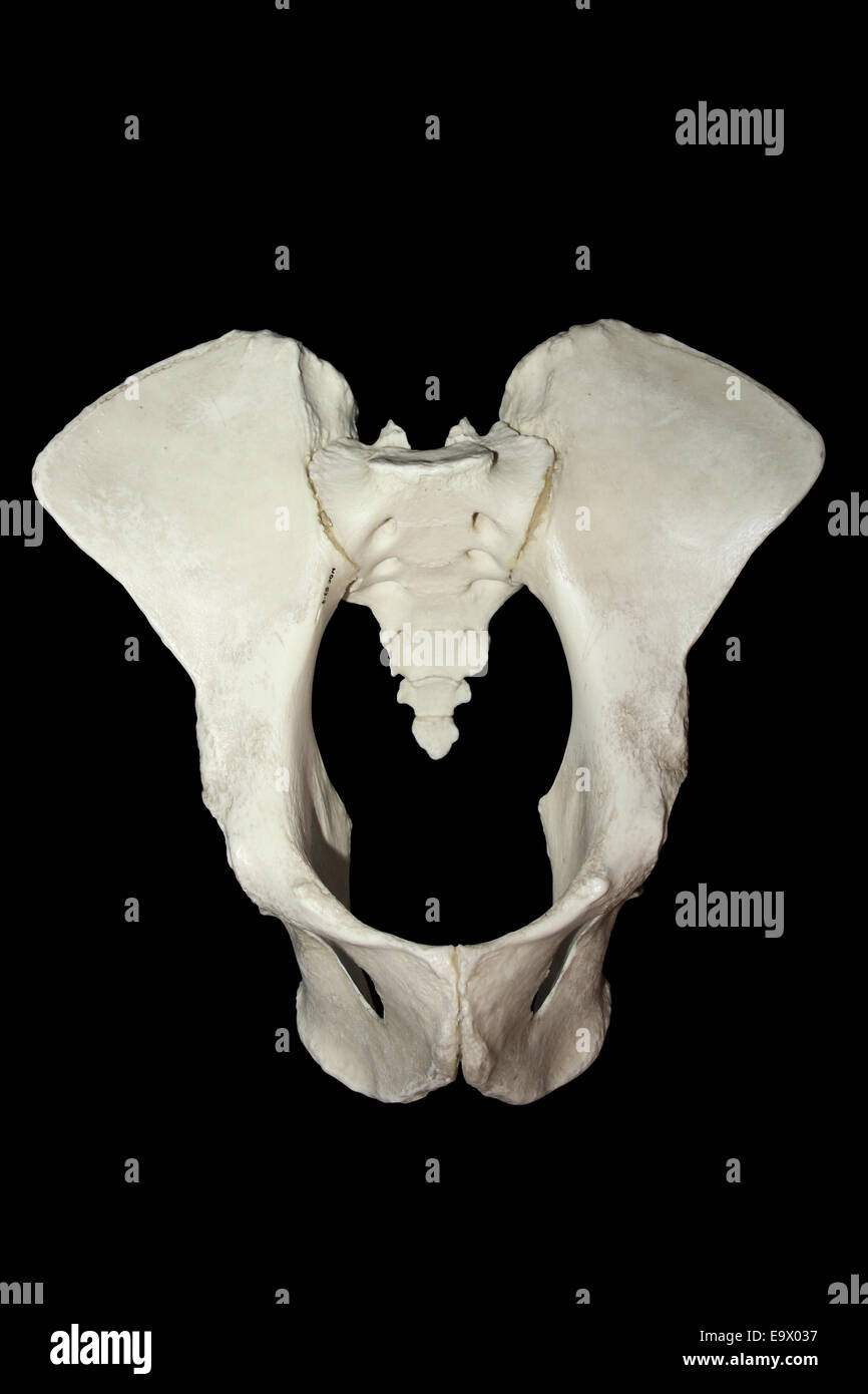 Female Chimpanzee Pelvis Stock Photohttps://www.alamy.com/image-license-details/?v=1https://www.alamy.com/stock-photo-female-chimpanzee-pelvis-74944219.html
RME9X037–Female Chimpanzee Pelvis
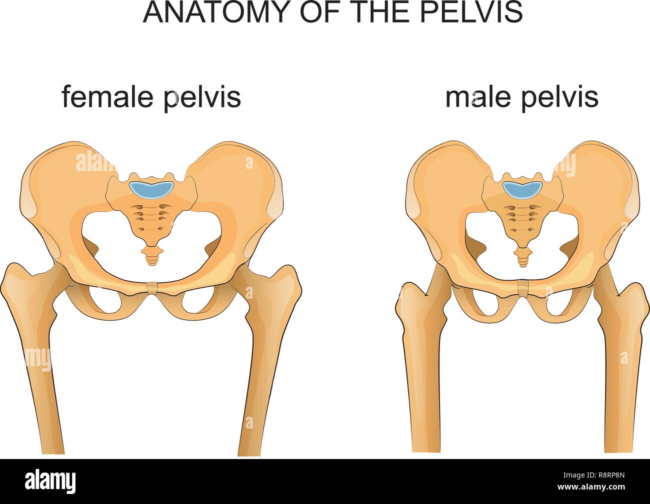 vector illustration of a comparison of the skeleton of the male and female pelvis Stock Vectorhttps://www.alamy.com/image-license-details/?v=1https://www.alamy.com/vector-illustration-of-a-comparison-of-the-skeleton-of-the-male-and-female-pelvis-image229174421.html
RFR8RP8N–vector illustration of a comparison of the skeleton of the male and female pelvis
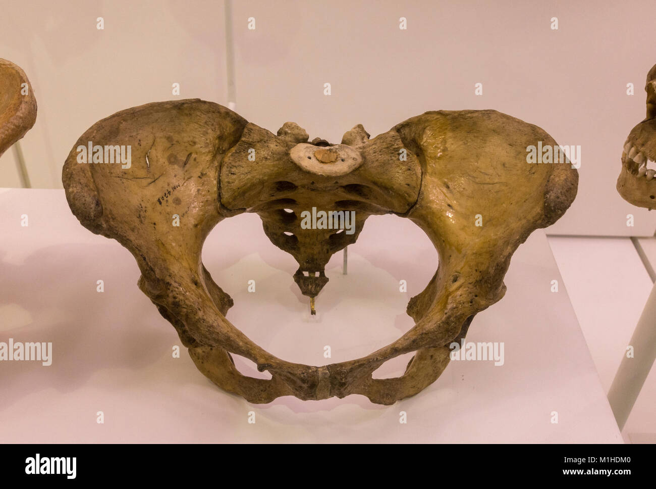 A female adult pelvis on display in the National Museum of Health and Medicine, Silver Spring, MD, USA. Stock Photohttps://www.alamy.com/image-license-details/?v=1https://www.alamy.com/stock-photo-a-female-adult-pelvis-on-display-in-the-national-museum-of-health-173102272.html
RMM1HDM0–A female adult pelvis on display in the National Museum of Health and Medicine, Silver Spring, MD, USA.
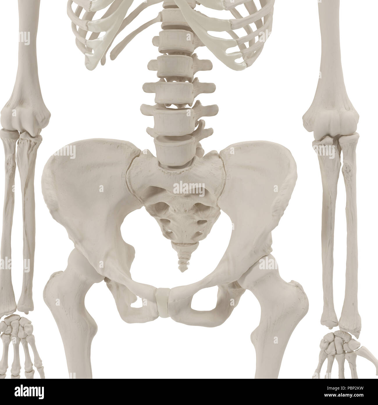 Female Pelvis Skeleton on white. 3D illustration Stock Photohttps://www.alamy.com/image-license-details/?v=1https://www.alamy.com/female-pelvis-skeleton-on-white-3d-illustration-image213770701.html
RFPBP2KW–Female Pelvis Skeleton on white. 3D illustration
 Gynecologist showing bones of female pelvis and giving consultation to woman Stock Photohttps://www.alamy.com/image-license-details/?v=1https://www.alamy.com/gynecologist-showing-bones-of-female-pelvis-and-giving-consultation-to-woman-image550534782.html
RF2PYK0ME–Gynecologist showing bones of female pelvis and giving consultation to woman
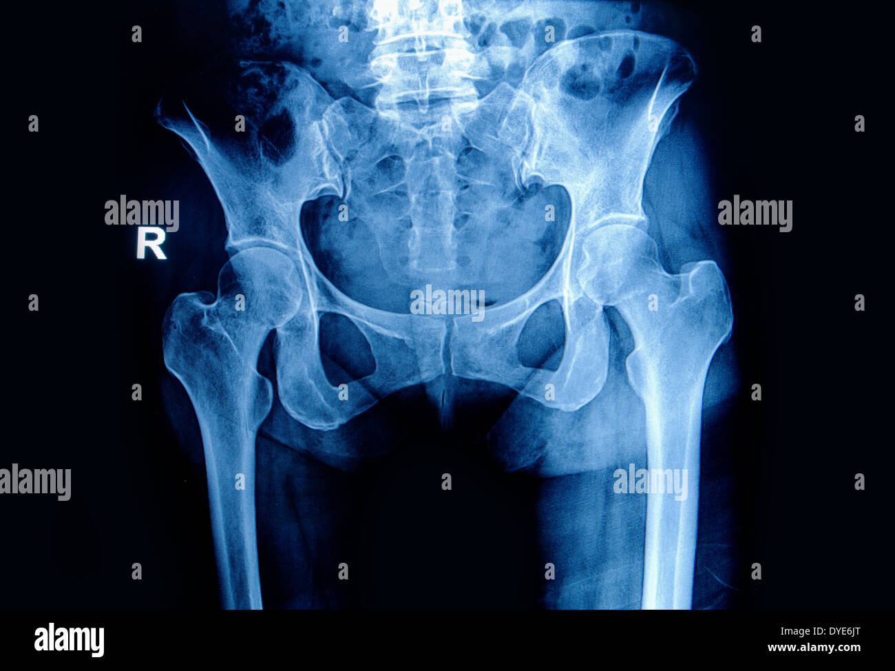 X-ray image pelvis and hip of a woman Stock Photohttps://www.alamy.com/image-license-details/?v=1https://www.alamy.com/x-ray-image-pelvis-and-hip-of-a-woman-image68539376.html
RFDYE6JT–X-ray image pelvis and hip of a woman
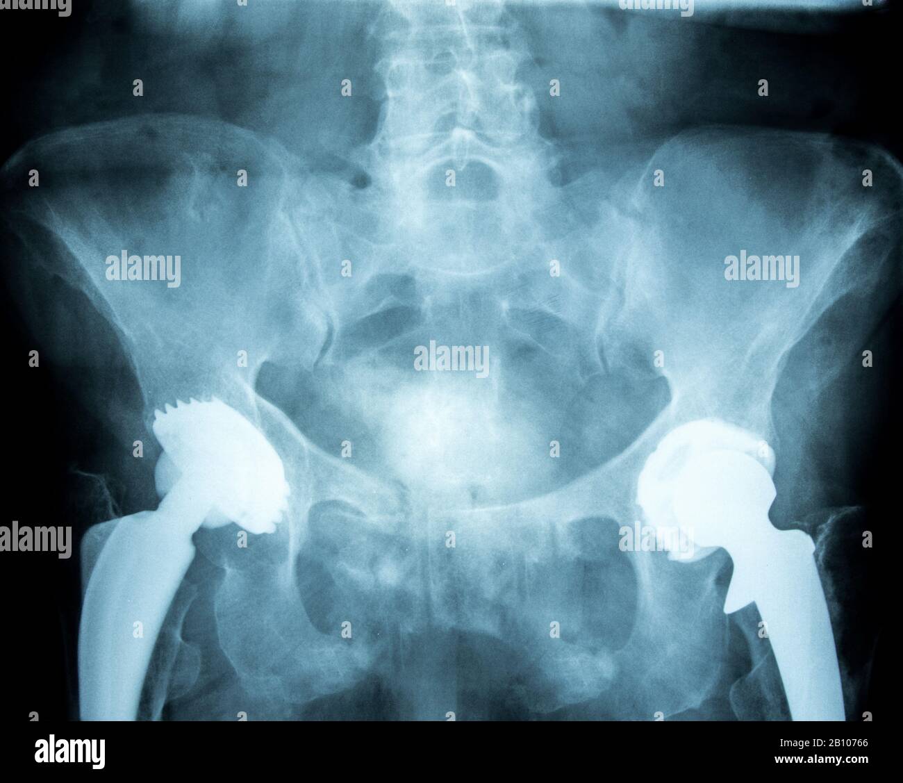 artificial hip joint Stock Photohttps://www.alamy.com/image-license-details/?v=1https://www.alamy.com/artificial-hip-joint-image344827678.html
RF2B10766–artificial hip joint
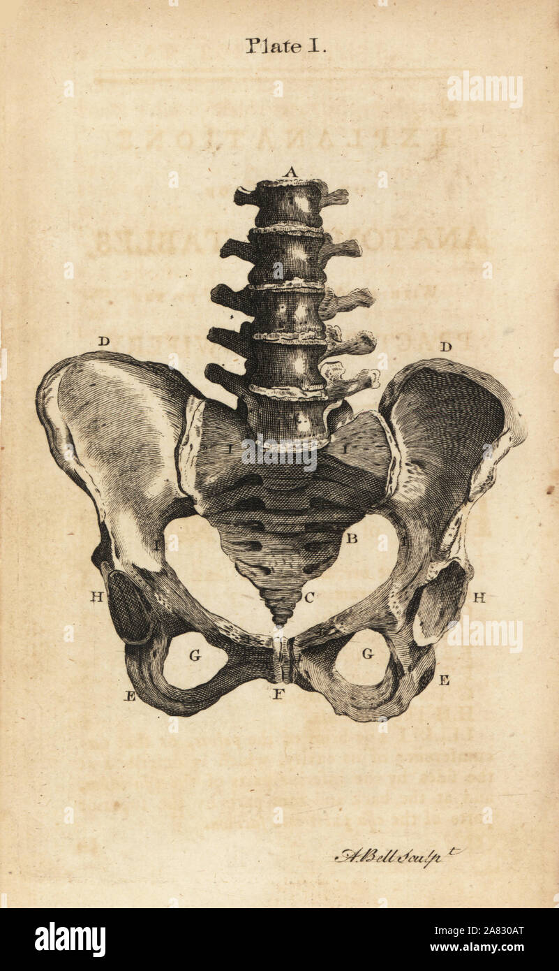 Front view of a healthy female pelvis. Copperplate engraving by Andrew Bell after an illustration by Jan van Rymsdyk from William Smellie's A Set of Anatomical Tables, Charles Elliot, Edinburgh, 1780. Stock Photohttps://www.alamy.com/image-license-details/?v=1https://www.alamy.com/front-view-of-a-healthy-female-pelvis-copperplate-engraving-by-andrew-bell-after-an-illustration-by-jan-van-rymsdyk-from-william-smellies-a-set-of-anatomical-tables-charles-elliot-edinburgh-1780-image331980400.html
RM2A830AT–Front view of a healthy female pelvis. Copperplate engraving by Andrew Bell after an illustration by Jan van Rymsdyk from William Smellie's A Set of Anatomical Tables, Charles Elliot, Edinburgh, 1780.
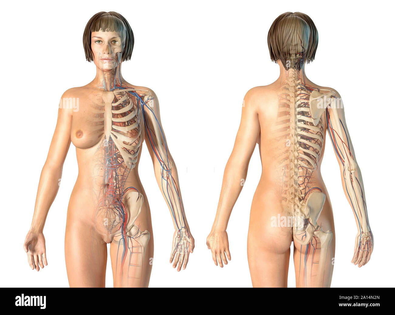 Female anatomy of cardiovascular system with skeleton. Stock Photohttps://www.alamy.com/image-license-details/?v=1https://www.alamy.com/female-anatomy-of-cardiovascular-system-with-skeleton-image327715997.html
RF2A14N2N–Female anatomy of cardiovascular system with skeleton.
 Doctor gynecologist showing bones of female pelvis closeup Stock Photohttps://www.alamy.com/image-license-details/?v=1https://www.alamy.com/doctor-gynecologist-showing-bones-of-female-pelvis-closeup-image495546016.html
RF2KP6200–Doctor gynecologist showing bones of female pelvis closeup
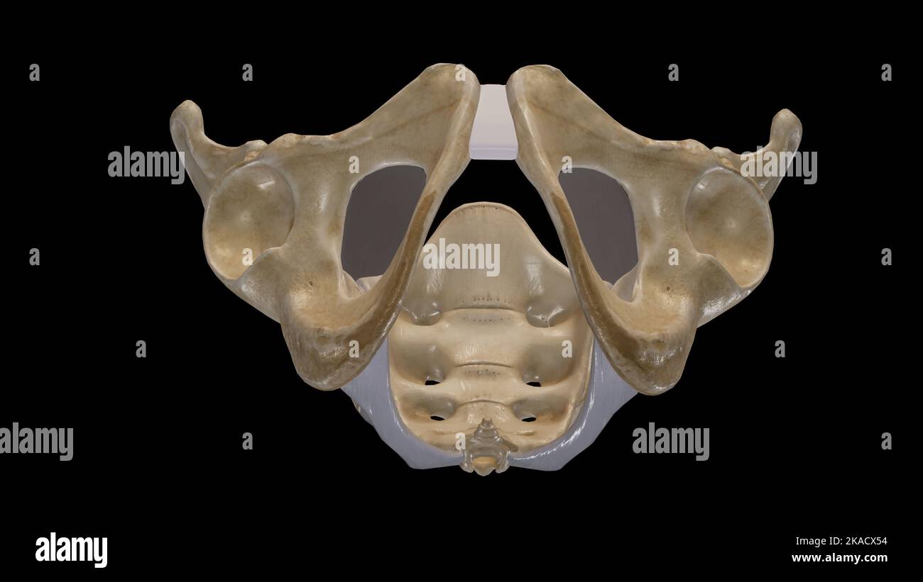 The Pelvic Girdle and Pelvic Outlet Stock Photohttps://www.alamy.com/image-license-details/?v=1https://www.alamy.com/the-pelvic-girdle-and-pelvic-outlet-image488320816.html
RF2KACX54–The Pelvic Girdle and Pelvic Outlet
 Swimmer Sarah charburner SG Frankfurt and swimmer Florian Wellbrock SC Magdeburg at the farewell for the Tokyo Olympics 2021, Swimmer Sarah charburner Stock Photohttps://www.alamy.com/image-license-details/?v=1https://www.alamy.com/swimmer-sarah-charburner-sg-frankfurt-and-swimmer-florian-wellbrock-sc-magdeburg-at-the-farewell-for-the-tokyo-olympics-2021-swimmer-sarah-charburner-image679015558.html
RM3BCKR7J–Swimmer Sarah charburner SG Frankfurt and swimmer Florian Wellbrock SC Magdeburg at the farewell for the Tokyo Olympics 2021, Swimmer Sarah charburner
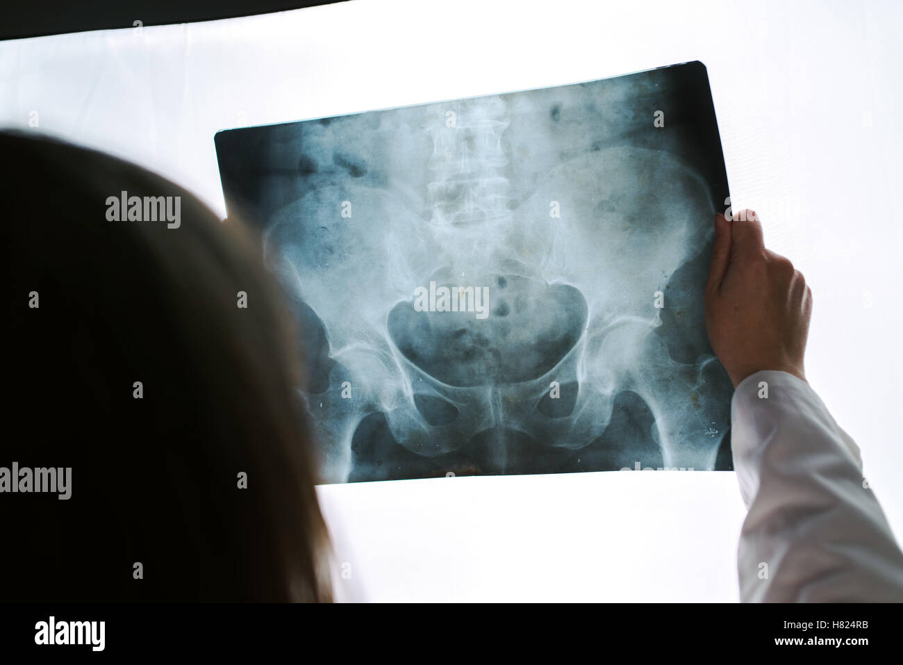 Female doctor examining pelvis x-ray in hospital office, medical professional in white uniform analyzing hip image in clinic. Stock Photohttps://www.alamy.com/image-license-details/?v=1https://www.alamy.com/stock-photo-female-doctor-examining-pelvis-x-ray-in-hospital-office-medical-professional-125437519.html
RFH824RB–Female doctor examining pelvis x-ray in hospital office, medical professional in white uniform analyzing hip image in clinic.
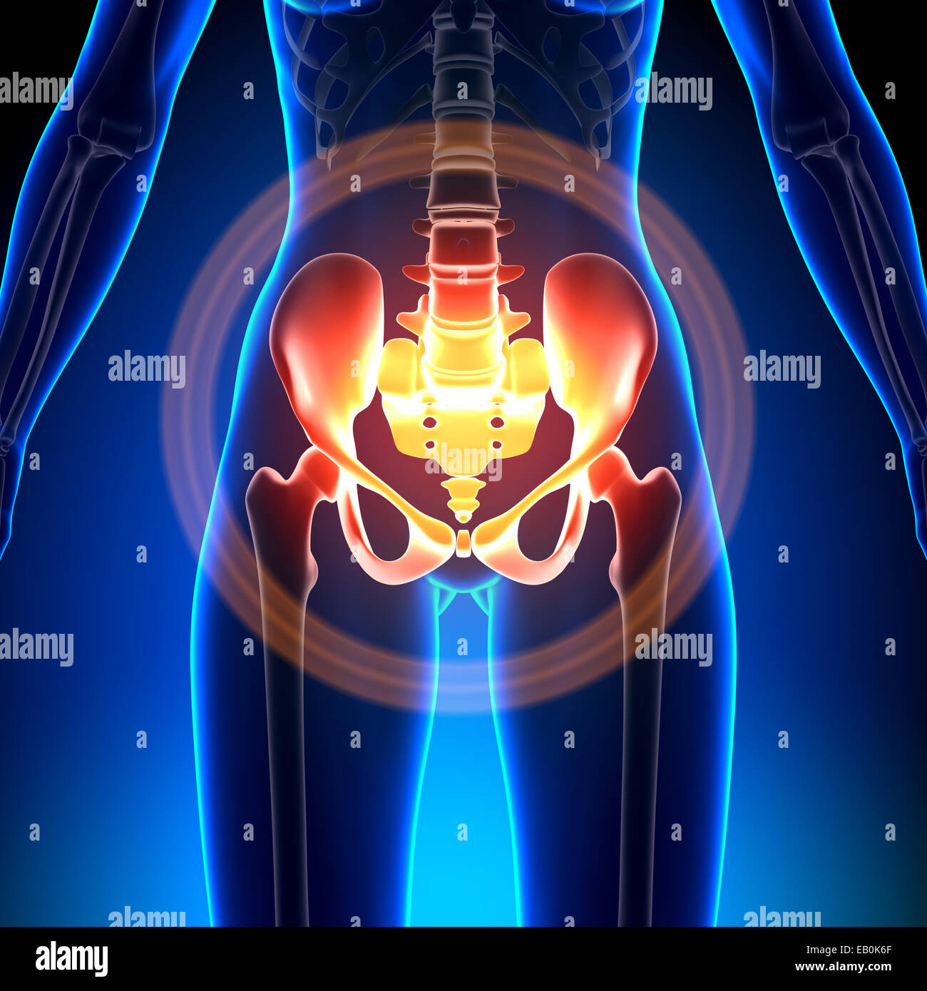 Female Hip / Sacrum / Pubis / Ischium / Ilium - Anatomy Bones Stock Photohttps://www.alamy.com/image-license-details/?v=1https://www.alamy.com/stock-photo-female-hip-sacrum-pubis-ischium-ilium-anatomy-bones-75617767.html
RFEB0K6F–Female Hip / Sacrum / Pubis / Ischium / Ilium - Anatomy Bones
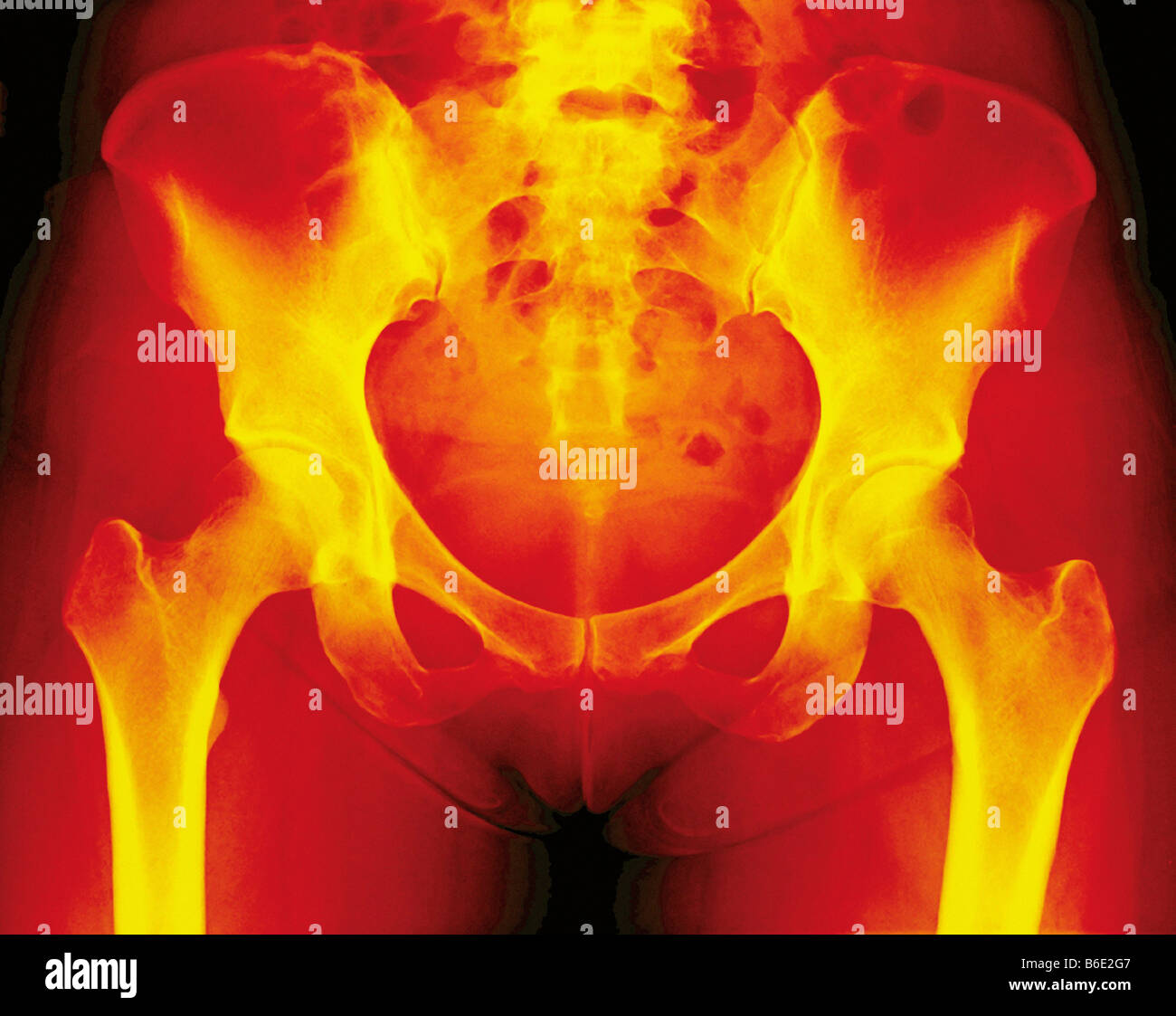 Normal female pelvis, coloured frontal X-ray. Thethigh (femur) bones of the leg are seen at bottomleft and right. Stock Photohttps://www.alamy.com/image-license-details/?v=1https://www.alamy.com/stock-photo-normal-female-pelvis-coloured-frontal-x-ray-thethigh-femur-bones-of-21207655.html
RFB6E2G7–Normal female pelvis, coloured frontal X-ray. Thethigh (femur) bones of the leg are seen at bottomleft and right.
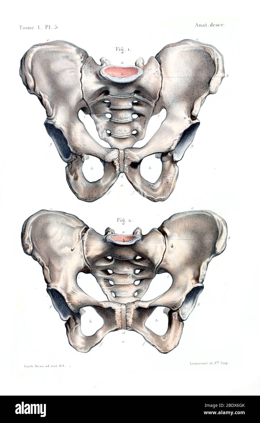 Human Female and Male Pelves, 1844 Stock Photohttps://www.alamy.com/image-license-details/?v=1https://www.alamy.com/human-female-and-male-pelves-1844-image352773811.html
RM2BDX6GK–Human Female and Male Pelves, 1844
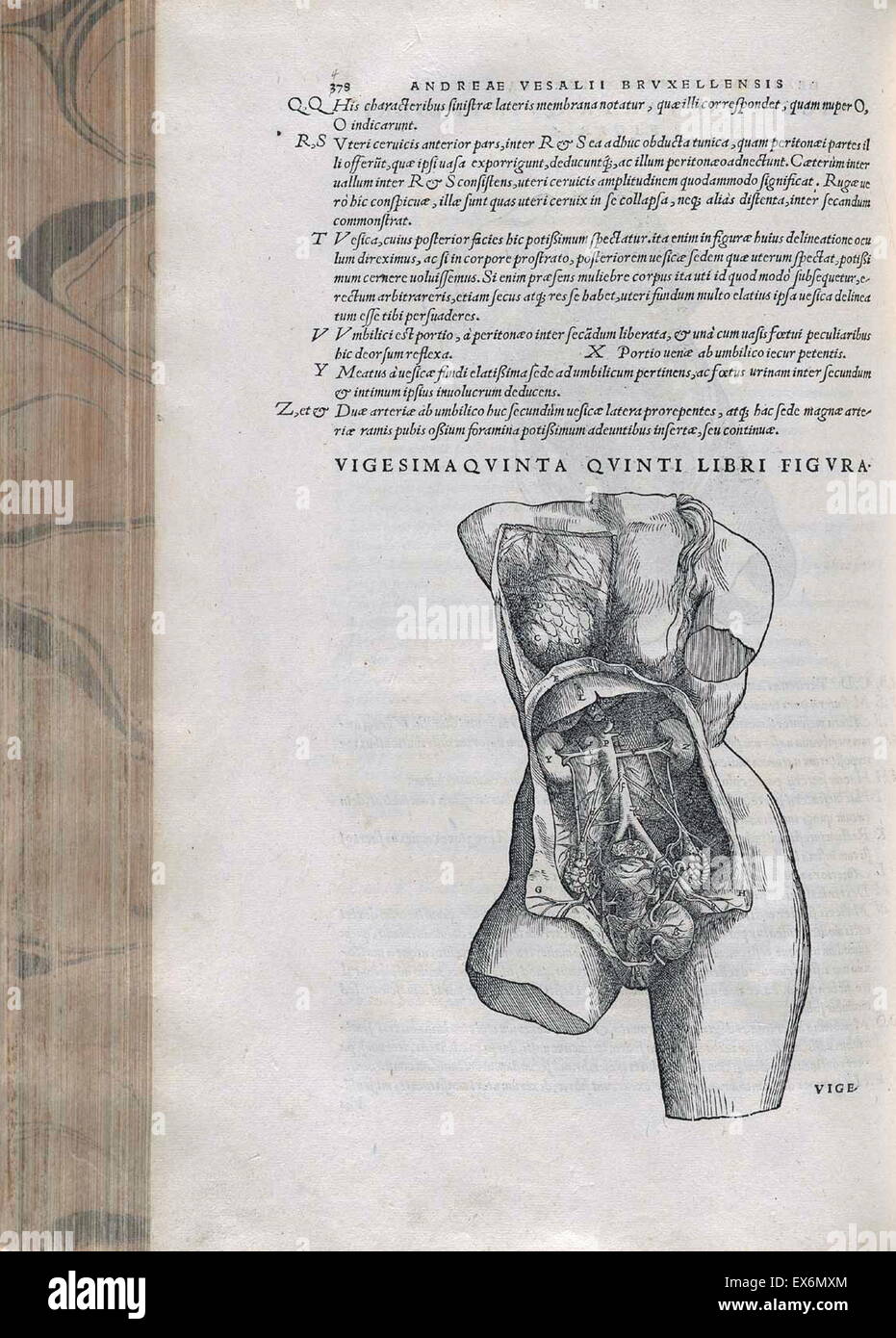 The female pelvic anatomy. From Andreas Vesalius' De Corporis Humani Fabrica, 1543 Stock Photohttps://www.alamy.com/image-license-details/?v=1https://www.alamy.com/stock-photo-the-female-pelvic-anatomy-from-andreas-vesalius-de-corporis-humani-84970668.html
RMEX6MXM–The female pelvic anatomy. From Andreas Vesalius' De Corporis Humani Fabrica, 1543
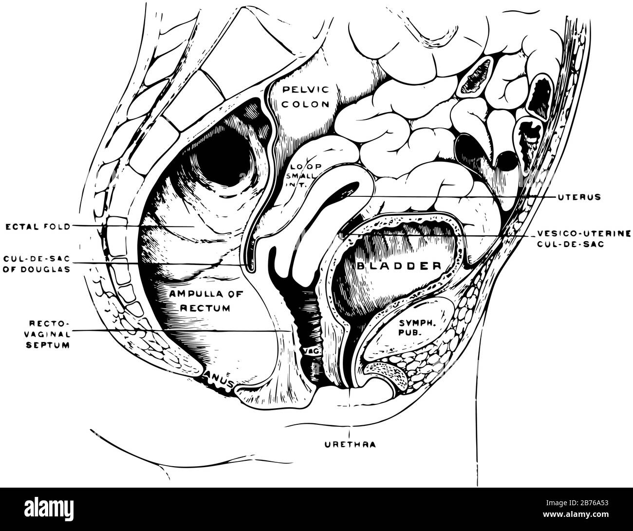 This illustration represents female Pelvis, vintage line drawing or engraving illustration. Stock Vectorhttps://www.alamy.com/image-license-details/?v=1https://www.alamy.com/this-illustration-represents-female-pelvis-vintage-line-drawing-or-engraving-illustration-image348649647.html
RF2B76A53–This illustration represents female Pelvis, vintage line drawing or engraving illustration.
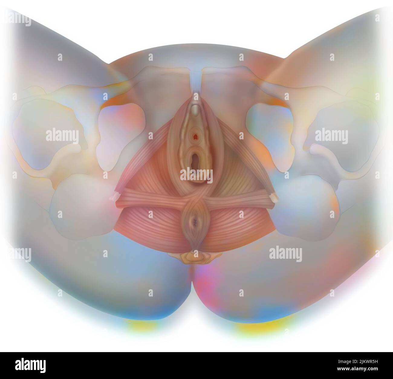 Inferior view of the perineum to highlight the external female genitalia. Stock Photohttps://www.alamy.com/image-license-details/?v=1https://www.alamy.com/inferior-view-of-the-perineum-to-highlight-the-external-female-genitalia-image476925389.html
RF2JKWR5H–Inferior view of the perineum to highlight the external female genitalia.
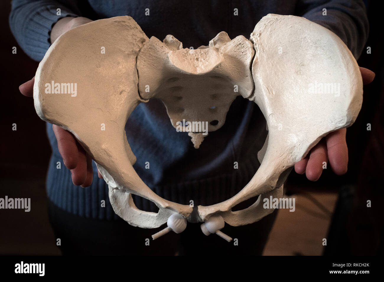 07 February 2019, Bavaria, Höhenkirchen-Siegertsbrunn: Midwife Sabine Pischinger holds the model of a female pelvis in her hands. The midwife has already helped more than 200 children into the world. Photo: Sina Schuldt/dpa - ATTENTION: Only for editorial use in connection with the current reporting Stock Photohttps://www.alamy.com/image-license-details/?v=1https://www.alamy.com/07-february-2019-bavaria-hhenkirchen-siegertsbrunn-midwife-sabine-pischinger-holds-the-model-of-a-female-pelvis-in-her-hands-the-midwife-has-already-helped-more-than-200-children-into-the-world-photo-sina-schuldtdpa-attention-only-for-editorial-use-in-connection-with-the-current-reporting-image235690075.html
RMRKCH2K–07 February 2019, Bavaria, Höhenkirchen-Siegertsbrunn: Midwife Sabine Pischinger holds the model of a female pelvis in her hands. The midwife has already helped more than 200 children into the world. Photo: Sina Schuldt/dpa - ATTENTION: Only for editorial use in connection with the current reporting
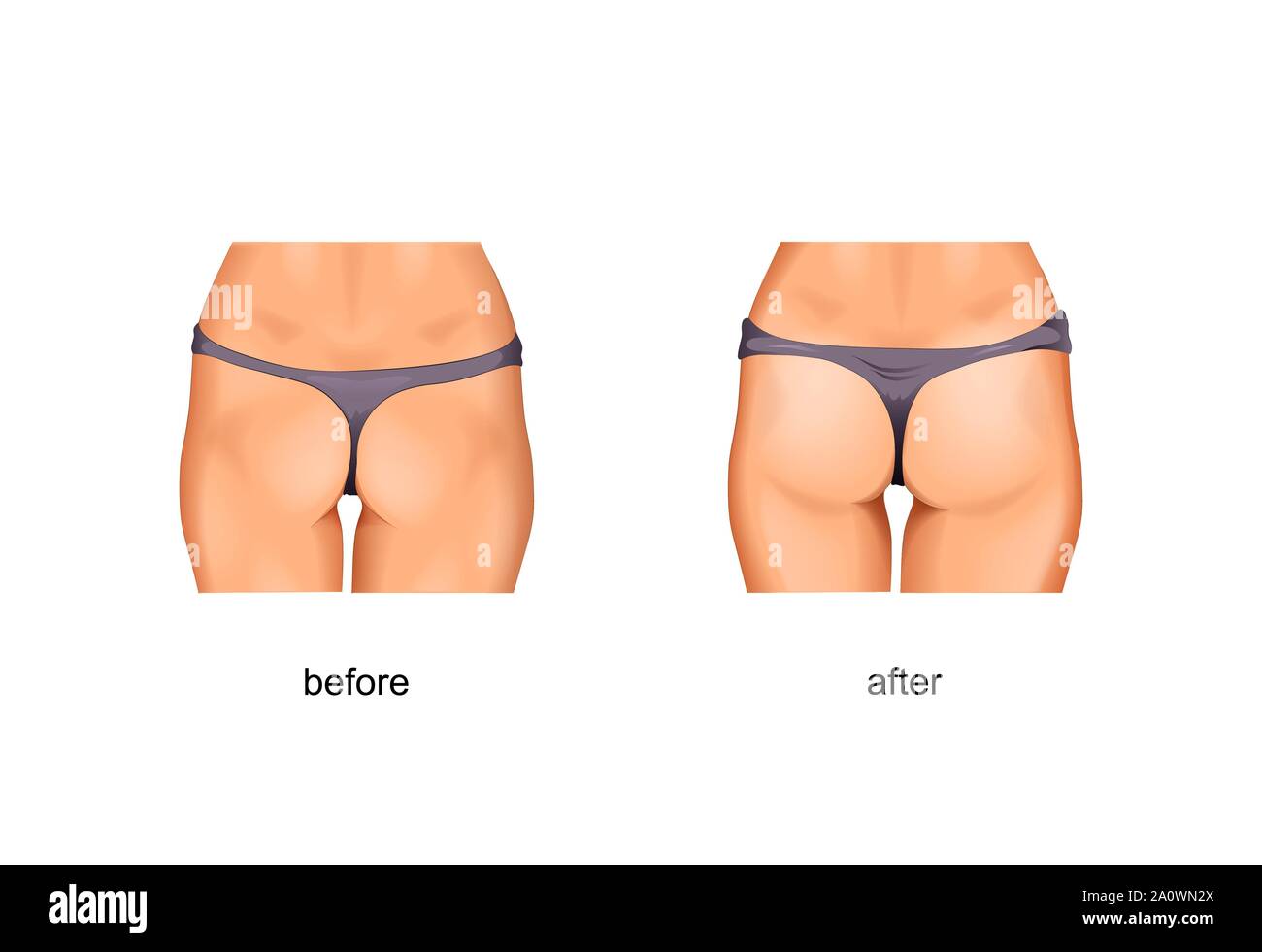 vector illustration of taut gluteal muscles. before and after treatments or fitness Stock Vectorhttps://www.alamy.com/image-license-details/?v=1https://www.alamy.com/vector-illustration-of-taut-gluteal-muscles-before-and-after-treatments-or-fitness-image327562338.html
RF2A0WN2X–vector illustration of taut gluteal muscles. before and after treatments or fitness
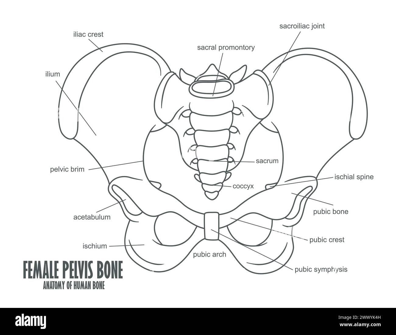 Female Pelvis Bone Anatomy, Vector Illustration Stock Vectorhttps://www.alamy.com/image-license-details/?v=1https://www.alamy.com/female-pelvis-bone-anatomy-vector-illustration-image601126641.html
RF2WWYK4H–Female Pelvis Bone Anatomy, Vector Illustration
 A Woman in a white satin nightgown has waist pain because the pelvis has a problem. Stock Photohttps://www.alamy.com/image-license-details/?v=1https://www.alamy.com/a-woman-in-a-white-satin-nightgown-has-waist-pain-because-the-pelvis-has-a-problem-image599733314.html
RF2WRM5XX–A Woman in a white satin nightgown has waist pain because the pelvis has a problem.
 Gynecologist showing bones of female pelvis and giving consultation to woman Stock Photohttps://www.alamy.com/image-license-details/?v=1https://www.alamy.com/gynecologist-showing-bones-of-female-pelvis-and-giving-consultation-to-woman-image550486172.html
RF2PYGPMC–Gynecologist showing bones of female pelvis and giving consultation to woman
 Doctor gynecologist showing layout of female pelvis with muscles closeup Stock Photohttps://www.alamy.com/image-license-details/?v=1https://www.alamy.com/doctor-gynecologist-showing-layout-of-female-pelvis-with-muscles-closeup-image485815892.html
RF2K6AR3G–Doctor gynecologist showing layout of female pelvis with muscles closeup
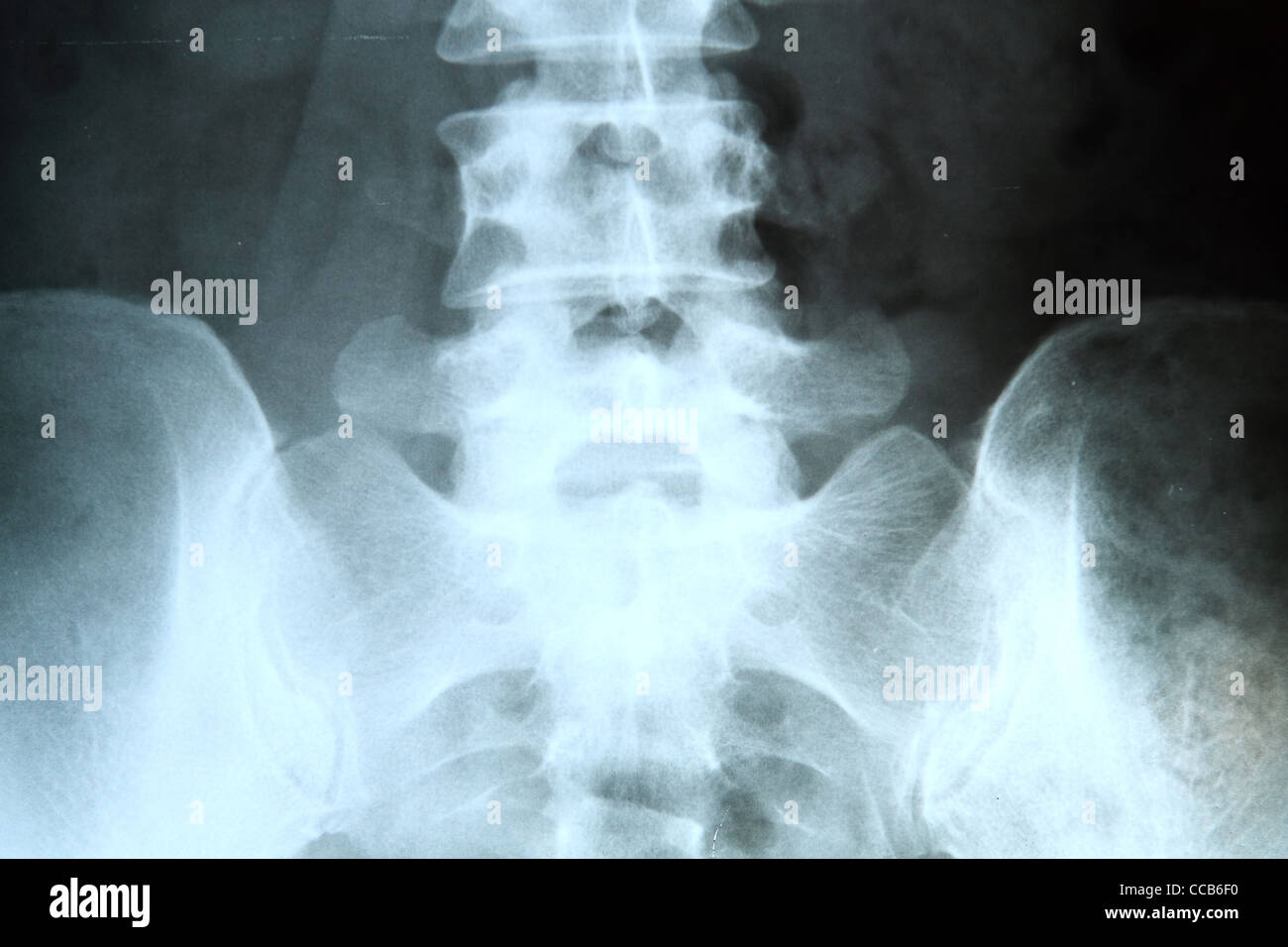 X-ray of the pelvis and spinal column. Stock Photohttps://www.alamy.com/image-license-details/?v=1https://www.alamy.com/stock-photo-x-ray-of-the-pelvis-and-spinal-column-42043204.html
RFCCB6F0–X-ray of the pelvis and spinal column.
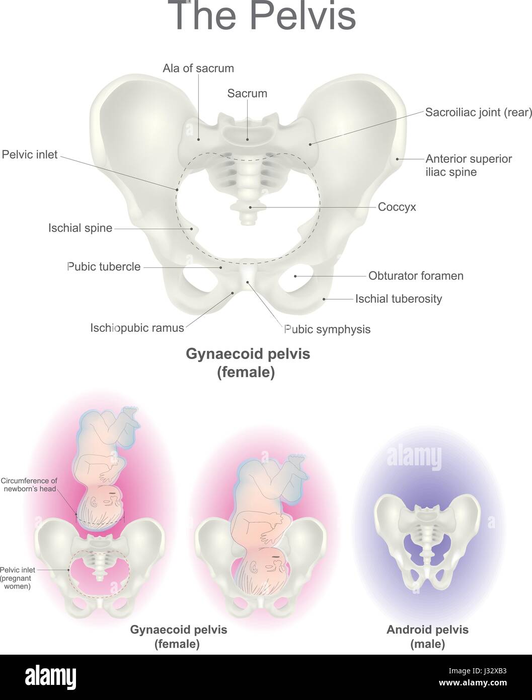 The pelvic region is the headquarters for reproductive organs and the end of the line for the digestive tract. It is also home to a collection of bone Stock Vectorhttps://www.alamy.com/image-license-details/?v=1https://www.alamy.com/stock-photo-the-pelvic-region-is-the-headquarters-for-reproductive-organs-and-139591511.html
RFJ32XB3–The pelvic region is the headquarters for reproductive organs and the end of the line for the digestive tract. It is also home to a collection of bone
 Osteopath performing a pelvis alignment assessment on a young female patient on a couch in his surgery or clinic in a healthcare and medical concept Stock Photohttps://www.alamy.com/image-license-details/?v=1https://www.alamy.com/osteopath-performing-a-pelvis-alignment-assessment-on-a-young-female-patient-on-a-couch-in-his-surgery-or-clinic-in-a-healthcare-and-medical-concept-image383004925.html
RF2D73AK9–Osteopath performing a pelvis alignment assessment on a young female patient on a couch in his surgery or clinic in a healthcare and medical concept
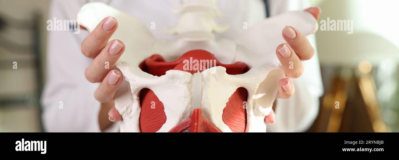 Gynecologist shows female pelvis with reproductive organs Stock Photohttps://www.alamy.com/image-license-details/?v=1https://www.alamy.com/gynecologist-shows-female-pelvis-with-reproductive-organs-image567797619.html
RF2RYNBJB–Gynecologist shows female pelvis with reproductive organs
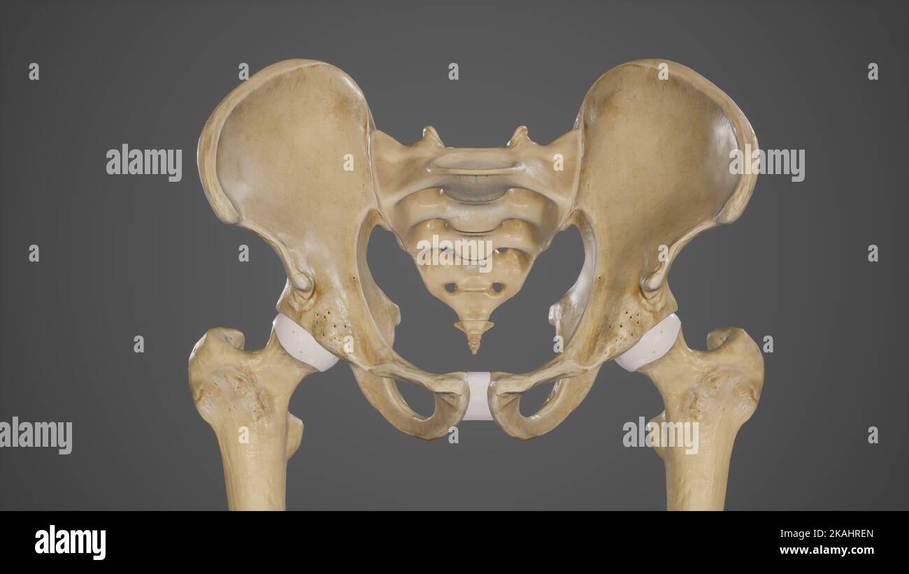 Medical Ilustration of Pelvic Bones-Hip Bone Stock Photohttps://www.alamy.com/image-license-details/?v=1https://www.alamy.com/medical-ilustration-of-pelvic-bones-hip-bone-image488428493.html
RF2KAHREN–Medical Ilustration of Pelvic Bones-Hip Bone
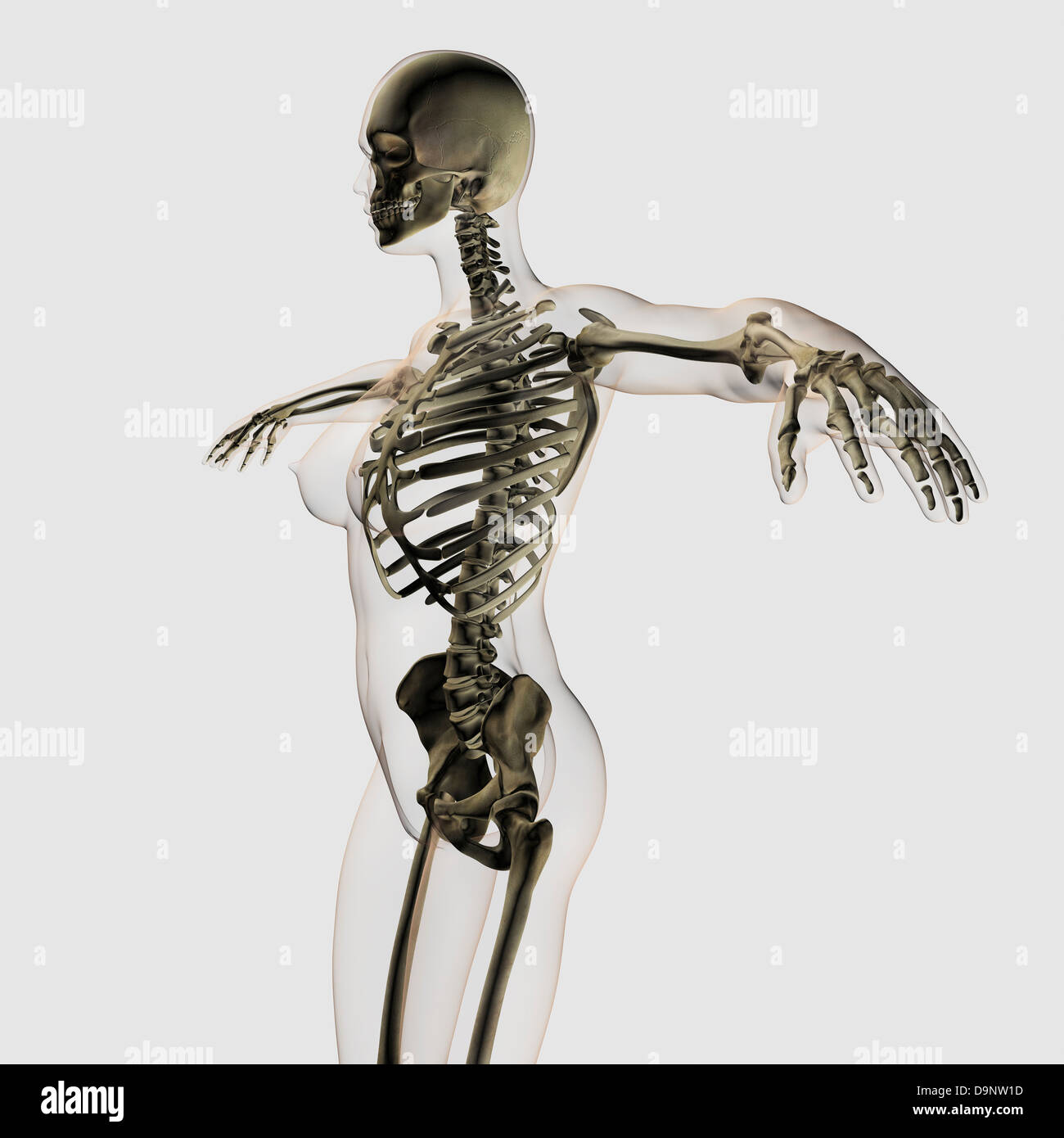 Three dimensional view of female skeletal system. Stock Photohttps://www.alamy.com/image-license-details/?v=1https://www.alamy.com/stock-photo-three-dimensional-view-of-female-skeletal-system-57643641.html
RFD9NW1D–Three dimensional view of female skeletal system.
 Female doctor examining pelvis x-ray in hospital office, medical professional in white uniform analyzing hip image in clinic. Stock Photohttps://www.alamy.com/image-license-details/?v=1https://www.alamy.com/stock-photo-female-doctor-examining-pelvis-x-ray-in-hospital-office-medical-professional-125437501.html
RFH824PN–Female doctor examining pelvis x-ray in hospital office, medical professional in white uniform analyzing hip image in clinic.
 Physiotherapist pressing patients pelvis Stock Photohttps://www.alamy.com/image-license-details/?v=1https://www.alamy.com/physiotherapist-pressing-patients-pelvis-image61939174.html
RFDGNG1A–Physiotherapist pressing patients pelvis
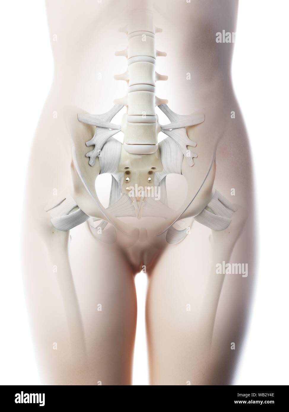 Female pelvis anatomy, computer illustration. Stock Photohttps://www.alamy.com/image-license-details/?v=1https://www.alamy.com/female-pelvis-anatomy-computer-illustration-image264981934.html
RFWB2Y4E–Female pelvis anatomy, computer illustration.
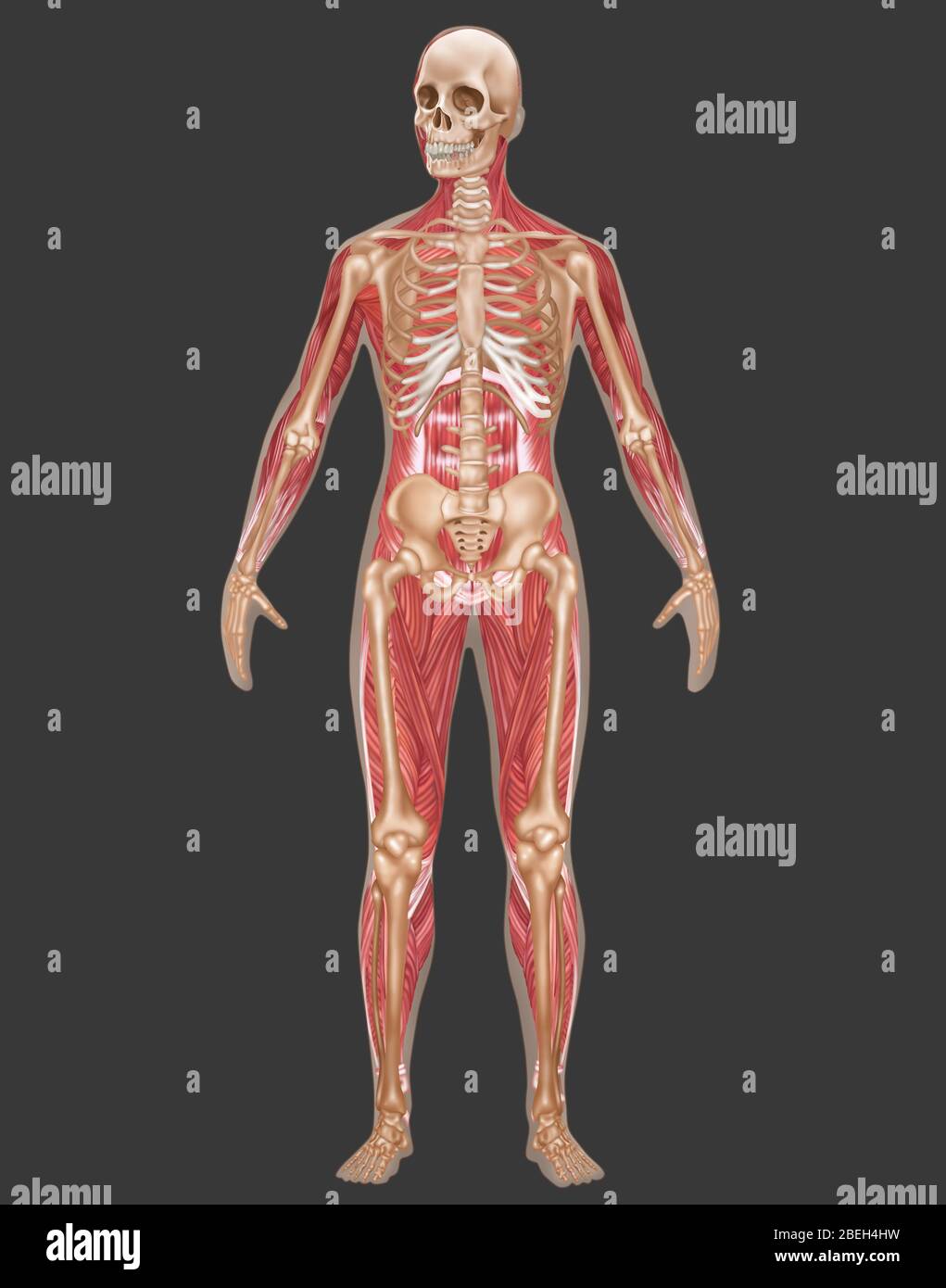 Skeletal & Muscular Systems, Female Anatomy Stock Photohttps://www.alamy.com/image-license-details/?v=1https://www.alamy.com/skeletal-muscular-systems-female-anatomy-image353189365.html
RF2BEH4HW–Skeletal & Muscular Systems, Female Anatomy
 Female physiotherapist pointing at spine model Stock Photohttps://www.alamy.com/image-license-details/?v=1https://www.alamy.com/stock-photo-female-physiotherapist-pointing-at-spine-model-122577972.html
RFH3BWCM–Female physiotherapist pointing at spine model
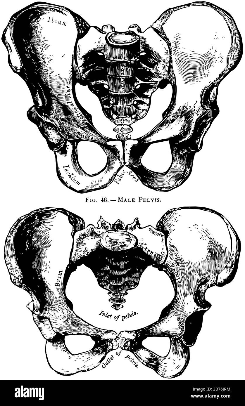 This illustration represents Human Pelvis Male and Female, vintage line drawing or engraving illustration. Stock Vectorhttps://www.alamy.com/image-license-details/?v=1https://www.alamy.com/this-illustration-represents-human-pelvis-male-and-female-vintage-line-drawing-or-engraving-illustration-image348656440.html
RF2B76JRM–This illustration represents Human Pelvis Male and Female, vintage line drawing or engraving illustration.
 Uterus cancer Stock Photohttps://www.alamy.com/image-license-details/?v=1https://www.alamy.com/uterus-cancer-image466107309.html
RM2J290HH–Uterus cancer
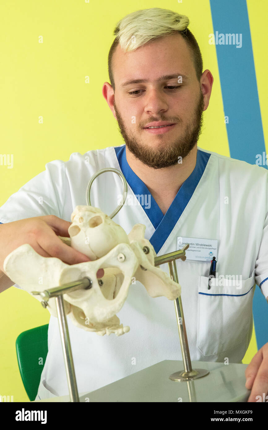 29 May 2018, Germany, Ansbach: Konstantin Wroblewski, male midwife in training at the Berufsfachschule für Hebammen und Entbindungspfleger am Klinikum Ansbach (lit. vocational school for midwifes and male midwifes at the Ansbach Clinic), sits next to a bone model of a female pelvis, in order to learn how the head of the baby has to be positioned during childbirth. Photo: Daniel Karmann/dpa Stock Photohttps://www.alamy.com/image-license-details/?v=1https://www.alamy.com/29-may-2018-germany-ansbach-konstantin-wroblewski-male-midwife-in-training-at-the-berufsfachschule-fr-hebammen-und-entbindungspfleger-am-klinikum-ansbach-lit-vocational-school-for-midwifes-and-male-midwifes-at-the-ansbach-clinic-sits-next-to-a-bone-model-of-a-female-pelvis-in-order-to-learn-how-the-head-of-the-baby-has-to-be-positioned-during-childbirth-photo-daniel-karmanndpa-image188451293.html
RMMXGKF9–29 May 2018, Germany, Ansbach: Konstantin Wroblewski, male midwife in training at the Berufsfachschule für Hebammen und Entbindungspfleger am Klinikum Ansbach (lit. vocational school for midwifes and male midwifes at the Ansbach Clinic), sits next to a bone model of a female pelvis, in order to learn how the head of the baby has to be positioned during childbirth. Photo: Daniel Karmann/dpa
 Female doctor examining a pelvis x-ray on white background. /// Woman examine looking pelvic x ray cut out cutout Spine hips femur Spinal backbone Stock Photohttps://www.alamy.com/image-license-details/?v=1https://www.alamy.com/stock-photo-female-doctor-examining-a-pelvis-x-ray-on-white-background-woman-examine-19271565.html
RFB39W25–Female doctor examining a pelvis x-ray on white background. /// Woman examine looking pelvic x ray cut out cutout Spine hips femur Spinal backbone
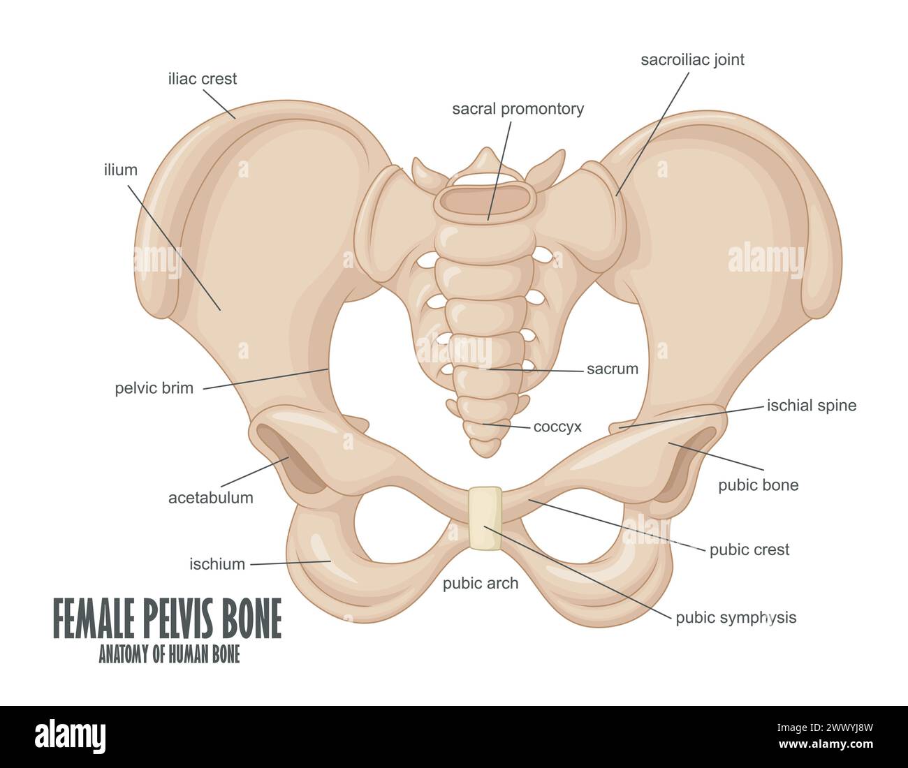 Female Pelvis Bone anatomy, Vector Illustration Stock Vectorhttps://www.alamy.com/image-license-details/?v=1https://www.alamy.com/female-pelvis-bone-anatomy-vector-illustration-image601125977.html
RF2WWYJ8W–Female Pelvis Bone anatomy, Vector Illustration
 A woman is putting on sunglasses beside the swimming pool Stock Photohttps://www.alamy.com/image-license-details/?v=1https://www.alamy.com/a-woman-is-putting-on-sunglasses-beside-the-swimming-pool-image605217535.html
RF2X4J13Y–A woman is putting on sunglasses beside the swimming pool
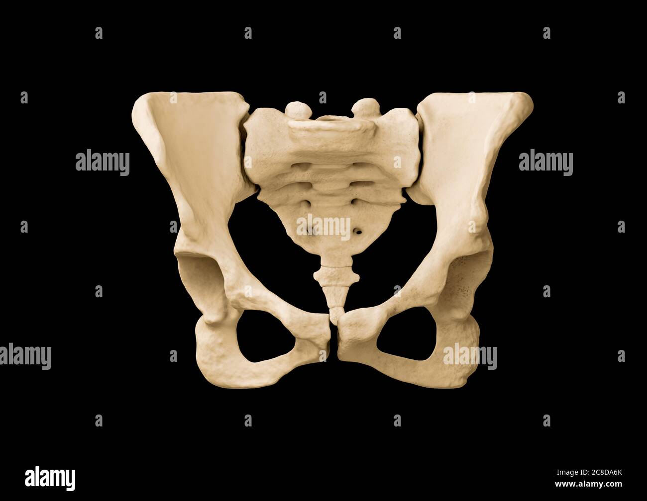 Pelvis, Human skeleton, Female Pelvic Bone anatomy, hip Stock Photohttps://www.alamy.com/image-license-details/?v=1https://www.alamy.com/pelvis-human-skeleton-female-pelvic-bone-anatomy-hip-image366628379.html
RF2C8DA6K–Pelvis, Human skeleton, Female Pelvic Bone anatomy, hip
 Doctor gynecologist showing layout of female pelvis with muscles closeup Stock Photohttps://www.alamy.com/image-license-details/?v=1https://www.alamy.com/doctor-gynecologist-showing-layout-of-female-pelvis-with-muscles-closeup-image460370631.html
RF2HMYKC7–Doctor gynecologist showing layout of female pelvis with muscles closeup
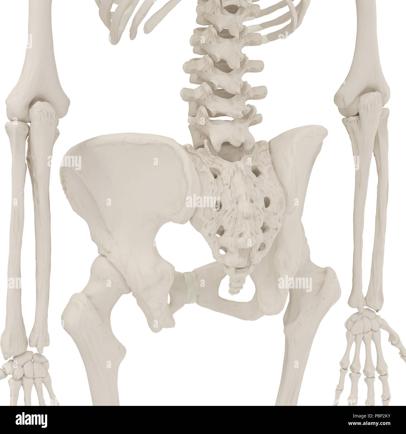 Female Pelvis Skeleton on white. 3D illustration Stock Photohttps://www.alamy.com/image-license-details/?v=1https://www.alamy.com/female-pelvis-skeleton-on-white-3d-illustration-image213770703.html
RFPBP2KY–Female Pelvis Skeleton on white. 3D illustration
 four oranges cut in half on a red cloth in front of a peach background as a symbol of the pelvic floor, no people Stock Photohttps://www.alamy.com/image-license-details/?v=1https://www.alamy.com/four-oranges-cut-in-half-on-a-red-cloth-in-front-of-a-peach-background-as-a-symbol-of-the-pelvic-floor-no-people-image503449587.html
RF2M7232B–four oranges cut in half on a red cloth in front of a peach background as a symbol of the pelvic floor, no people
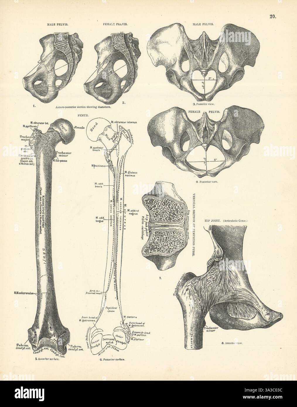 Anatomy. Male & Female Pelvis, Femur, Hip Joint 1880 old antique print picture Stock Photohttps://www.alamy.com/image-license-details/?v=1https://www.alamy.com/anatomy-male-female-pelvis-femur-hip-joint-1880-old-antique-print-picture-image656101472.html
RF3A3C03C–Anatomy. Male & Female Pelvis, Femur, Hip Joint 1880 old antique print picture
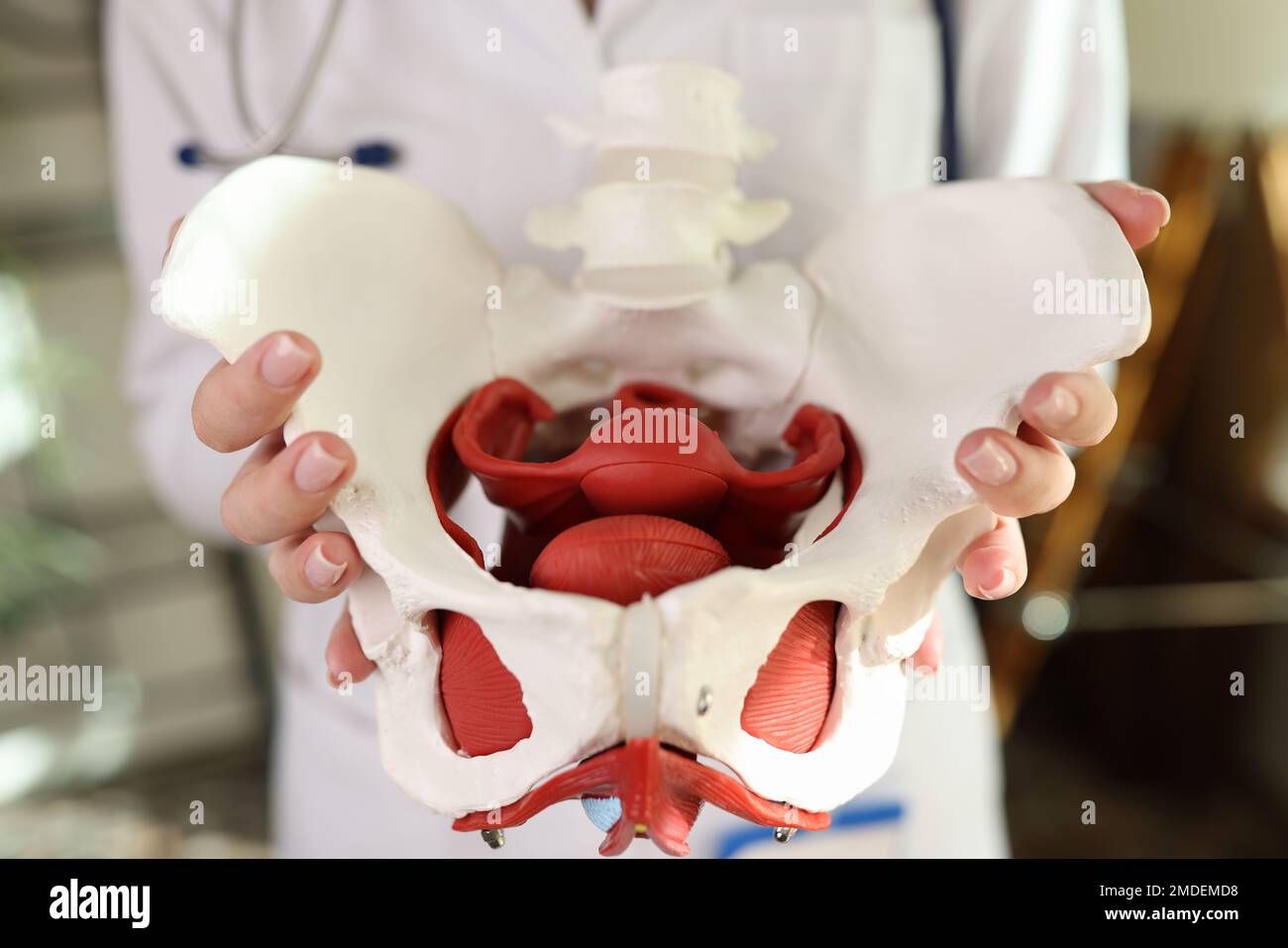 Close-up of model of female pelvis with muscles in hands of gynecologist. Stock Photohttps://www.alamy.com/image-license-details/?v=1https://www.alamy.com/close-up-of-model-of-female-pelvis-with-muscles-in-hands-of-gynecologist-image507414580.html
RF2MDEMD8–Close-up of model of female pelvis with muscles in hands of gynecologist.
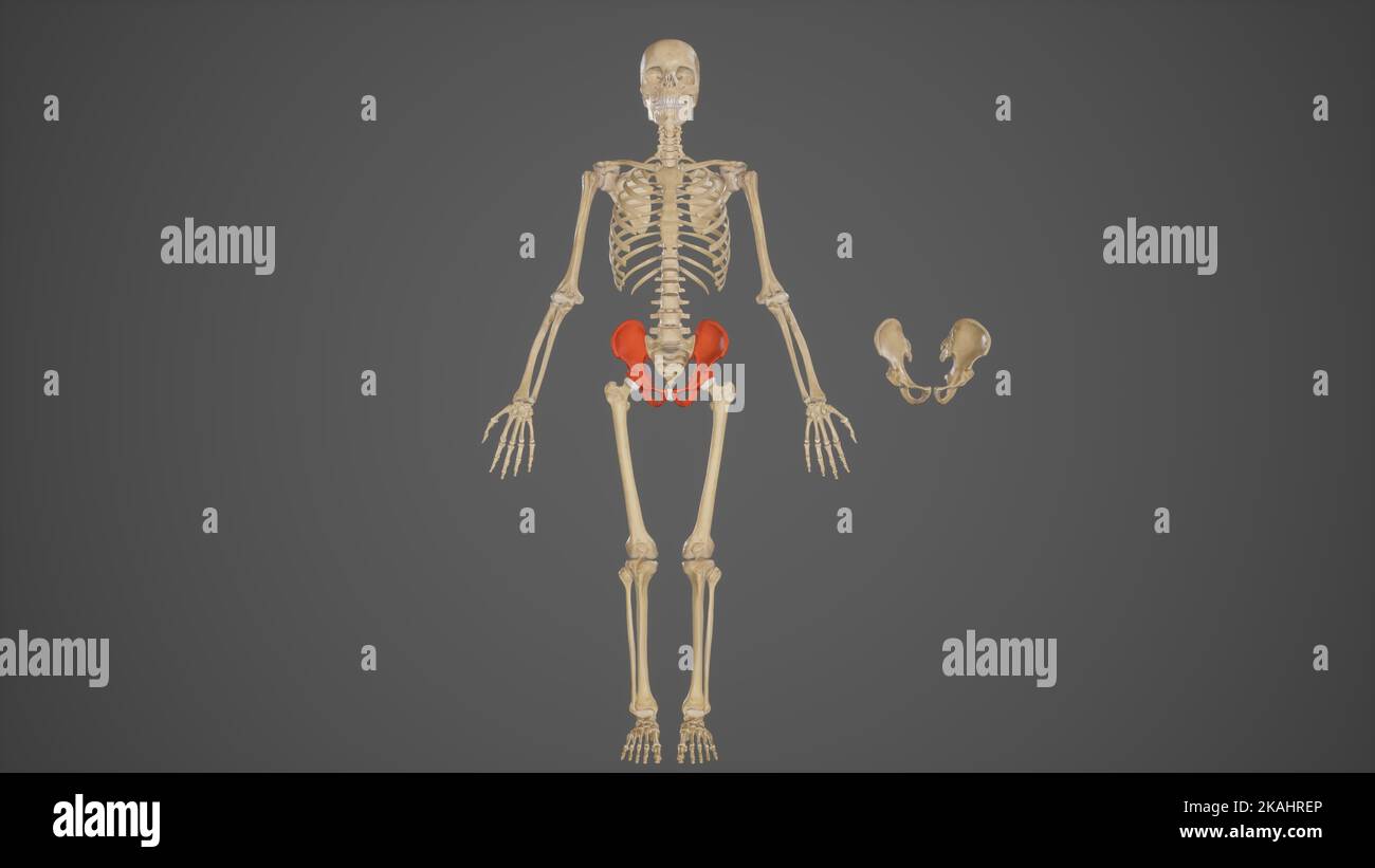 Medical Ilustration of Pelvic Bones Stock Photohttps://www.alamy.com/image-license-details/?v=1https://www.alamy.com/medical-ilustration-of-pelvic-bones-image488428494.html
RF2KAHREP–Medical Ilustration of Pelvic Bones
 . English: Fleuron from book: A course of lectures, on the Theory and Practice of midwifry, by William Rowley, Surgeon, in which Every thing material to the True Knowledge and Practice of the Art will be fully explained and clearly demonstrated, by a proper Apparatus on a new Construction, so formed as in every Case to imitate real Nature. Of the Form, Structure, and Parts of the Female Pelvis. Of the Organs of Generation and the Doctrine of Conception. The Pathology and Treatment of the various Diseases of Child-Bearing Women and Children. A Comparative View of the Antient, and Modern Practic Stock Photohttps://www.alamy.com/image-license-details/?v=1https://www.alamy.com/english-fleuron-from-book-a-course-of-lectures-on-the-theory-and-practice-of-midwifry-by-william-rowley-surgeon-in-which-every-thing-material-to-the-true-knowledge-and-practice-of-the-art-will-be-fully-explained-and-clearly-demonstrated-by-a-proper-apparatus-on-a-new-construction-so-formed-as-in-every-case-to-imitate-real-nature-of-the-form-structure-and-parts-of-the-female-pelvis-of-the-organs-of-generation-and-the-doctrine-of-conception-the-pathology-and-treatment-of-the-various-diseases-of-child-bearing-women-and-children-a-comparative-view-of-the-antient-and-modern-practic-image206613584.html
RMP041MG–. English: Fleuron from book: A course of lectures, on the Theory and Practice of midwifry, by William Rowley, Surgeon, in which Every thing material to the True Knowledge and Practice of the Art will be fully explained and clearly demonstrated, by a proper Apparatus on a new Construction, so formed as in every Case to imitate real Nature. Of the Form, Structure, and Parts of the Female Pelvis. Of the Organs of Generation and the Doctrine of Conception. The Pathology and Treatment of the various Diseases of Child-Bearing Women and Children. A Comparative View of the Antient, and Modern Practic
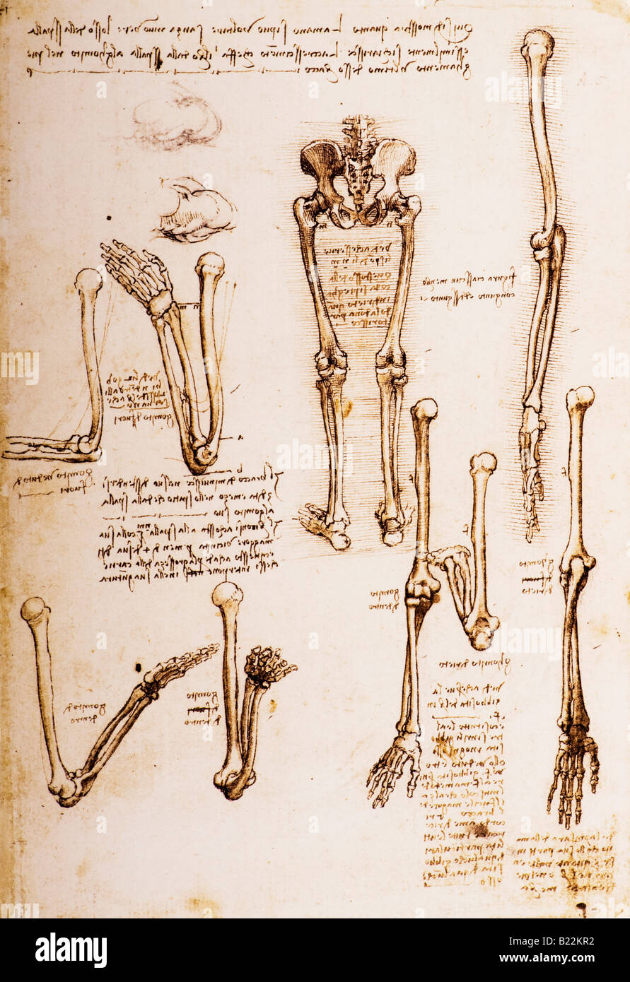 Anatomical Studies of the Female Pelvis, the Coccyx and the Lower Limbs and Rotation of Lower Arm by Leonardo da Vinci 1509 Stock Photohttps://www.alamy.com/image-license-details/?v=1https://www.alamy.com/stock-photo-anatomical-studies-of-the-female-pelvis-the-coccyx-and-the-lower-limbs-18499126.html
RMB22KR2–Anatomical Studies of the Female Pelvis, the Coccyx and the Lower Limbs and Rotation of Lower Arm by Leonardo da Vinci 1509
 Physiotherapist checking patients pelvis Stock Photohttps://www.alamy.com/image-license-details/?v=1https://www.alamy.com/physiotherapist-checking-patients-pelvis-image61939175.html
RFDGNG1B–Physiotherapist checking patients pelvis
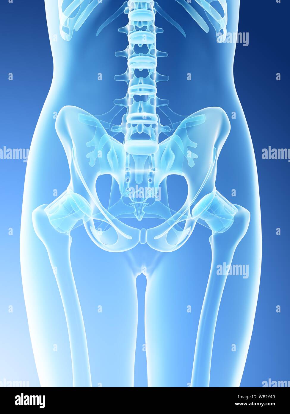 Female pelvis anatomy, computer illustration. Stock Photohttps://www.alamy.com/image-license-details/?v=1https://www.alamy.com/female-pelvis-anatomy-computer-illustration-image264981943.html
RFWB2Y4R–Female pelvis anatomy, computer illustration.
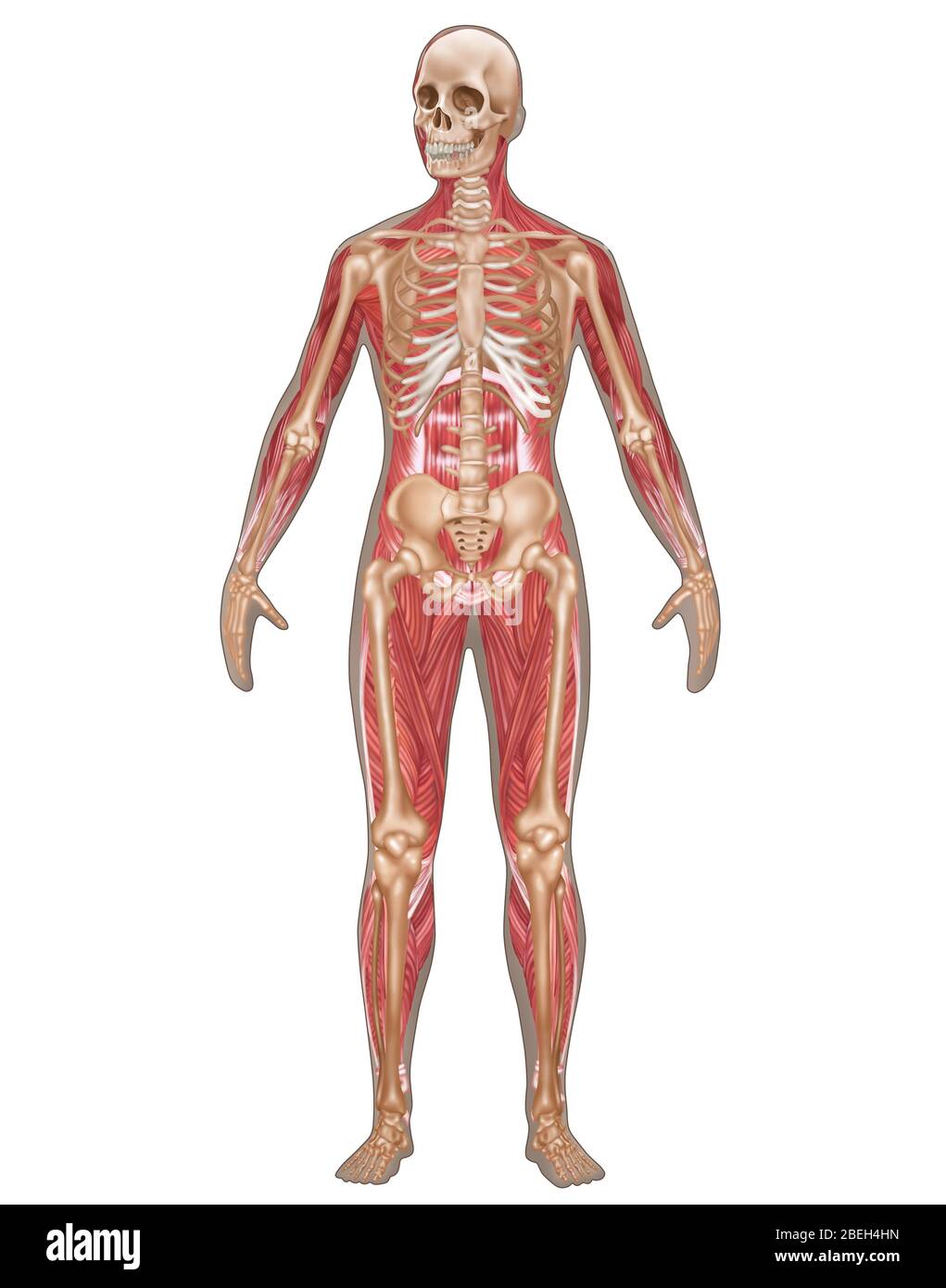 Skeletal & Muscular Systems, Female Anatomy Stock Photohttps://www.alamy.com/image-license-details/?v=1https://www.alamy.com/skeletal-muscular-systems-female-anatomy-image353189361.html
RF2BEH4HN–Skeletal & Muscular Systems, Female Anatomy
 Female Human Skeleton Stock Photohttps://www.alamy.com/image-license-details/?v=1https://www.alamy.com/stock-photo-female-human-skeleton-173207808.html
RMM1P894–Female Human Skeleton
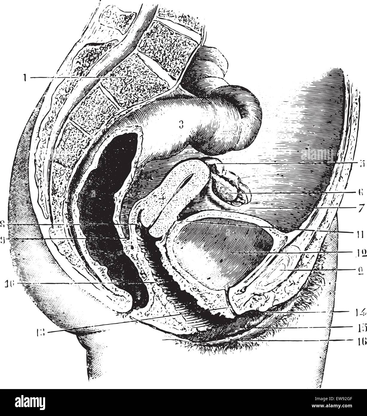 Female pelvis (anteroposterior section), vintage engraved illustration. Usual Medicine Dictionary by Dr Labarthe - 1885. Stock Vectorhttps://www.alamy.com/image-license-details/?v=1https://www.alamy.com/stock-photo-female-pelvis-anteroposterior-section-vintage-engraved-illustration-84407471.html
RFEW92GF–Female pelvis (anteroposterior section), vintage engraved illustration. Usual Medicine Dictionary by Dr Labarthe - 1885.
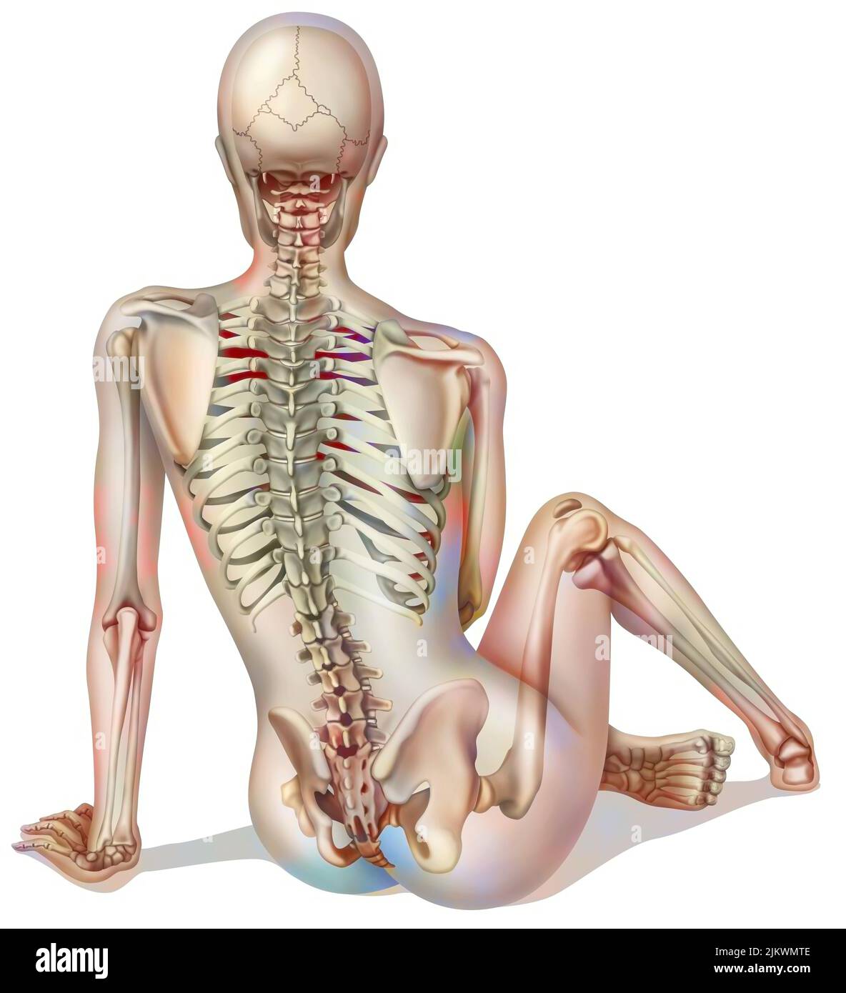 Bone system: female skeleton seen from the back. Stock Photohttps://www.alamy.com/image-license-details/?v=1https://www.alamy.com/bone-system-female-skeleton-seen-from-the-back-image476923566.html
RF2JKWMTE–Bone system: female skeleton seen from the back.
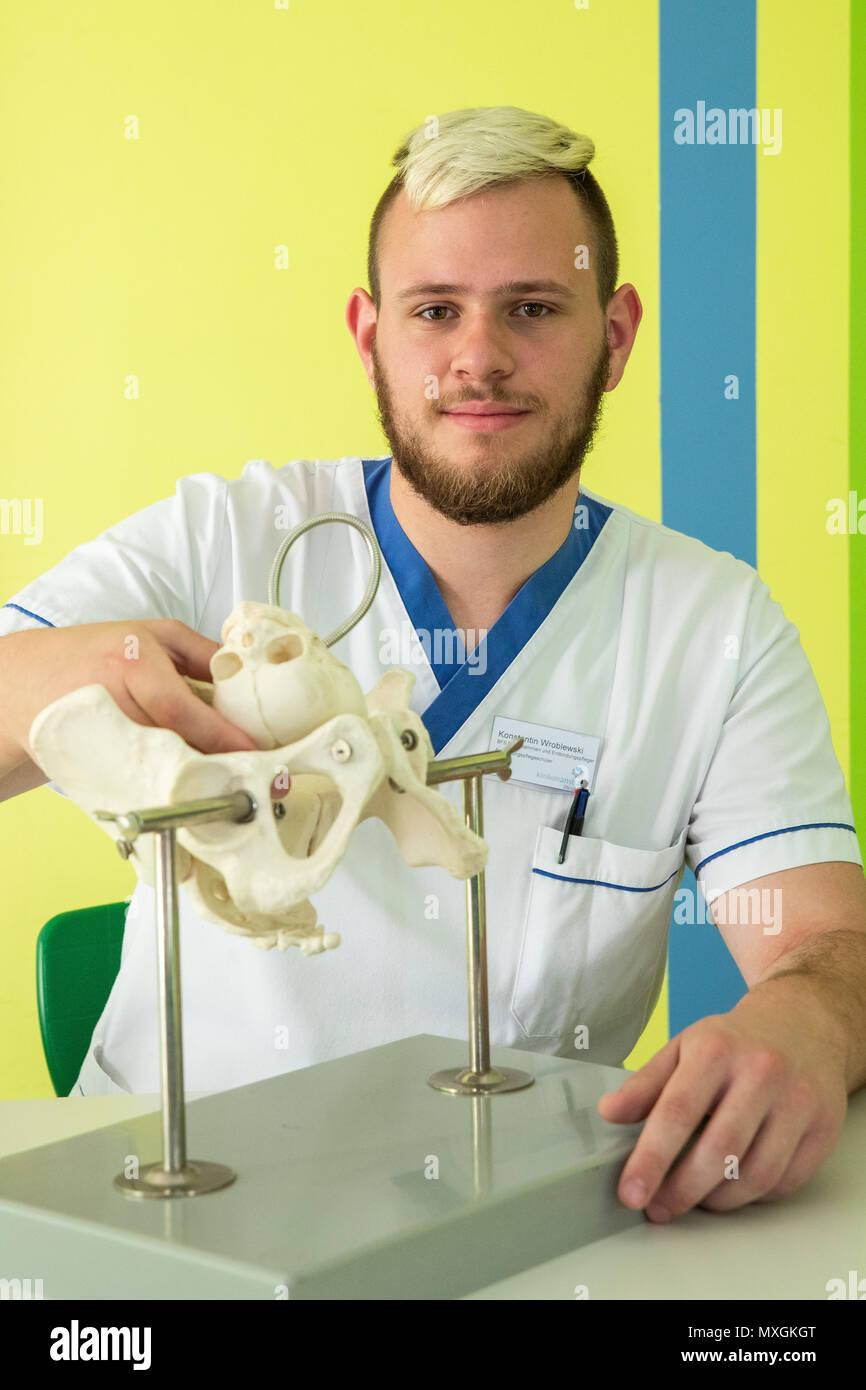 29 May 2018, Germany, Ansbach: Konstantin Wroblewski, male midwife in training at the Berufsfachschule für Hebammen und Entbindungspfleger am Klinikum Ansbach (lit. vocational school for midwifes and male midwifes at the Ansbach Clinic), sits next to a bone model of a female pelvis, in order to learn how the head of the baby has to be positioned during childbirth. Photo: Daniel Karmann/dpa Stock Photohttps://www.alamy.com/image-license-details/?v=1https://www.alamy.com/29-may-2018-germany-ansbach-konstantin-wroblewski-male-midwife-in-training-at-the-berufsfachschule-fr-hebammen-und-entbindungspfleger-am-klinikum-ansbach-lit-vocational-school-for-midwifes-and-male-midwifes-at-the-ansbach-clinic-sits-next-to-a-bone-model-of-a-female-pelvis-in-order-to-learn-how-the-head-of-the-baby-has-to-be-positioned-during-childbirth-photo-daniel-karmanndpa-image188451336.html
RMMXGKGT–29 May 2018, Germany, Ansbach: Konstantin Wroblewski, male midwife in training at the Berufsfachschule für Hebammen und Entbindungspfleger am Klinikum Ansbach (lit. vocational school for midwifes and male midwifes at the Ansbach Clinic), sits next to a bone model of a female pelvis, in order to learn how the head of the baby has to be positioned during childbirth. Photo: Daniel Karmann/dpa
 Female brunette hair tied into ponytail, wearing a green camisole and blue shorts, standing in bad posture, hunched shoulders, Stock Photohttps://www.alamy.com/image-license-details/?v=1https://www.alamy.com/stock-photo-female-brunette-hair-tied-into-ponytail-wearing-a-green-camisole-and-43514782.html
RMCEP7FA–Female brunette hair tied into ponytail, wearing a green camisole and blue shorts, standing in bad posture, hunched shoulders,
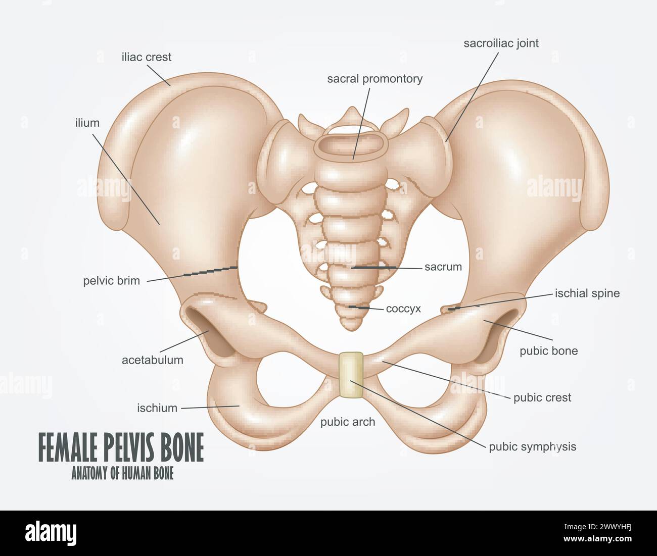 Female Pelvis Bone Anatomy, Vector Illustration Stock Vectorhttps://www.alamy.com/image-license-details/?v=1https://www.alamy.com/female-pelvis-bone-anatomy-vector-illustration-image601125382.html
RF2WWYHFJ–Female Pelvis Bone Anatomy, Vector Illustration
 Woman sitting on the edge of swimming pool with her legs into the water Stock Photohttps://www.alamy.com/image-license-details/?v=1https://www.alamy.com/woman-sitting-on-the-edge-of-swimming-pool-with-her-legs-into-the-water-image605217503.html
RF2X4J12R–Woman sitting on the edge of swimming pool with her legs into the water
 Diet and exercise, slim weight and healthy and beautiful female skin Stock Photohttps://www.alamy.com/image-license-details/?v=1https://www.alamy.com/diet-and-exercise-slim-weight-and-healthy-and-beautiful-female-skin-image475530054.html
RF2JHJ7C6–Diet and exercise, slim weight and healthy and beautiful female skin
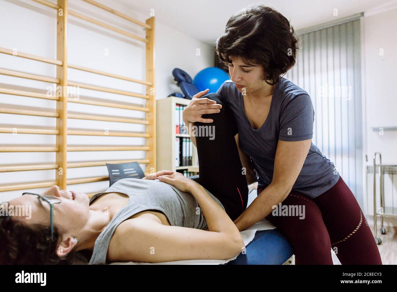 Female physiotherapist stretching leg of client Stock Photohttps://www.alamy.com/image-license-details/?v=1https://www.alamy.com/female-physiotherapist-stretching-leg-of-client-image366652471.html
RF2C8ECY3–Female physiotherapist stretching leg of client
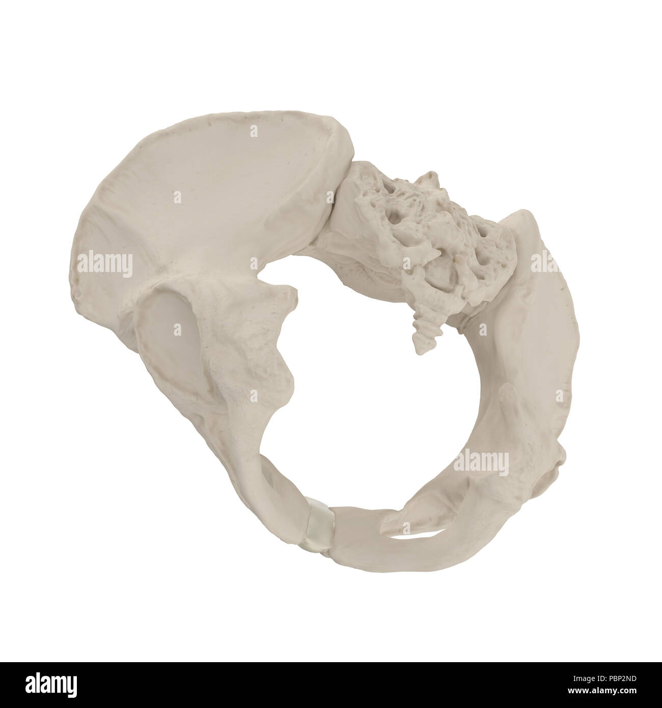 Female Pelvis Skeleton on white. 3D illustration Stock Photohttps://www.alamy.com/image-license-details/?v=1https://www.alamy.com/female-pelvis-skeleton-on-white-3d-illustration-image213770745.html
RFPBP2ND–Female Pelvis Skeleton on white. 3D illustration
 two oranges and a papaya cut in half on a red cloth in front of a peach background as a symbol of the pelvic floor, no people Stock Photohttps://www.alamy.com/image-license-details/?v=1https://www.alamy.com/two-oranges-and-a-papaya-cut-in-half-on-a-red-cloth-in-front-of-a-peach-background-as-a-symbol-of-the-pelvic-floor-no-people-image503449590.html
RF2M7232E–two oranges and a papaya cut in half on a red cloth in front of a peach background as a symbol of the pelvic floor, no people
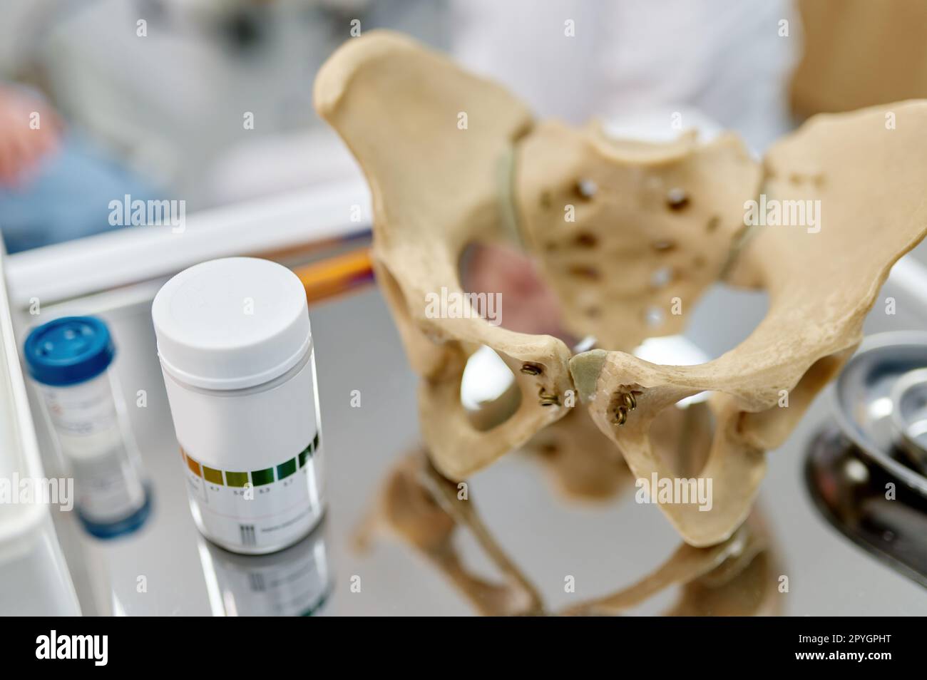 Pelvis anatomical skeleton structure model and pills on table Stock Photohttps://www.alamy.com/image-license-details/?v=1https://www.alamy.com/pelvis-anatomical-skeleton-structure-model-and-pills-on-table-image550486100.html
RF2PYGPHT–Pelvis anatomical skeleton structure model and pills on table
 Doctor gynecologist shows location of female pelvis with muscles. Stock Photohttps://www.alamy.com/image-license-details/?v=1https://www.alamy.com/doctor-gynecologist-shows-location-of-female-pelvis-with-muscles-image507414581.html
RF2MDEMD9–Doctor gynecologist shows location of female pelvis with muscles.
 Composite image of midsection of female doctor showing digital tablet Stock Photohttps://www.alamy.com/image-license-details/?v=1https://www.alamy.com/stock-photo-composite-image-of-midsection-of-female-doctor-showing-digital-tablet-126415507.html
RFH9JM7F–Composite image of midsection of female doctor showing digital tablet
 Female doctor checking xray image, isolated on white background. Stock Photohttps://www.alamy.com/image-license-details/?v=1https://www.alamy.com/stock-photo-female-doctor-checking-xray-image-isolated-on-white-background-51727965.html
RFD04BF9–Female doctor checking xray image, isolated on white background.
 Anatomy of female muscular system, back view. Stock Photohttps://www.alamy.com/image-license-details/?v=1https://www.alamy.com/anatomy-of-female-muscular-system-back-view-image64844283.html
RFDNDWF7–Anatomy of female muscular system, back view.
 Physiotherapist checking patients pelvis alignment Stock Photohttps://www.alamy.com/image-license-details/?v=1https://www.alamy.com/physiotherapist-checking-patients-pelvis-alignment-image61939176.html
RFDGNG1C–Physiotherapist checking patients pelvis alignment
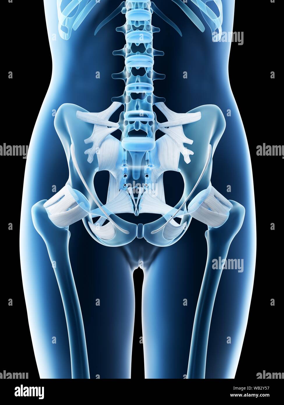 Female pelvis anatomy, computer illustration. Stock Photohttps://www.alamy.com/image-license-details/?v=1https://www.alamy.com/female-pelvis-anatomy-computer-illustration-image264981955.html
RFWB2Y57–Female pelvis anatomy, computer illustration.
 female doctor looking at x-ray on smartphone Stock Photohttps://www.alamy.com/image-license-details/?v=1https://www.alamy.com/stock-photo-female-doctor-looking-at-x-ray-on-smartphone-51395159.html
RMCYH71B–female doctor looking at x-ray on smartphone
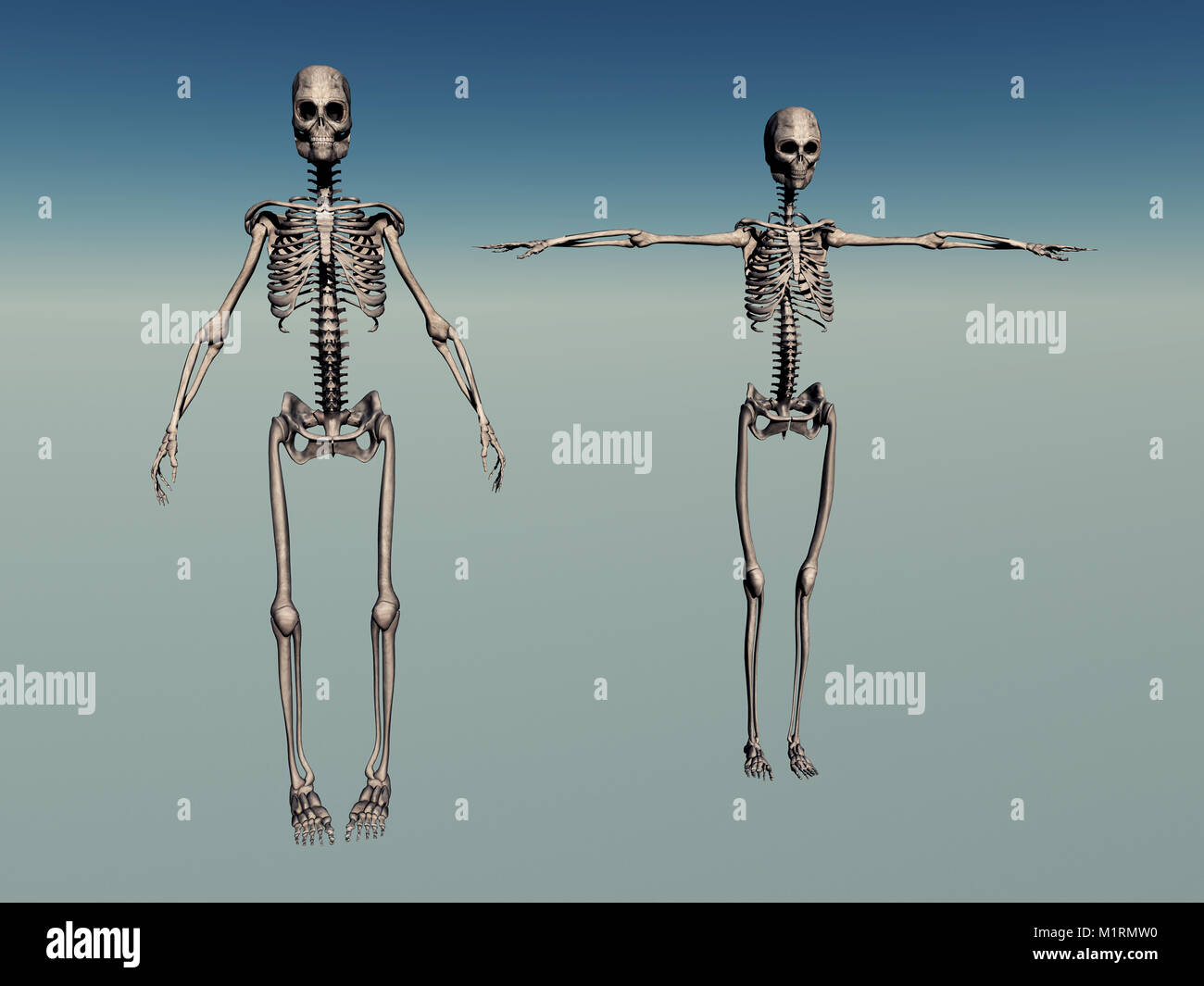 Male & Female Human Skeletons Stock Photohttps://www.alamy.com/image-license-details/?v=1https://www.alamy.com/stock-photo-male-female-human-skeletons-173239612.html
RMM1RMW0–Male & Female Human Skeletons
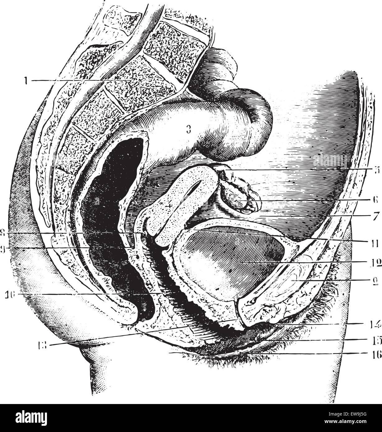 Female pelvis (anteroposterior section), vintage engraved illustration. Usual Medicine Dictionary by Dr Labarthe - 1885. Stock Vectorhttps://www.alamy.com/image-license-details/?v=1https://www.alamy.com/stock-photo-female-pelvis-anteroposterior-section-vintage-engraved-illustration-84419708.html
RFEW9J5G–Female pelvis (anteroposterior section), vintage engraved illustration. Usual Medicine Dictionary by Dr Labarthe - 1885.
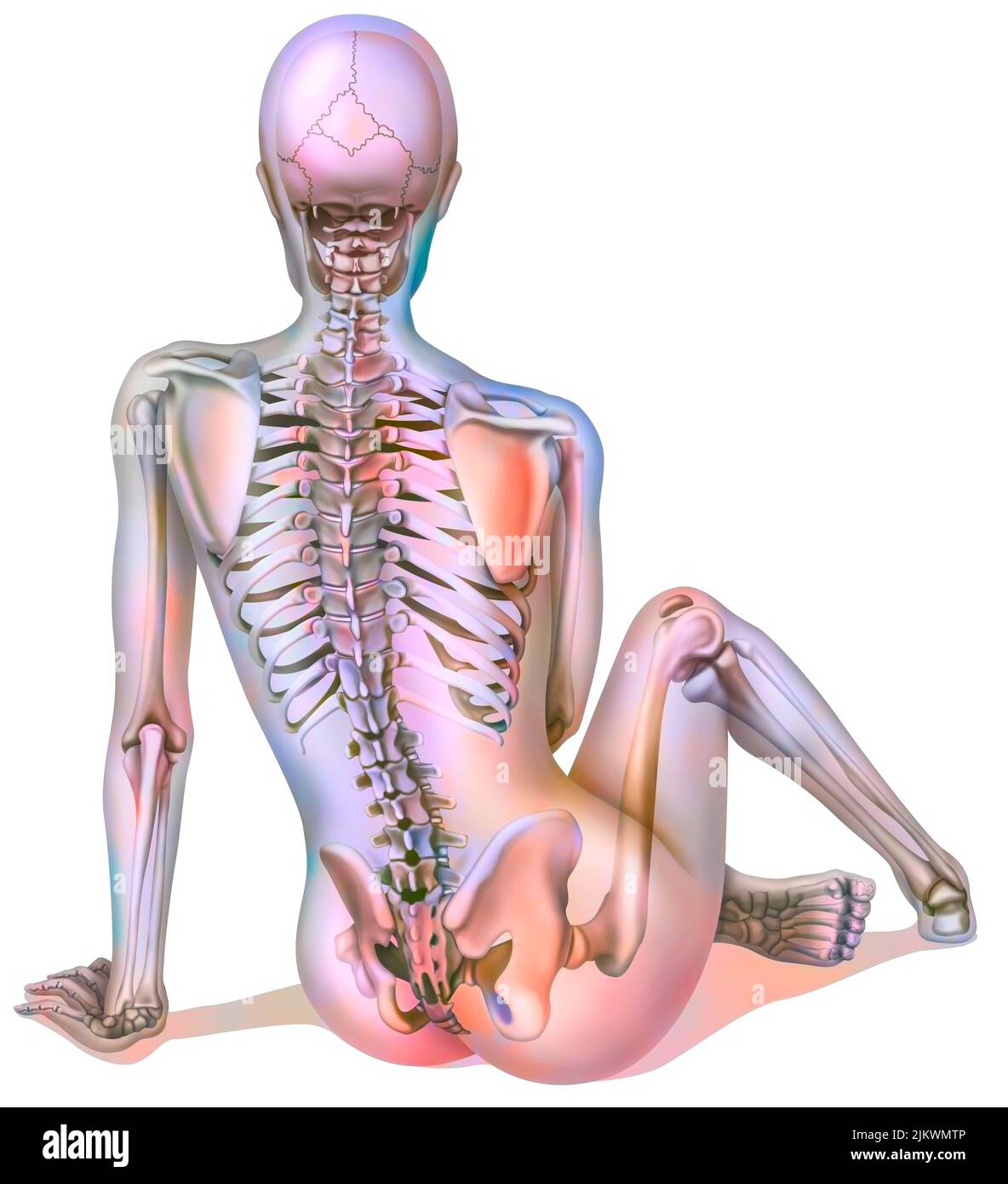 Bone system: female skeleton seen from the back. Stock Photohttps://www.alamy.com/image-license-details/?v=1https://www.alamy.com/bone-system-female-skeleton-seen-from-the-back-image476923574.html
RF2JKWMTP–Bone system: female skeleton seen from the back.
 Female doctor examining a pelvis x-ray Stock Photohttps://www.alamy.com/image-license-details/?v=1https://www.alamy.com/stock-photo-female-doctor-examining-a-pelvis-x-ray-18989345.html
RFB2W12W–Female doctor examining a pelvis x-ray
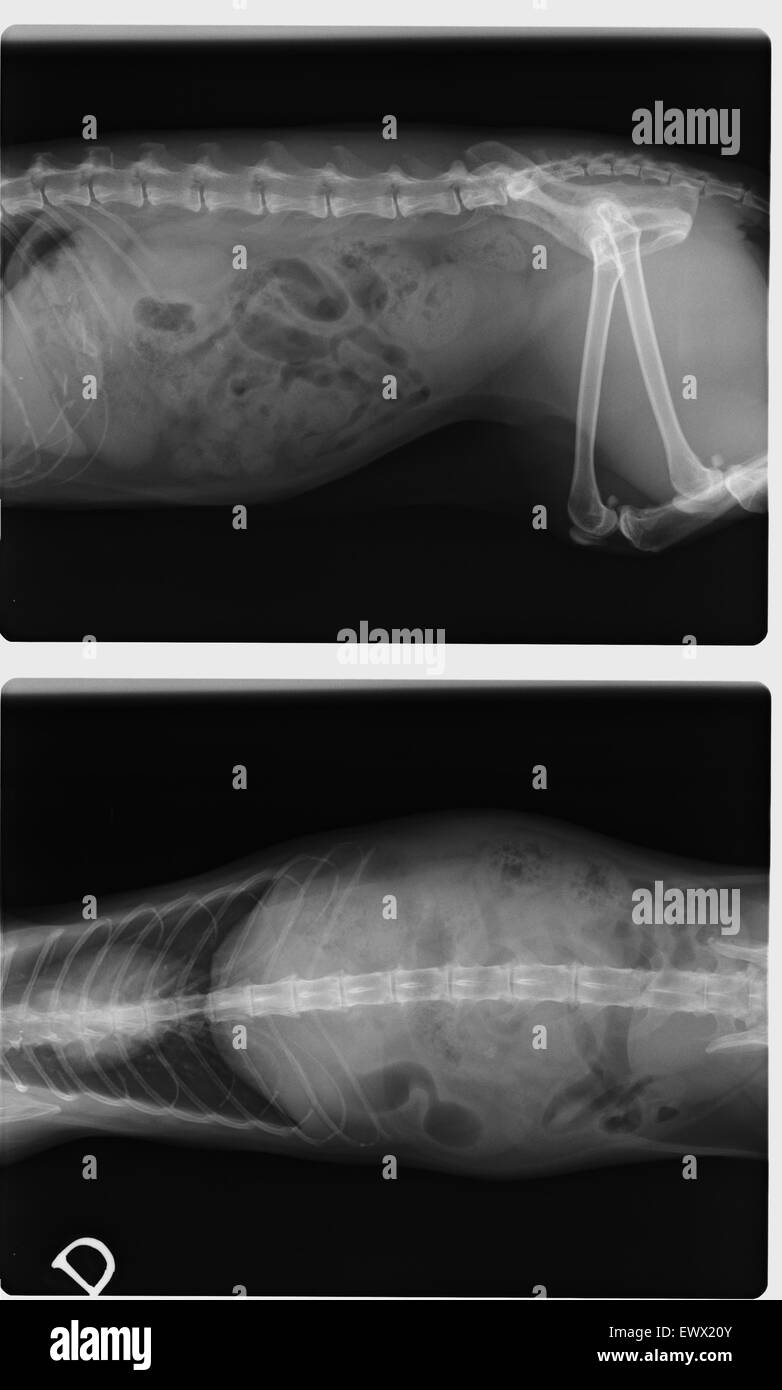 Side and top view of negative X-Ray of spinal column, chest, abdomen, pelvis and thigh bone of a female 13 years old cat Stock Photohttps://www.alamy.com/image-license-details/?v=1https://www.alamy.com/stock-photo-side-and-top-view-of-negative-x-ray-of-spinal-column-chest-abdomen-84780219.html
RFEWX20Y–Side and top view of negative X-Ray of spinal column, chest, abdomen, pelvis and thigh bone of a female 13 years old cat
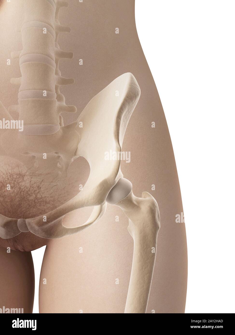 female hip bone Stock Photohttps://www.alamy.com/image-license-details/?v=1https://www.alamy.com/female-hip-bone-image343650229.html
RM2AY2HAD–female hip bone
 Swimmer in swimming pool, swimming, woman, water, water sports, pool, leisure, sport, breaststroke Stock Photohttps://www.alamy.com/image-license-details/?v=1https://www.alamy.com/swimmer-in-swimming-pool-swimming-woman-water-water-sports-pool-leisure-sport-breaststroke-image575822532.html
RF2TCPYEC–Swimmer in swimming pool, swimming, woman, water, water sports, pool, leisure, sport, breaststroke
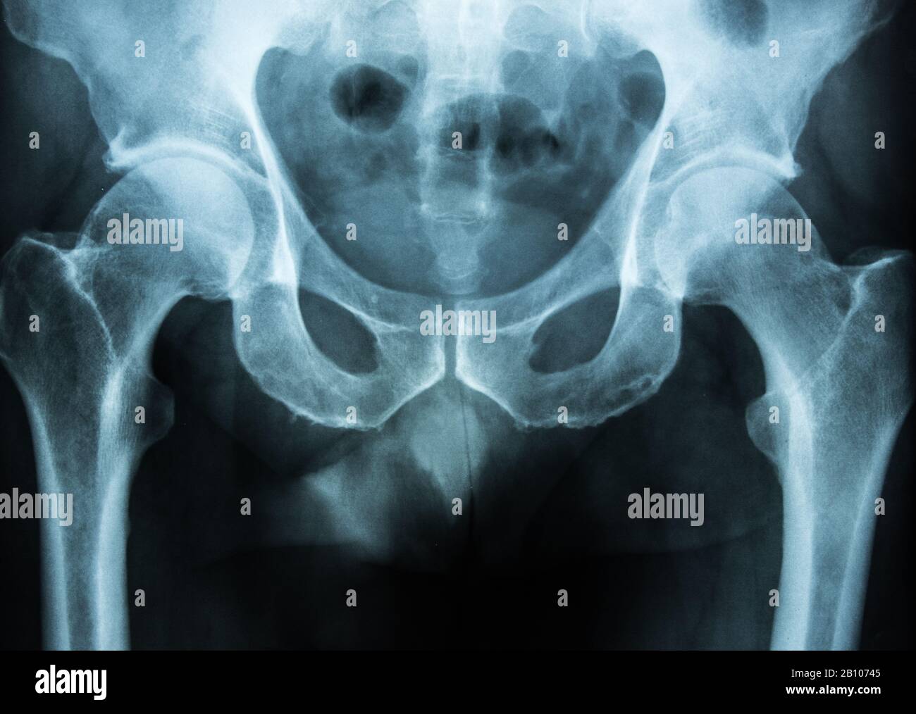 pelvis xray scan Stock Photohttps://www.alamy.com/image-license-details/?v=1https://www.alamy.com/pelvis-xray-scan-image344827621.html
RF2B10745–pelvis xray scan
RF2HM6J47–Pelvis Simple vector icon.
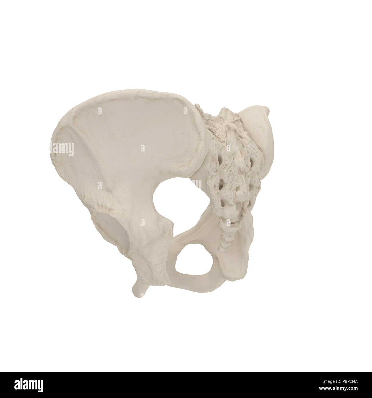 Female Pelvis Skeleton on white. 3D illustration Stock Photohttps://www.alamy.com/image-license-details/?v=1https://www.alamy.com/female-pelvis-skeleton-on-white-3d-illustration-image213770742.html
RFPBP2NA–Female Pelvis Skeleton on white. 3D illustration
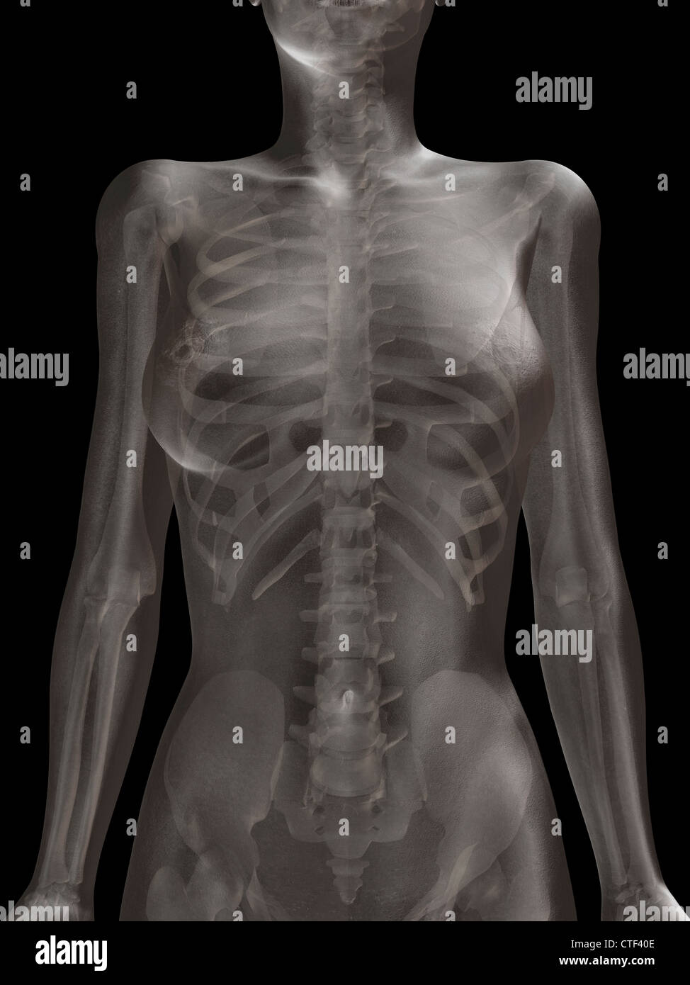 Digitally generated image of female representation with human skeleton visible Stock Photohttps://www.alamy.com/image-license-details/?v=1https://www.alamy.com/stock-photo-digitally-generated-image-of-female-representation-with-human-skeleton-49504910.html
RFCTF40E–Digitally generated image of female representation with human skeleton visible
 Gynecologist doctor measuring female patient hip volume during checkup Stock Photohttps://www.alamy.com/image-license-details/?v=1https://www.alamy.com/gynecologist-doctor-measuring-female-patient-hip-volume-during-checkup-image550486192.html
RF2PYGPN4–Gynecologist doctor measuring female patient hip volume during checkup
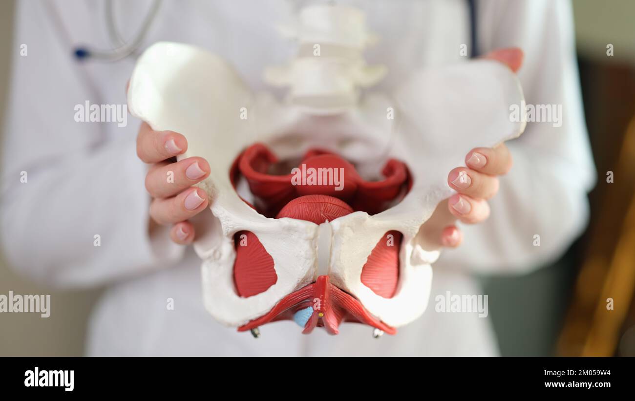 Female gynecologist showing model of female pelvis with muscles Stock Photohttps://www.alamy.com/image-license-details/?v=1https://www.alamy.com/female-gynecologist-showing-model-of-female-pelvis-with-muscles-image499218192.html
RF2M059W4–Female gynecologist showing model of female pelvis with muscles
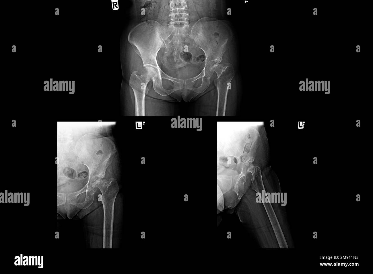 Film x-ray of human pelvis and hip joints Stock Photohttps://www.alamy.com/image-license-details/?v=1https://www.alamy.com/film-x-ray-of-human-pelvis-and-hip-joints-image504655903.html
RF2M911N3–Film x-ray of human pelvis and hip joints
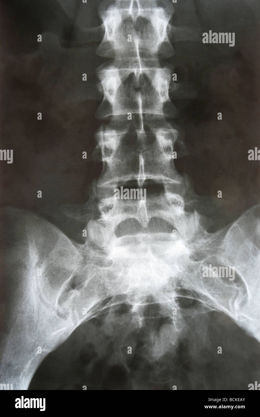 X-RAY film of lower thorax and pelvis. Stock Photohttps://www.alamy.com/image-license-details/?v=1https://www.alamy.com/stock-photo-x-ray-film-of-lower-thorax-and-pelvis-25014611.html
RMBCKEAY–X-RAY film of lower thorax and pelvis.
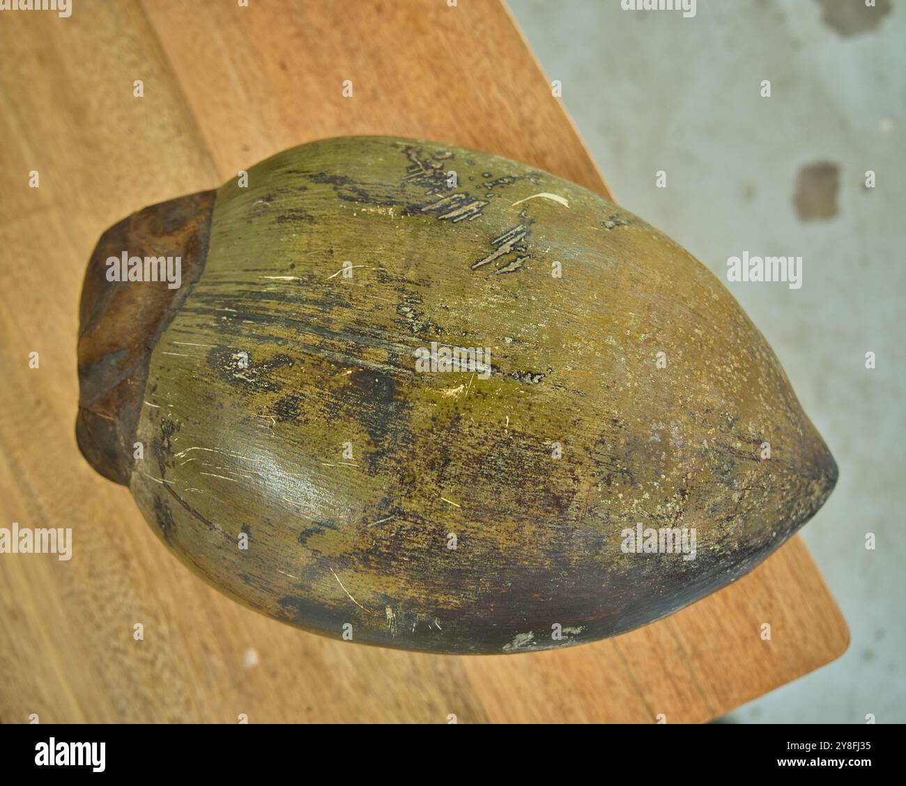 One coco de mer female nut on the table as display, Mahe, Seychelles Stock Photohttps://www.alamy.com/image-license-details/?v=1https://www.alamy.com/one-coco-de-mer-female-nut-on-the-table-as-display-mahe-seychelles-image624833977.html
RF2Y8FJ35–One coco de mer female nut on the table as display, Mahe, Seychelles
 Physiotherapist examining patients pelvis Stock Photohttps://www.alamy.com/image-license-details/?v=1https://www.alamy.com/physiotherapist-examining-patients-pelvis-image61939178.html
RFDGNG1E–Physiotherapist examining patients pelvis
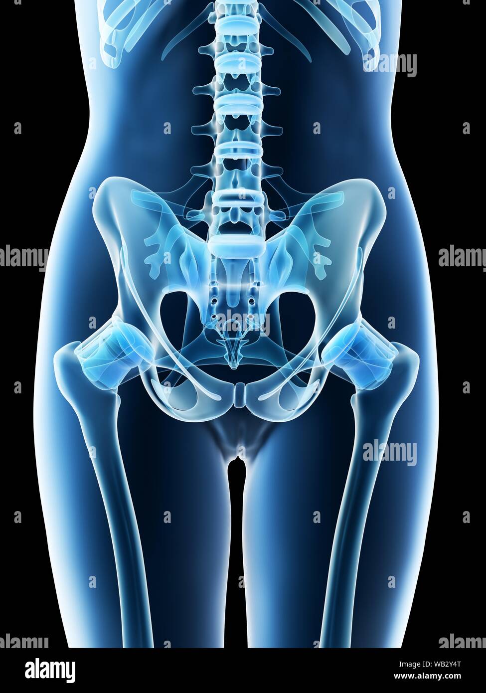 Female pelvis anatomy, computer illustration. Stock Photohttps://www.alamy.com/image-license-details/?v=1https://www.alamy.com/female-pelvis-anatomy-computer-illustration-image264981944.html
RFWB2Y4T–Female pelvis anatomy, computer illustration.
 female doctor looking at x-ray image on smartphone Stock Photohttps://www.alamy.com/image-license-details/?v=1https://www.alamy.com/stock-photo-female-doctor-looking-at-x-ray-image-on-smartphone-50887866.html
RMCXP3YP–female doctor looking at x-ray image on smartphone
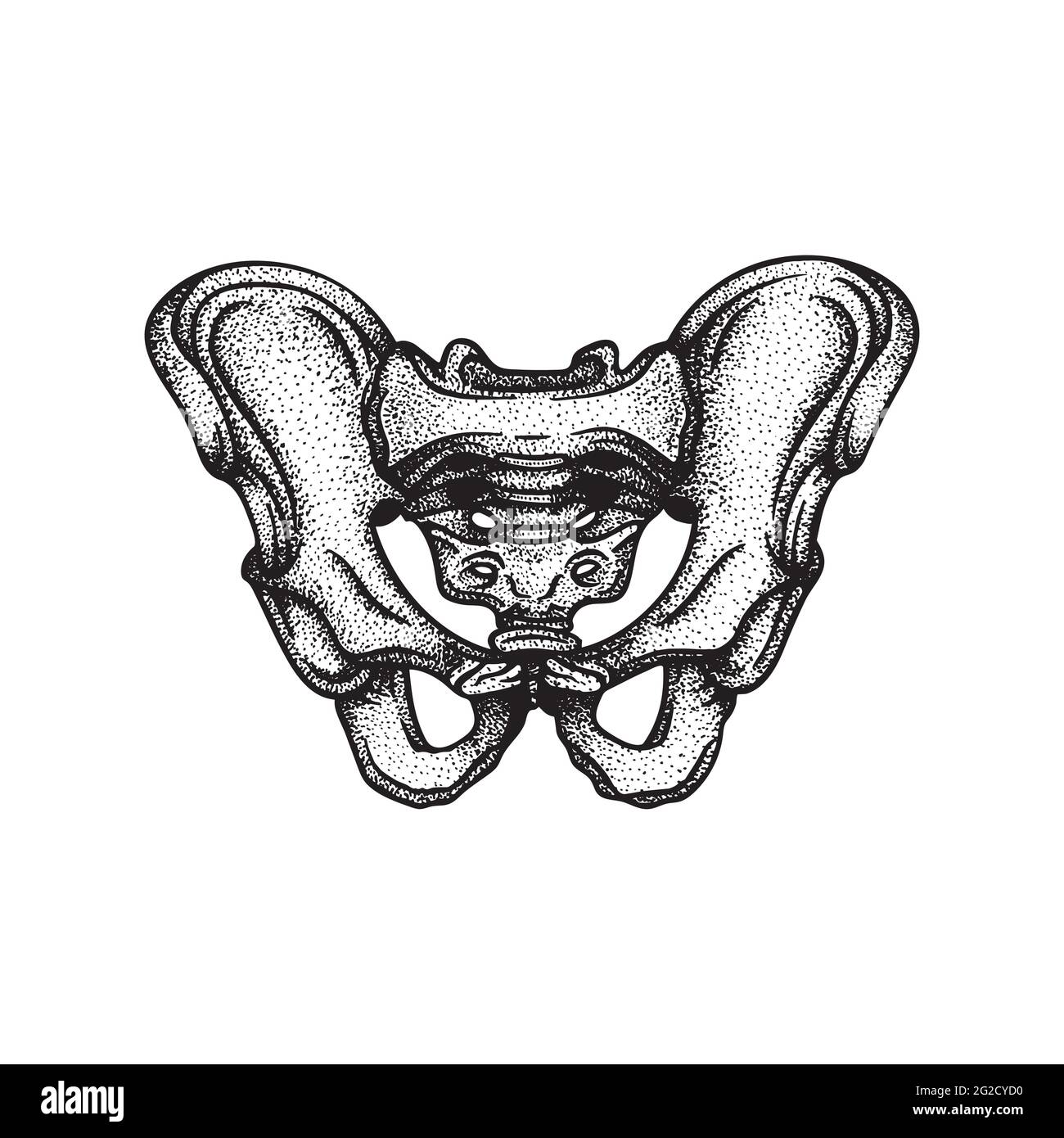 Pelvis. Human pelvis bone hand drawn vector illustration. Part of human skeleton graphic. Stock Vectorhttps://www.alamy.com/image-license-details/?v=1https://www.alamy.com/pelvis-human-pelvis-bone-hand-drawn-vector-illustration-part-of-human-skeleton-graphic-image431773468.html
RF2G2CYD0–Pelvis. Human pelvis bone hand drawn vector illustration. Part of human skeleton graphic.
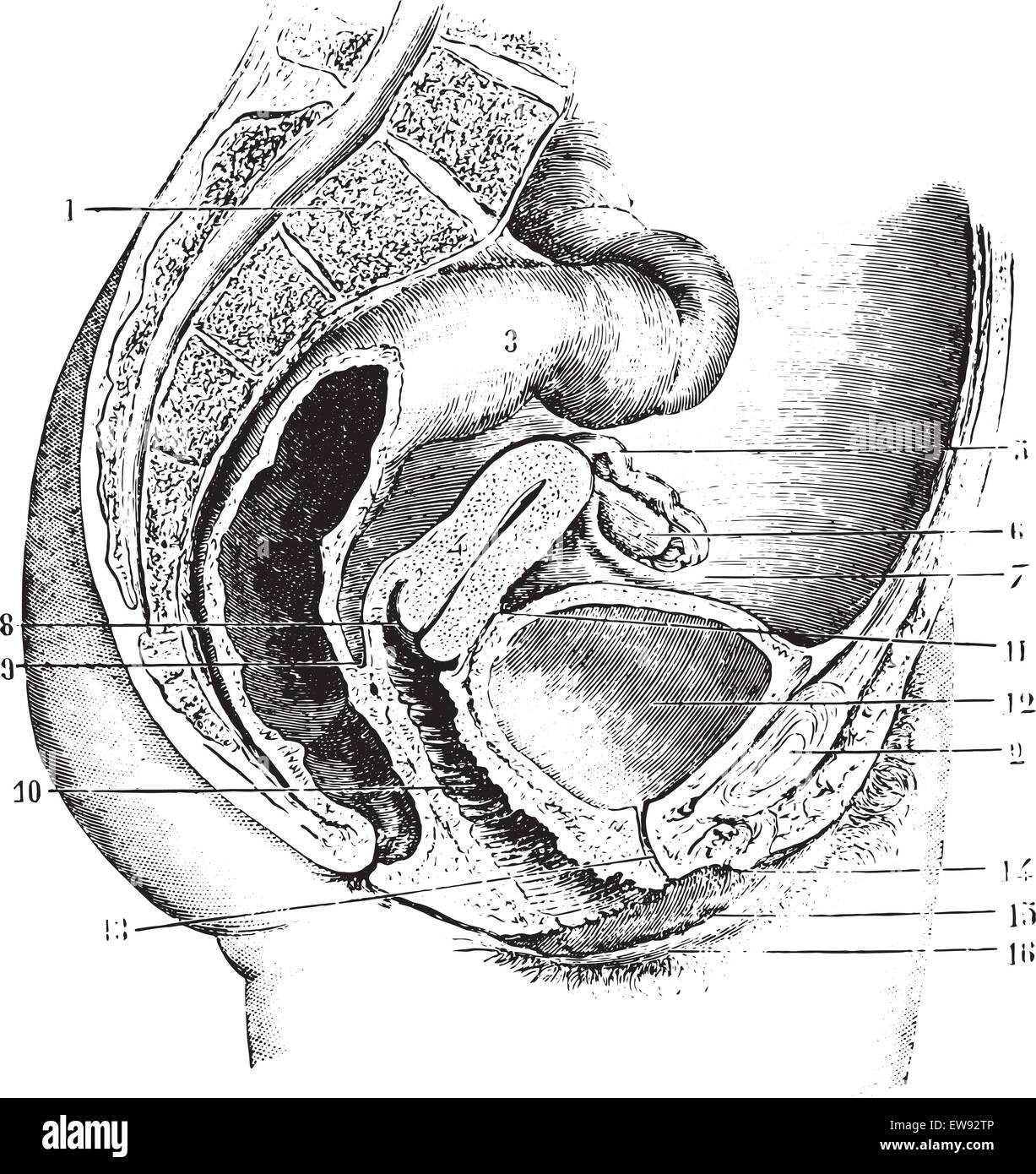 Female pelvis (antero-posterior section), vintage engraved illustration. Usual Medicine Dictionary by Dr Labarthe - 1885. Stock Vectorhttps://www.alamy.com/image-license-details/?v=1https://www.alamy.com/stock-photo-female-pelvis-antero-posterior-section-vintage-engraved-illustration-84407702.html
RFEW92TP–Female pelvis (antero-posterior section), vintage engraved illustration. Usual Medicine Dictionary by Dr Labarthe - 1885.
