Quick filters:
Fasciae Stock Photos and Images
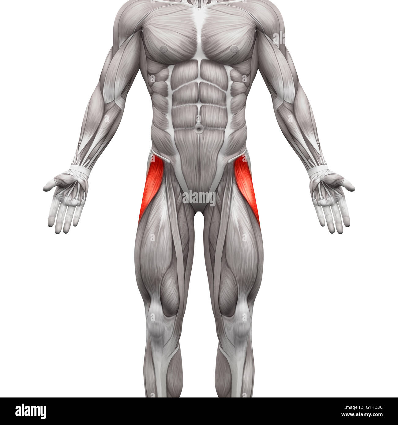 Tensor Fasciae Latae Muscle - Anatomy Muscles isolated on white - 3D illustration Stock Photohttps://www.alamy.com/image-license-details/?v=1https://www.alamy.com/stock-photo-tensor-fasciae-latae-muscle-anatomy-muscles-isolated-on-white-3d-illustration-104260336.html
RFG1HD3C–Tensor Fasciae Latae Muscle - Anatomy Muscles isolated on white - 3D illustration
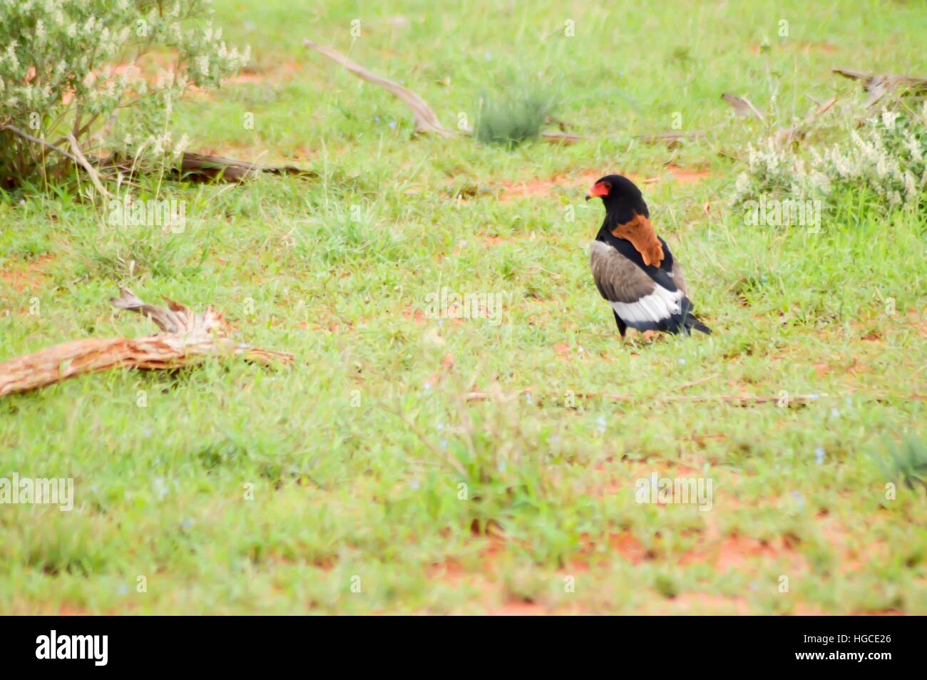 Eagle fasciae posed in the savanna in the park of Tsavo East Stock Photohttps://www.alamy.com/image-license-details/?v=1https://www.alamy.com/stock-photo-eagle-fasciae-posed-in-the-savanna-in-the-park-of-tsavo-east-130581534.html
RFHGCE26–Eagle fasciae posed in the savanna in the park of Tsavo East
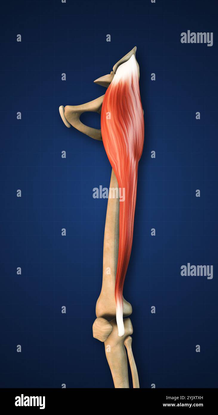 Tensor Fasciae Muscle with Lower Limb Stock Photohttps://www.alamy.com/image-license-details/?v=1https://www.alamy.com/tensor-fasciae-muscle-with-lower-limb-image631227369.html
RF2YJXTXH–Tensor Fasciae Muscle with Lower Limb
 Tensor fasciae latae: Myofascial trigger points and associated pain locations Stock Photohttps://www.alamy.com/image-license-details/?v=1https://www.alamy.com/tensor-fasciae-latae-myofascial-trigger-points-and-associated-pain-locations-image592977838.html
RF2WCMD7X–Tensor fasciae latae: Myofascial trigger points and associated pain locations
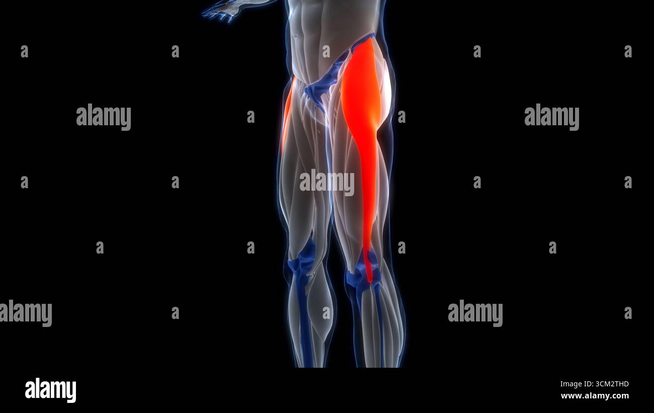 Human Muscular System Leg Muscles Tensor Fasciae Latae Muscles Anatomy Stock Photohttps://www.alamy.com/image-license-details/?v=1https://www.alamy.com/human-muscular-system-leg-muscles-tensor-fasciae-latae-muscles-anatomy-image700771049.html
RF3CM2THD–Human Muscular System Leg Muscles Tensor Fasciae Latae Muscles Anatomy
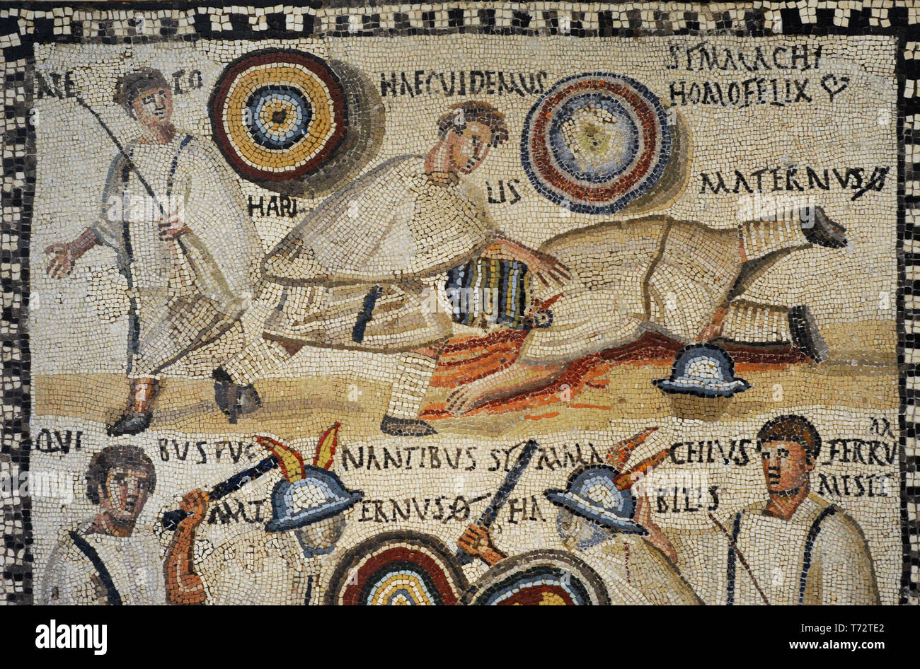 Roman mosaic depicting a Gladiator fight. In the lower part, the murmillones Symmanchus and Maternus are fighting in the arena cheered by the lanistae. At the top, Maternus lies slain by Symmachus. Detail. 3rd century AD. Limestone and vitreous paste. From Rome (Italy). National Archaeological Museum. Madrid. Spain. Stock Photohttps://www.alamy.com/image-license-details/?v=1https://www.alamy.com/roman-mosaic-depicting-a-gladiator-fight-in-the-lower-part-the-murmillones-symmanchus-and-maternus-are-fighting-in-the-arena-cheered-by-the-lanistae-at-the-top-maternus-lies-slain-by-symmachus-detail-3rd-century-ad-limestone-and-vitreous-paste-from-rome-italy-national-archaeological-museum-madrid-spain-image245310858.html
RMT72TE2–Roman mosaic depicting a Gladiator fight. In the lower part, the murmillones Symmanchus and Maternus are fighting in the arena cheered by the lanistae. At the top, Maternus lies slain by Symmachus. Detail. 3rd century AD. Limestone and vitreous paste. From Rome (Italy). National Archaeological Museum. Madrid. Spain.
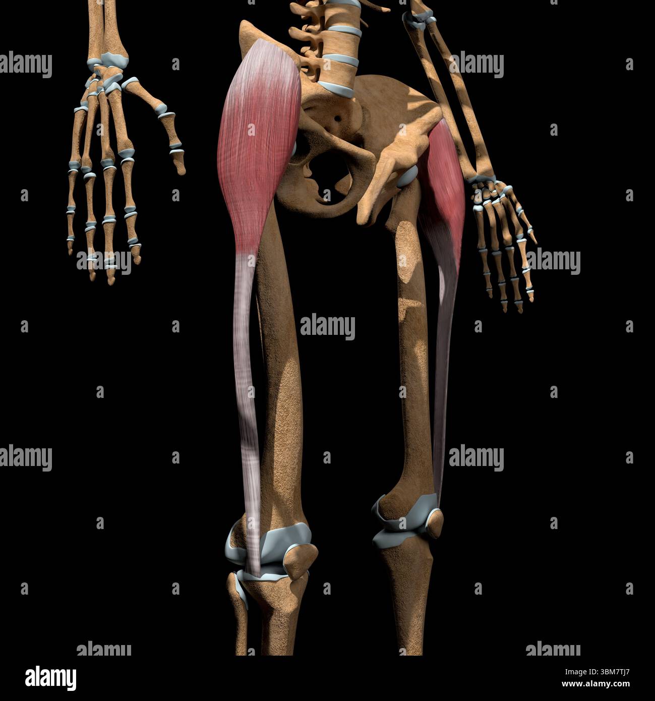 This 3d illustration shows the tensor fasciae latae muscles on skeleton. Stock Photohttps://www.alamy.com/image-license-details/?v=1https://www.alamy.com/this-3d-illustration-shows-the-tensor-fasciae-latae-muscles-on-skeleton-image683670463.html
RF3BM7TJ7–This 3d illustration shows the tensor fasciae latae muscles on skeleton.
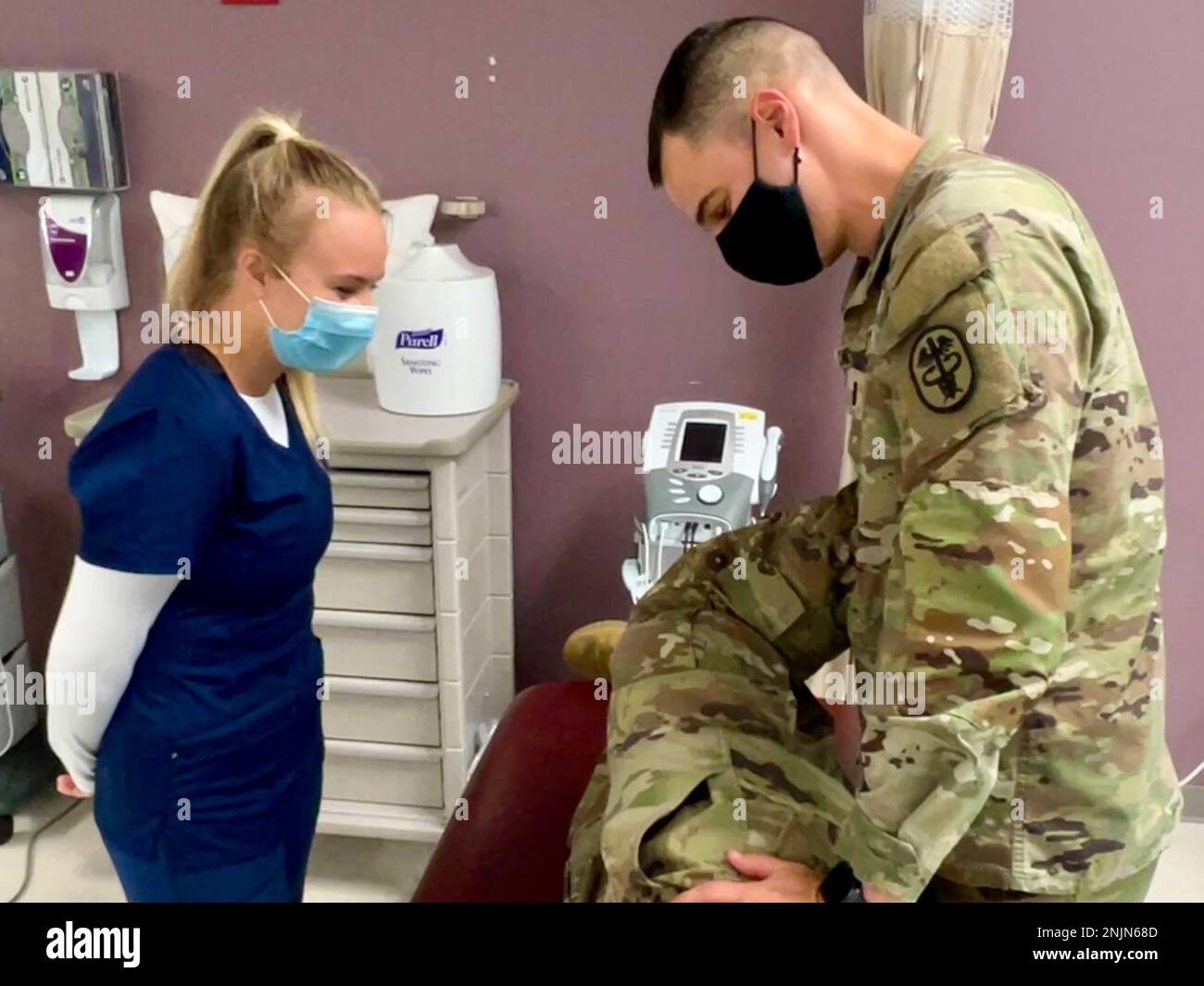 1st Lt. Steven Candeto demonstrates the Obers Test, which evaluates a tight, contracted or inflamed Tensor Fasciae Latae (TFL) and Iliotibial band (ITB) for Christina McDonald, a member of the Crimson Tide Reserve Officer Training Corps (ROTC) Battalion and a kinesiology major from the University of Alabama. McDonald spent two weeks shadowing the physical therapists as an American Red Cross volunteer for Bayne-Jones Army Community Hospital at the Joint Readiness Training Center and Fort Polk, Louisiana. Stock Photohttps://www.alamy.com/image-license-details/?v=1https://www.alamy.com/1st-lt-steven-candeto-demonstrates-the-obers-test-which-evaluates-a-tight-contracted-or-inflamed-tensor-fasciae-latae-tfl-and-iliotibial-band-itb-for-christina-mcdonald-a-member-of-the-crimson-tide-reserve-officer-training-corps-rotc-battalion-and-a-kinesiology-major-from-the-university-of-alabama-mcdonald-spent-two-weeks-shadowing-the-physical-therapists-as-an-american-red-cross-volunteer-for-bayne-jones-army-community-hospital-at-the-joint-readiness-training-center-and-fort-polk-louisiana-image527840781.html
RM2NJN68D–1st Lt. Steven Candeto demonstrates the Obers Test, which evaluates a tight, contracted or inflamed Tensor Fasciae Latae (TFL) and Iliotibial band (ITB) for Christina McDonald, a member of the Crimson Tide Reserve Officer Training Corps (ROTC) Battalion and a kinesiology major from the University of Alabama. McDonald spent two weeks shadowing the physical therapists as an American Red Cross volunteer for Bayne-Jones Army Community Hospital at the Joint Readiness Training Center and Fort Polk, Louisiana.
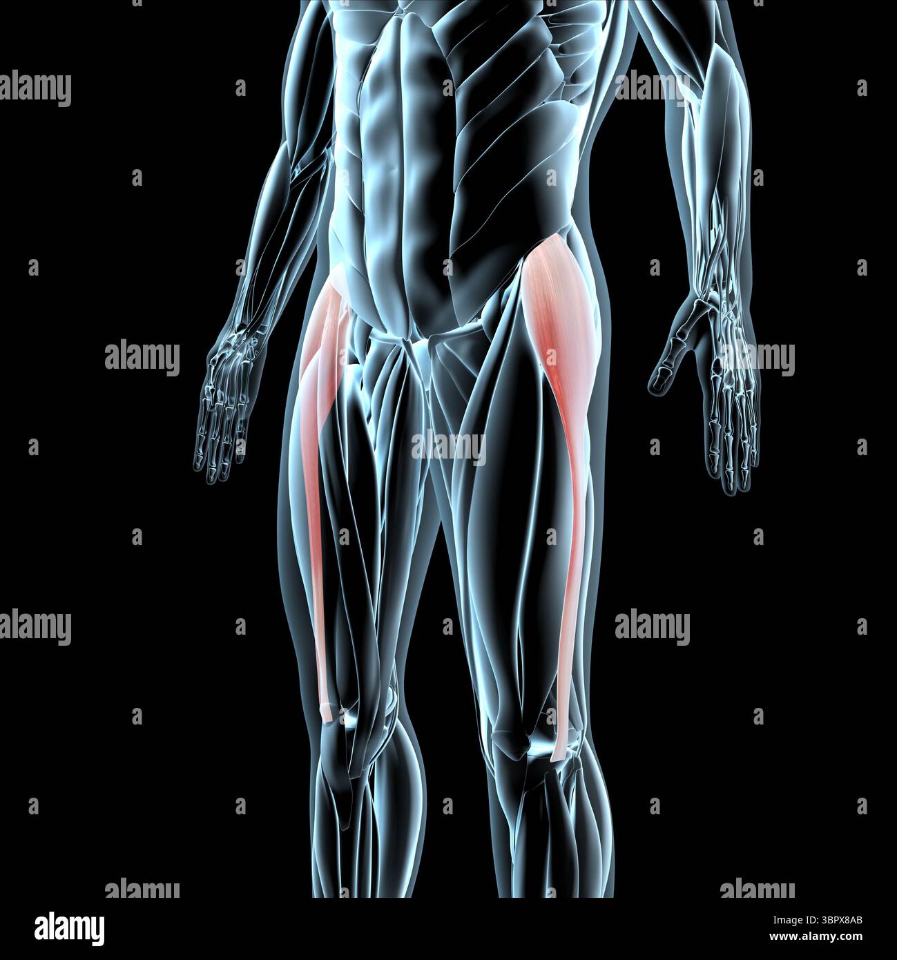 This 3d illustration shows a view of the tensor fasciae latae muscles on xray musculature Stock Photohttps://www.alamy.com/image-license-details/?v=1https://www.alamy.com/this-3d-illustration-shows-a-view-of-the-tensor-fasciae-latae-muscles-on-xray-musculature-image685304099.html
RF3BPX8AB–This 3d illustration shows a view of the tensor fasciae latae muscles on xray musculature
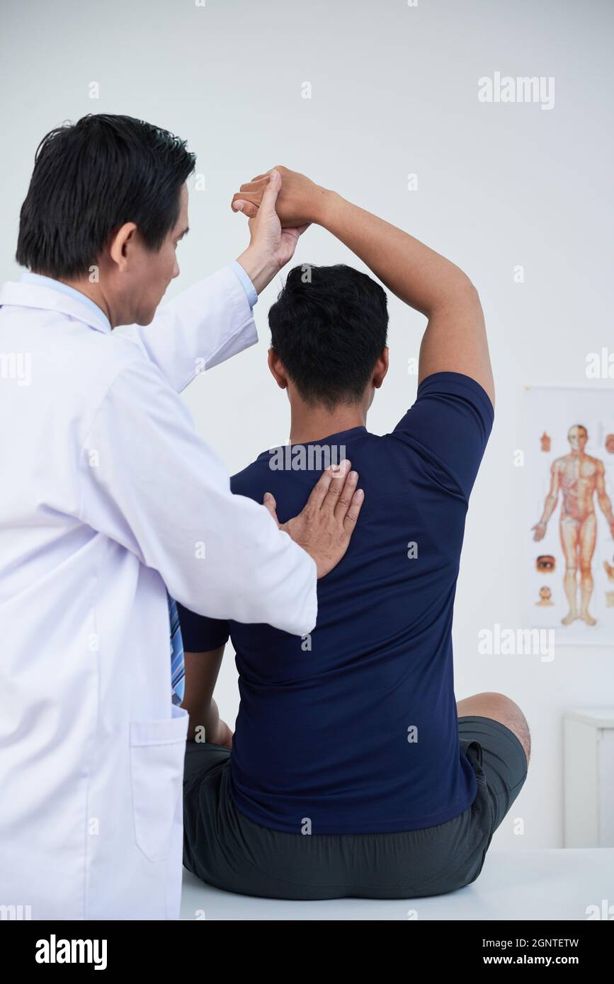 Vertical medium back view shot of modern mature osteopathic therapist treating young man manipulating his bones, muscles and fasciae Stock Photohttps://www.alamy.com/image-license-details/?v=1https://www.alamy.com/vertical-medium-back-view-shot-of-modern-mature-osteopathic-therapist-treating-young-man-manipulating-his-bones-muscles-and-fasciae-image443705497.html
RF2GNTETW–Vertical medium back view shot of modern mature osteopathic therapist treating young man manipulating his bones, muscles and fasciae
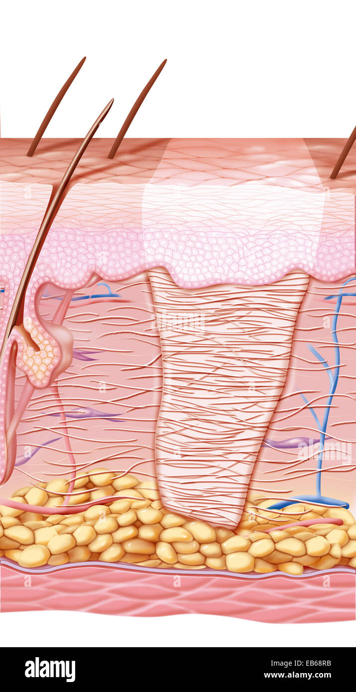 SCAR DRAWING Stock Photohttps://www.alamy.com/image-license-details/?v=1https://www.alamy.com/stock-photo-scar-drawing-75741327.html
RMEB68RB–SCAR DRAWING
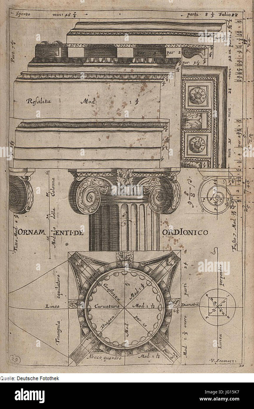 Fotothek df tg 0007925 Architektur 5E Gebälk 5E Gesims 5E Faszie 5E Kapitell 5E Volute 5E Ornament 5E Rosette Stock Photohttps://www.alamy.com/image-license-details/?v=1https://www.alamy.com/stock-photo-fotothek-df-tg-0007925-architektur-5e-geblk-5e-gesims-5e-faszie-5e-147543851.html
RMJG15K7–Fotothek df tg 0007925 Architektur 5E Gebälk 5E Gesims 5E Faszie 5E Kapitell 5E Volute 5E Ornament 5E Rosette
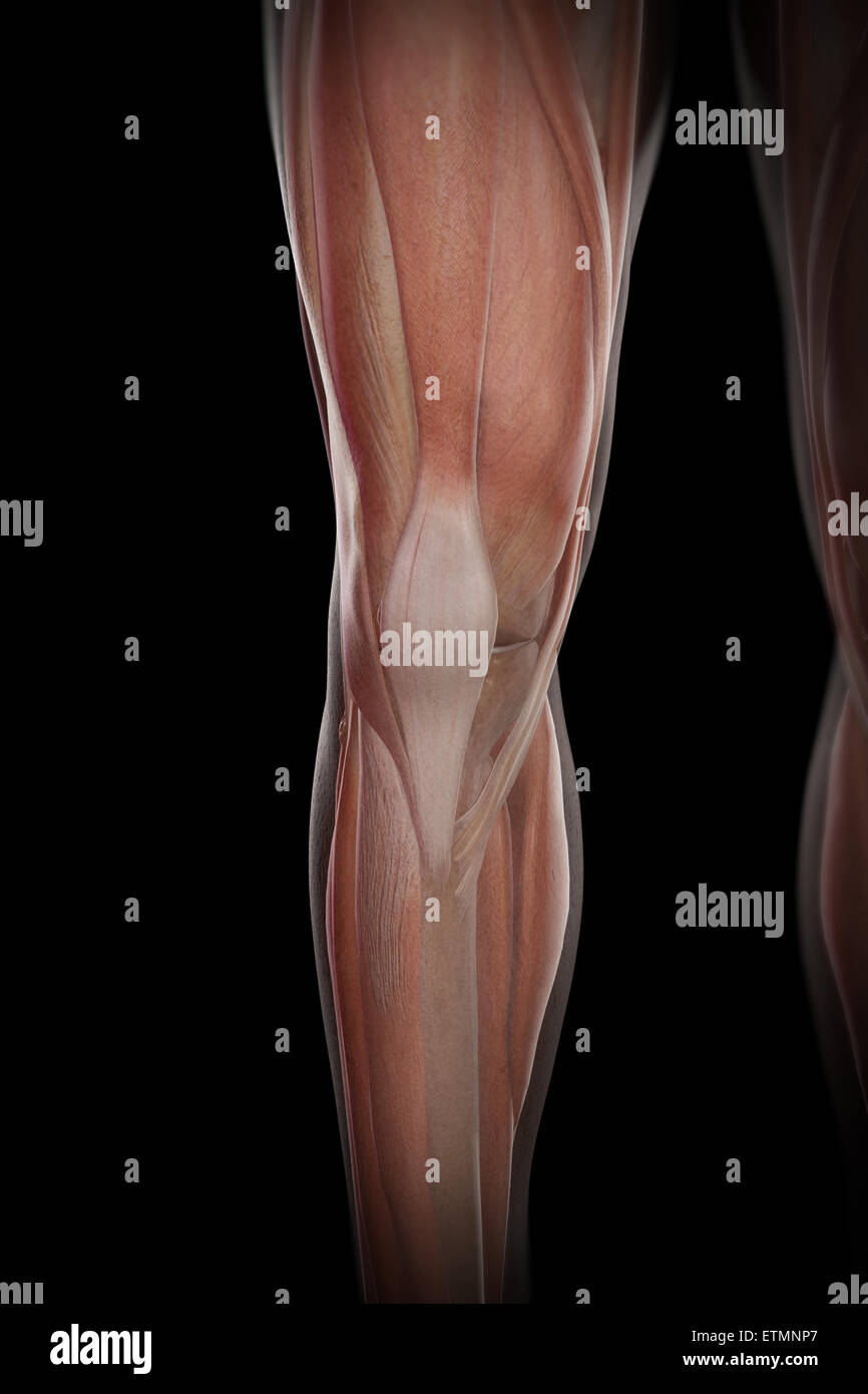 Illustration of the musculature and skeletal structure of the lower legs, visible through skin. Stock Photohttps://www.alamy.com/image-license-details/?v=1https://www.alamy.com/stock-photo-illustration-of-the-musculature-and-skeletal-structure-of-the-lower-84049343.html
RMETMNP7–Illustration of the musculature and skeletal structure of the lower legs, visible through skin.
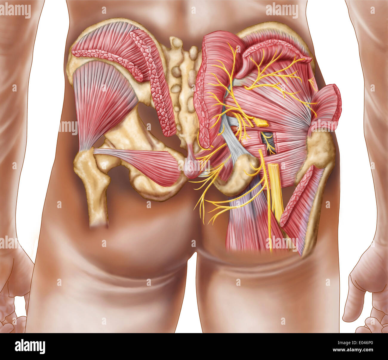 Anatomy of the gluteal muscles in the human buttocks. Stock Photohttps://www.alamy.com/image-license-details/?v=1https://www.alamy.com/anatomy-of-the-gluteal-muscles-in-the-human-buttocks-image68934600.html
RFE046P0–Anatomy of the gluteal muscles in the human buttocks.
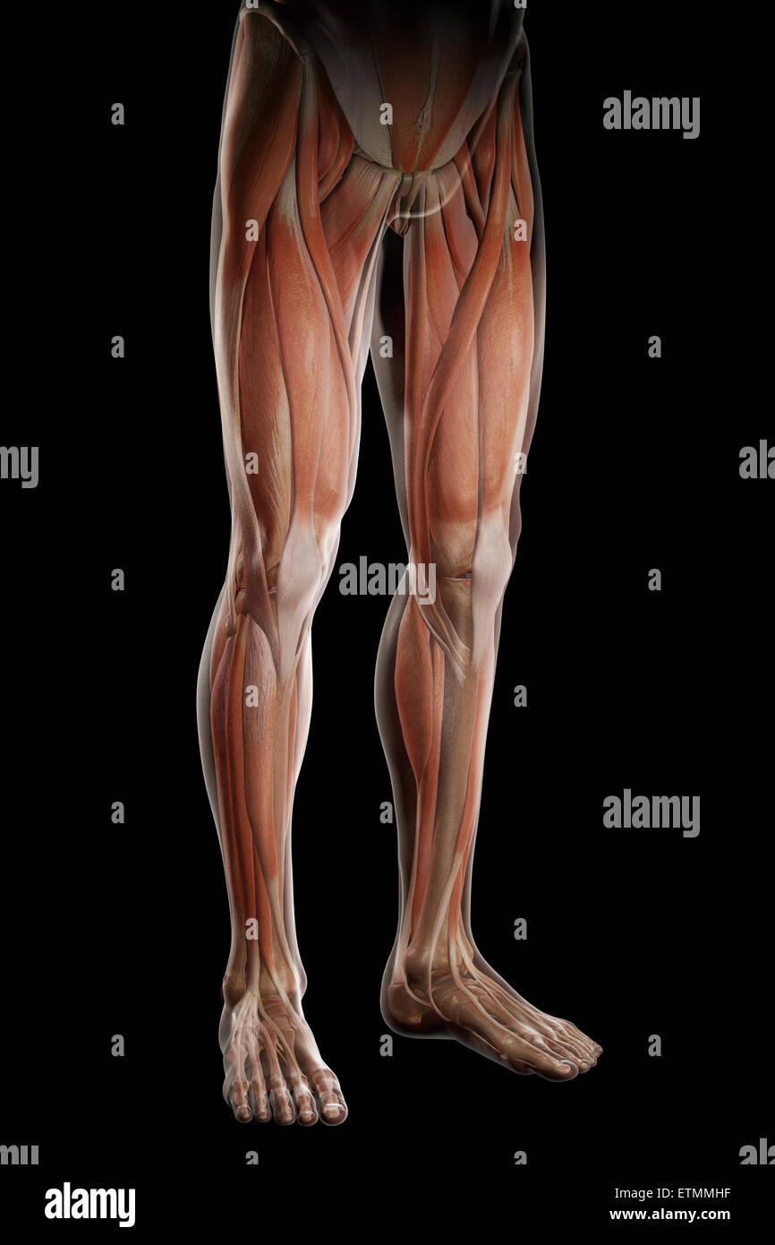 Illustration of the musculature and skeletal structure of the legs, visible through skin. Stock Photohttps://www.alamy.com/image-license-details/?v=1https://www.alamy.com/stock-photo-illustration-of-the-musculature-and-skeletal-structure-of-the-legs-84048427.html
RMETMMHF–Illustration of the musculature and skeletal structure of the legs, visible through skin.
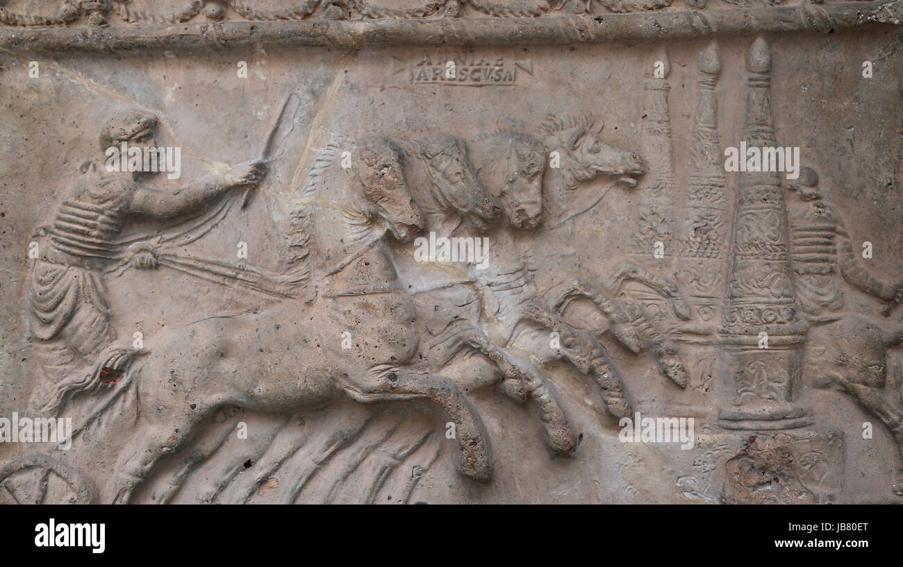 Terracotta decorative relief. Chariot race. c. 70 AD. British Museum. London. UK Stock Photohttps://www.alamy.com/image-license-details/?v=1https://www.alamy.com/stock-photo-terracotta-decorative-relief-chariot-race-c-70-ad-british-museum-london-144620192.html
RMJB80ET–Terracotta decorative relief. Chariot race. c. 70 AD. British Museum. London. UK
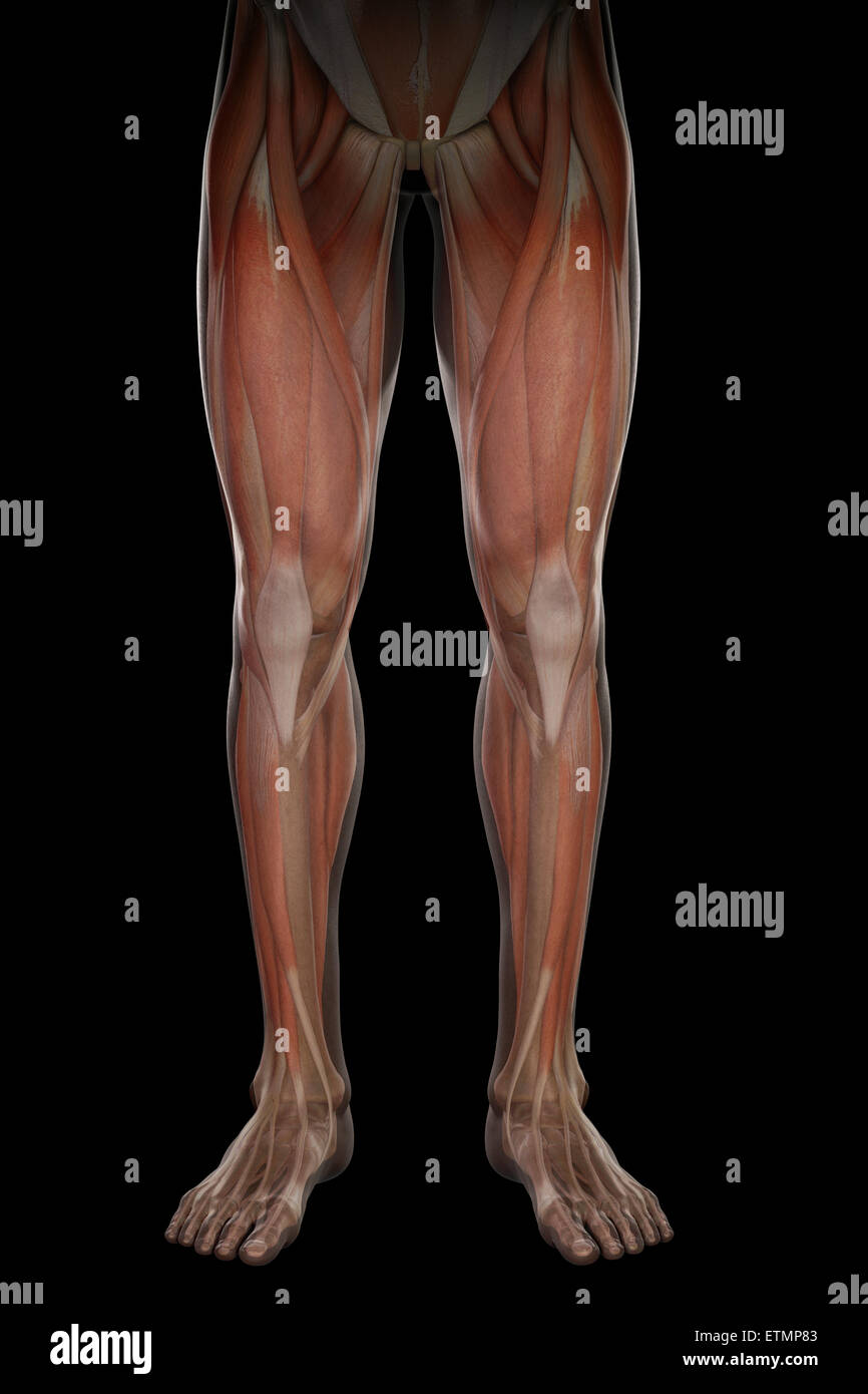 Illustration showing the musculature and skeletal structure of the legs, visible through skin. Stock Photohttps://www.alamy.com/image-license-details/?v=1https://www.alamy.com/stock-photo-illustration-showing-the-musculature-and-skeletal-structure-of-the-84049731.html
RMETMP83–Illustration showing the musculature and skeletal structure of the legs, visible through skin.
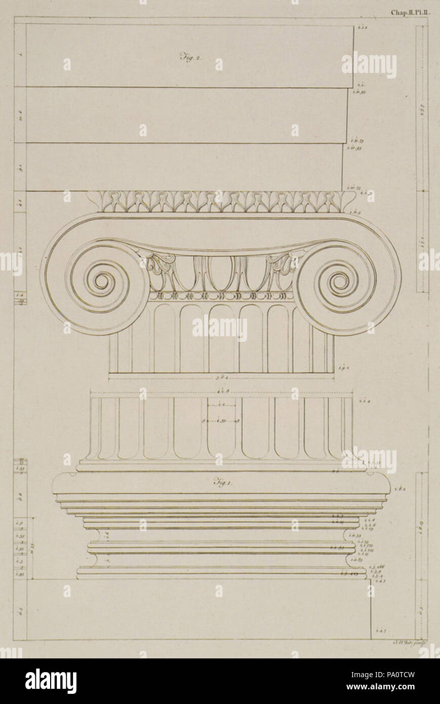 647 Fig I- The uppermost Step and Base, with the lower part of the Shaft of the Column,Fig II- The Capital and Fasciae of th - Society Of Dilettanti - 1769 Stock Photohttps://www.alamy.com/image-license-details/?v=1https://www.alamy.com/647-fig-i-the-uppermost-step-and-base-with-the-lower-part-of-the-shaft-of-the-columnfig-ii-the-capital-and-fasciae-of-th-society-of-dilettanti-1769-image212690153.html
RMPA0TCW–647 Fig I- The uppermost Step and Base, with the lower part of the Shaft of the Column,Fig II- The Capital and Fasciae of th - Society Of Dilettanti - 1769
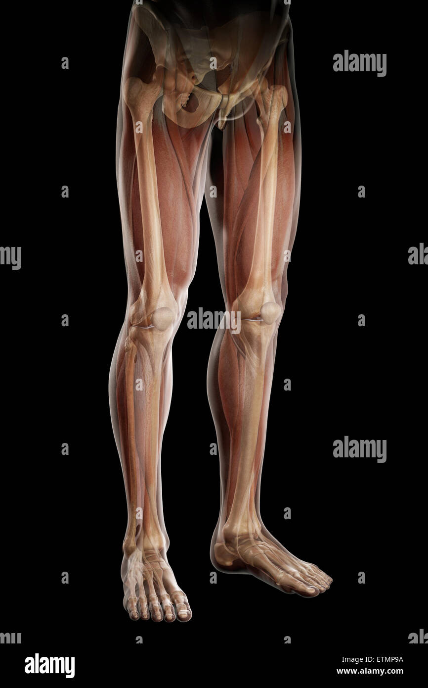 Illustration of the musculature and skeletal structure of the legs, visible through skin. Stock Photohttps://www.alamy.com/image-license-details/?v=1https://www.alamy.com/stock-photo-illustration-of-the-musculature-and-skeletal-structure-of-the-legs-84049766.html
RMETMP9A–Illustration of the musculature and skeletal structure of the legs, visible through skin.
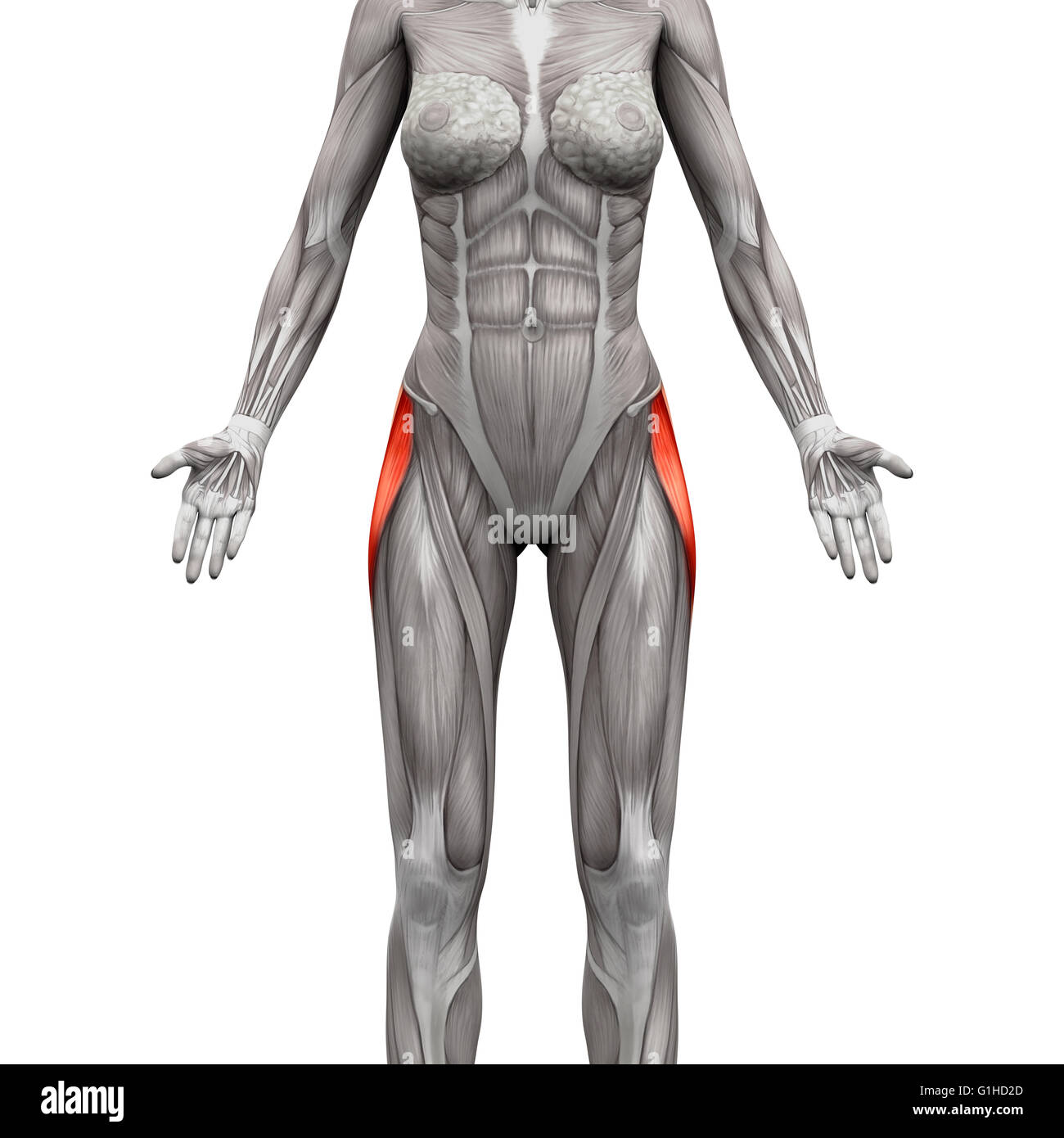 Tensor Fasciae Latae Muscle - Anatomy Muscles isolated on white - 3D illustration Stock Photohttps://www.alamy.com/image-license-details/?v=1https://www.alamy.com/stock-photo-tensor-fasciae-latae-muscle-anatomy-muscles-isolated-on-white-3d-illustration-104260309.html
RFG1HD2D–Tensor Fasciae Latae Muscle - Anatomy Muscles isolated on white - 3D illustration
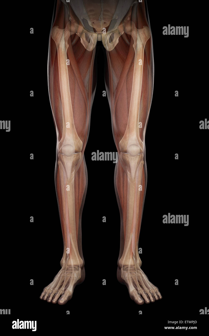 Illustration showing the musculature and skeletal structure of the legs, visible through skin. Stock Photohttps://www.alamy.com/image-license-details/?v=1https://www.alamy.com/stock-photo-illustration-showing-the-musculature-and-skeletal-structure-of-the-84050021.html
RMETMPJD–Illustration showing the musculature and skeletal structure of the legs, visible through skin.
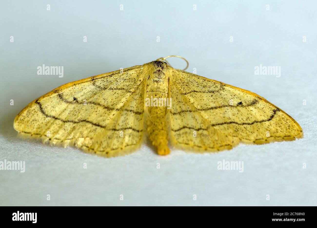 Idaea aversata Riband Wave Moth Stock Photohttps://www.alamy.com/image-license-details/?v=1https://www.alamy.com/idaea-aversata-riband-wave-moth-image365858892.html
RF2C768N0–Idaea aversata Riband Wave Moth
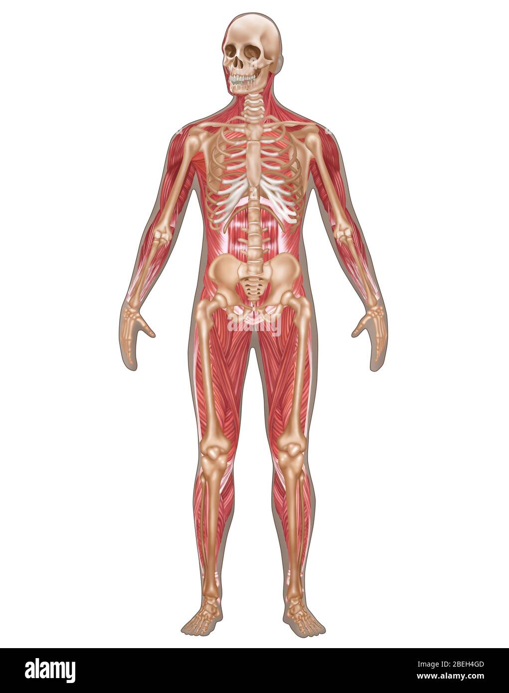 Skeletal & Muscular Systems, Male Anatomy Stock Photohttps://www.alamy.com/image-license-details/?v=1https://www.alamy.com/skeletal-muscular-systems-male-anatomy-image353189325.html
RF2BEH4GD–Skeletal & Muscular Systems, Male Anatomy
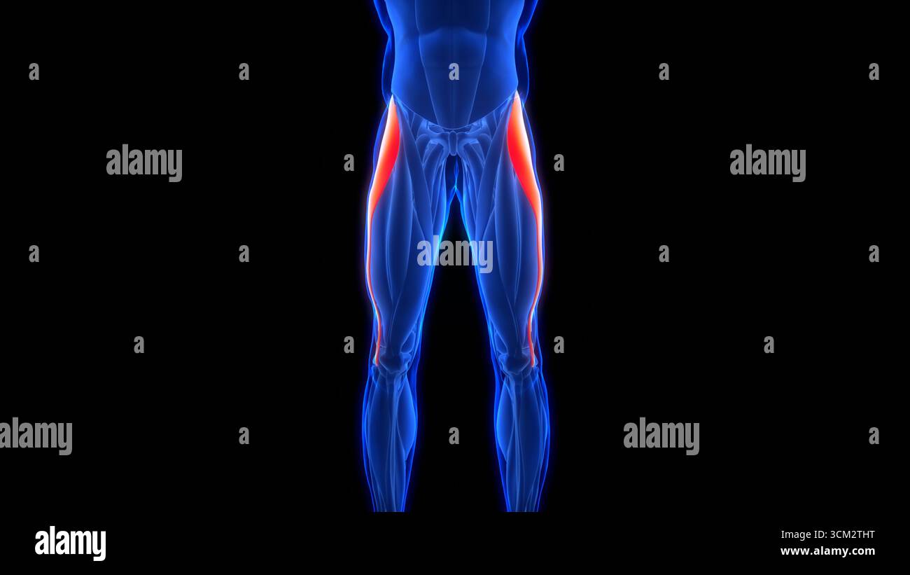 Human Muscular System Leg Muscles Tensor Fasciae Latae Muscles Anatomy Stock Photohttps://www.alamy.com/image-license-details/?v=1https://www.alamy.com/human-muscular-system-leg-muscles-tensor-fasciae-latae-muscles-anatomy-image700771060.html
RF3CM2THT–Human Muscular System Leg Muscles Tensor Fasciae Latae Muscles Anatomy
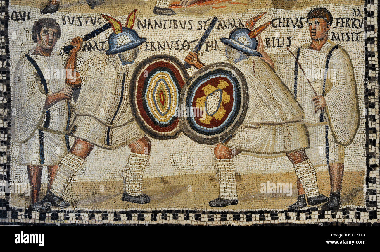 Roman mosaic depicting a Gladiator fight. Detail of the lower part with the murmillones Symmanchus and Maternus are fighting in the arena cheered by the lanistae. 3rd century AD. Limestone and vitreous paste. From Rome (Italy). National Archaeological Museum. Madrid. Spain. Stock Photohttps://www.alamy.com/image-license-details/?v=1https://www.alamy.com/roman-mosaic-depicting-a-gladiator-fight-detail-of-the-lower-part-with-the-murmillones-symmanchus-and-maternus-are-fighting-in-the-arena-cheered-by-the-lanistae-3rd-century-ad-limestone-and-vitreous-paste-from-rome-italy-national-archaeological-museum-madrid-spain-image245310857.html
RMT72TE1–Roman mosaic depicting a Gladiator fight. Detail of the lower part with the murmillones Symmanchus and Maternus are fighting in the arena cheered by the lanistae. 3rd century AD. Limestone and vitreous paste. From Rome (Italy). National Archaeological Museum. Madrid. Spain.
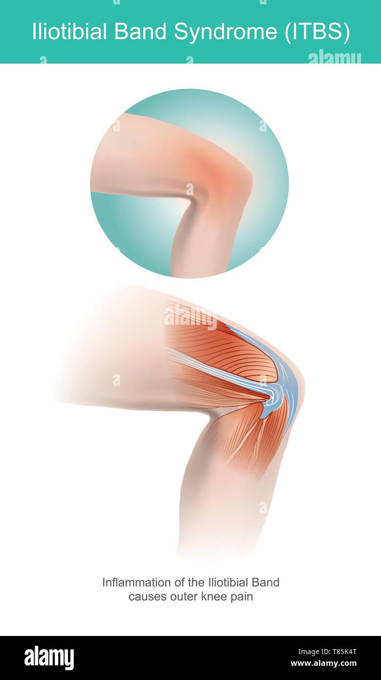 The Iliotibial Band is a longitudinal fibrous reinforcement of the fascia lata in a knee muscle. Part of anatomy human body. Illustration. Stock Vectorhttps://www.alamy.com/image-license-details/?v=1https://www.alamy.com/the-iliotibial-band-is-a-longitudinal-fibrous-reinforcement-of-the-fascia-lata-in-a-knee-muscle-part-of-anatomy-human-body-illustration-image245987192.html
RFT85K4T–The Iliotibial Band is a longitudinal fibrous reinforcement of the fascia lata in a knee muscle. Part of anatomy human body. Illustration.
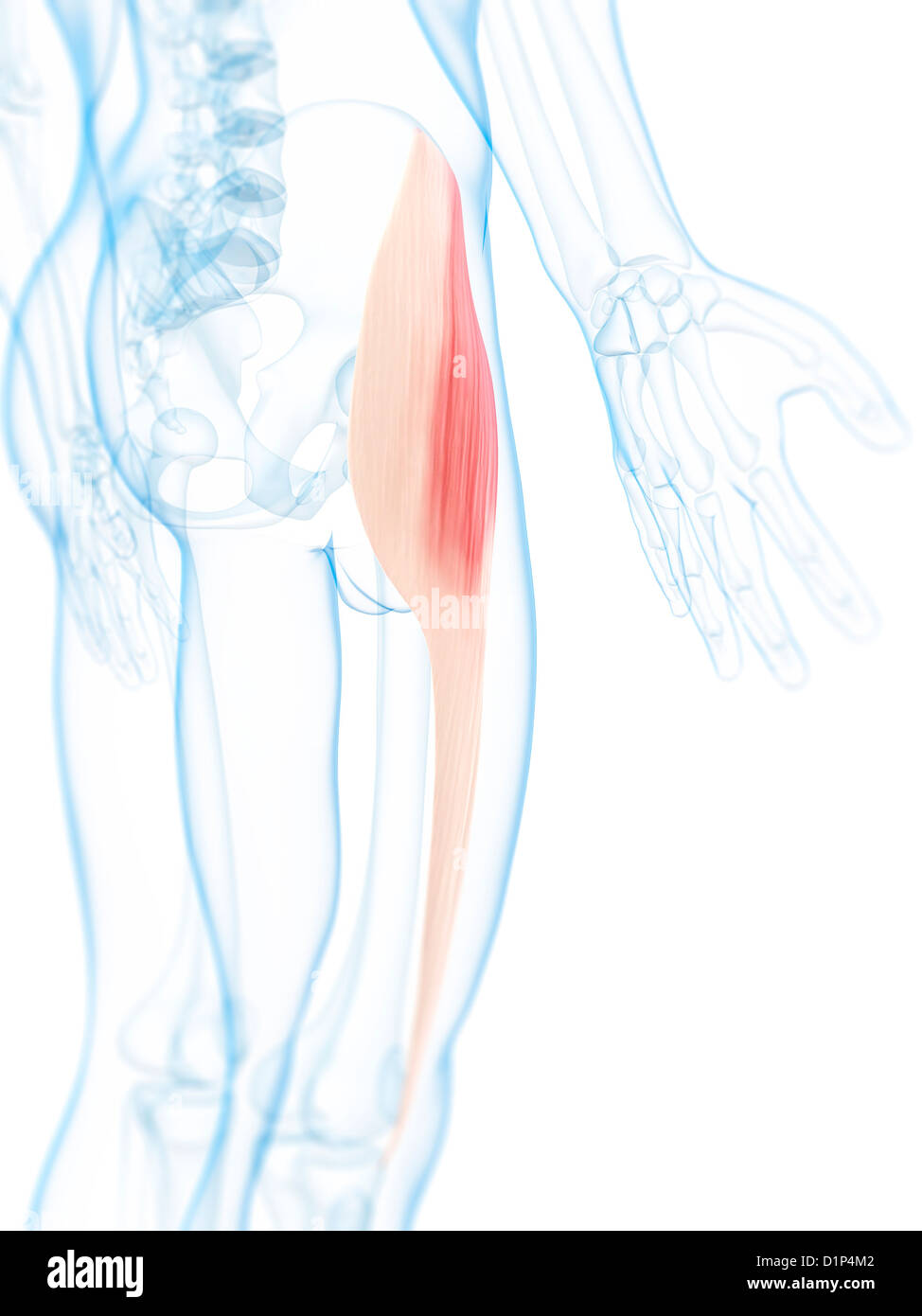 Thigh muscle, artwork Stock Photohttps://www.alamy.com/image-license-details/?v=1https://www.alamy.com/stock-photo-thigh-muscle-artwork-52732402.html
RFD1P4M2–Thigh muscle, artwork
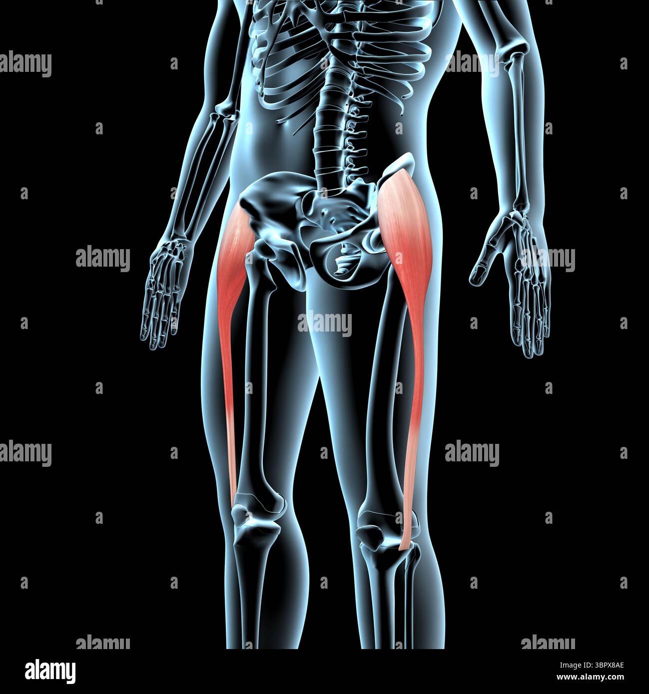 This 3d illustration shows the tensor fasciae latae muscles anatomical position on xray body Stock Photohttps://www.alamy.com/image-license-details/?v=1https://www.alamy.com/this-3d-illustration-shows-the-tensor-fasciae-latae-muscles-anatomical-position-on-xray-body-image685304102.html
RF3BPX8AE–This 3d illustration shows the tensor fasciae latae muscles anatomical position on xray body
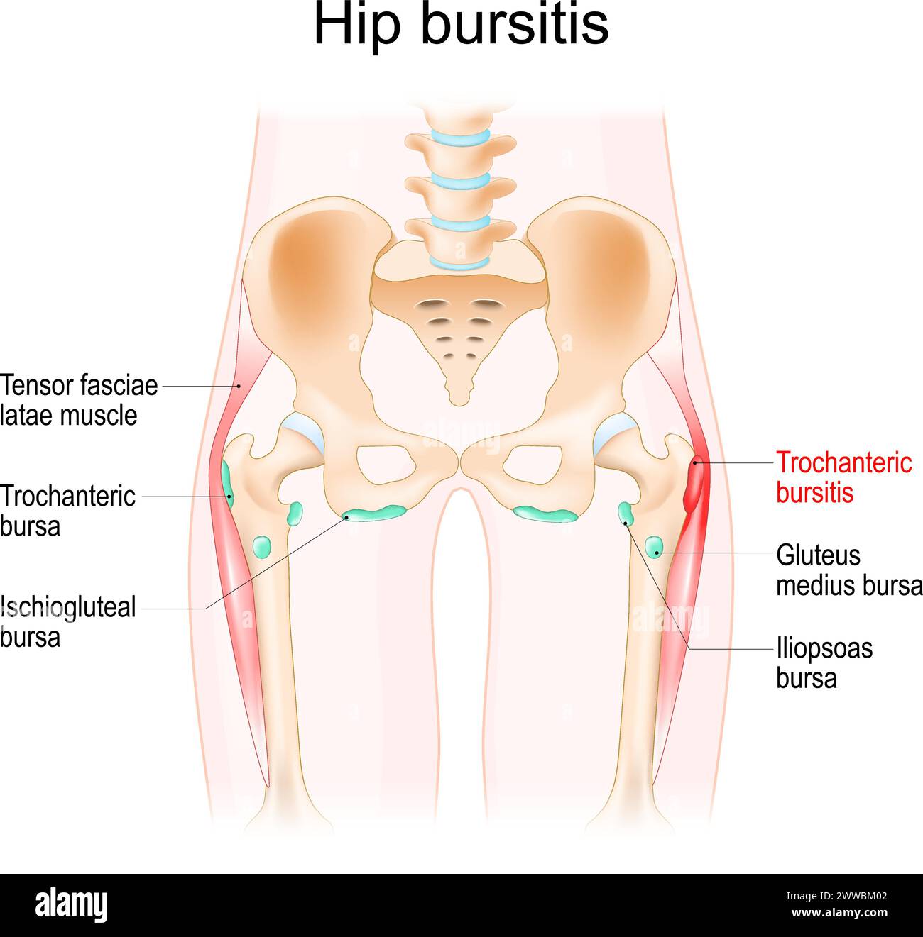 Hip bursitis. Muscles, Synovial bursas and bones of a human hip. Trochanteric bursitis. Realistic vector illustration Stock Vectorhttps://www.alamy.com/image-license-details/?v=1https://www.alamy.com/hip-bursitis-muscles-synovial-bursas-and-bones-of-a-human-hip-trochanteric-bursitis-realistic-vector-illustration-image600776066.html
RF2WWBM02–Hip bursitis. Muscles, Synovial bursas and bones of a human hip. Trochanteric bursitis. Realistic vector illustration
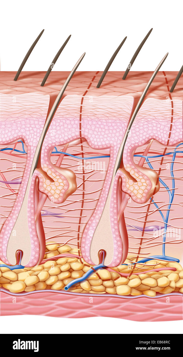 SCAR DRAWING Stock Photohttps://www.alamy.com/image-license-details/?v=1https://www.alamy.com/stock-photo-scar-drawing-75741328.html
RMEB68RC–SCAR DRAWING
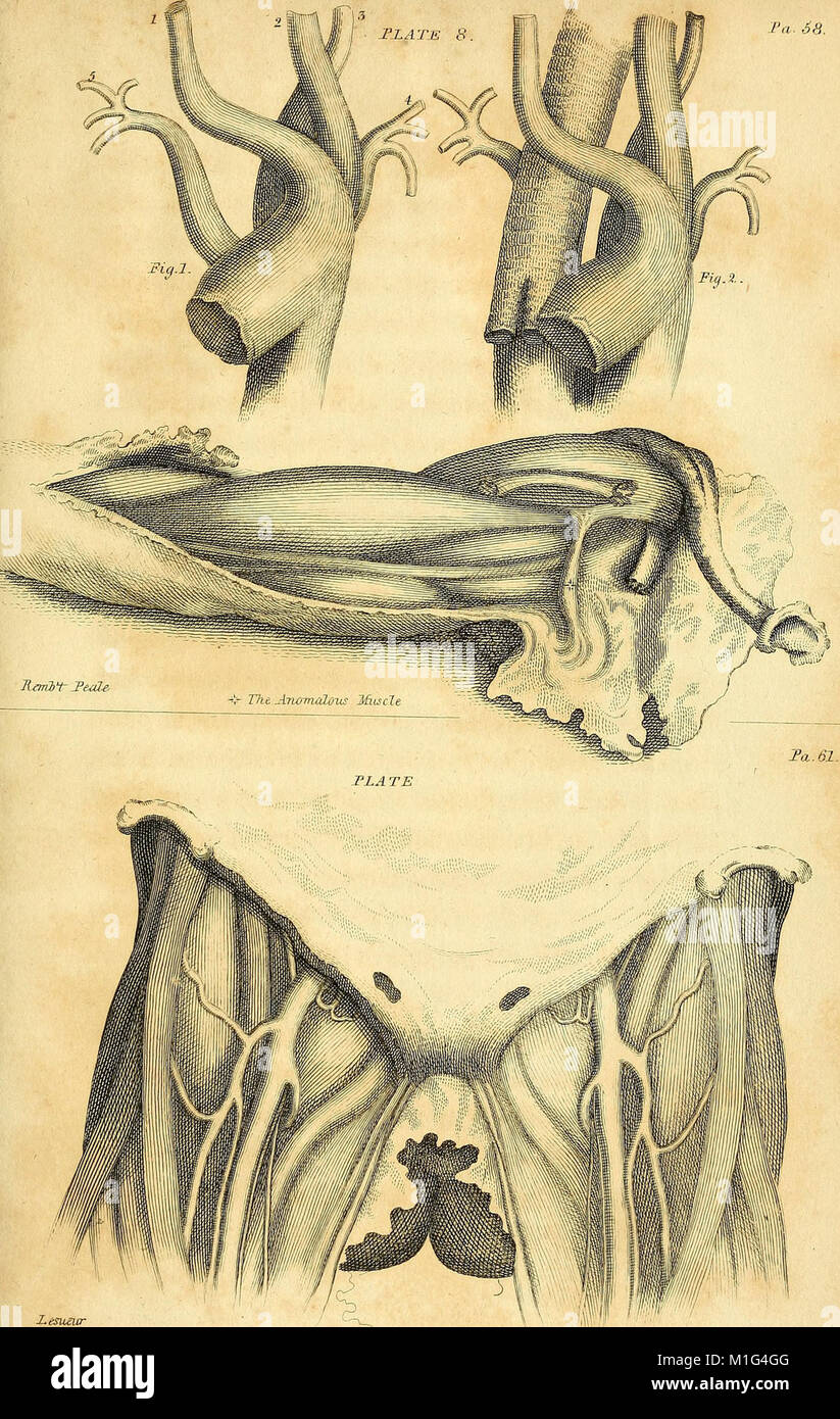 Anatomical investigations, comprising descriptions of various fasciae of the human body to which is added an account of some irregularities of structure and morbid anatomy; with a description of a new (18170837441) Stock Photohttps://www.alamy.com/image-license-details/?v=1https://www.alamy.com/stock-photo-anatomical-investigations-comprising-descriptions-of-various-fasciae-173073168.html
RMM1G4GG–Anatomical investigations, comprising descriptions of various fasciae of the human body to which is added an account of some irregularities of structure and morbid anatomy; with a description of a new (18170837441)
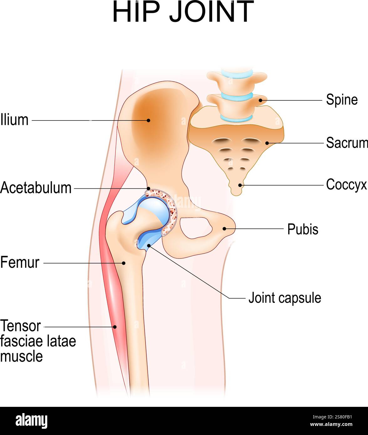 Hip joint anatomy. Ball-and-socket joint. Vector poster Stock Vectorhttps://www.alamy.com/image-license-details/?v=1https://www.alamy.com/hip-joint-anatomy-ball-and-socket-joint-vector-poster-image641712933.html
RF2S80FB1–Hip joint anatomy. Ball-and-socket joint. Vector poster
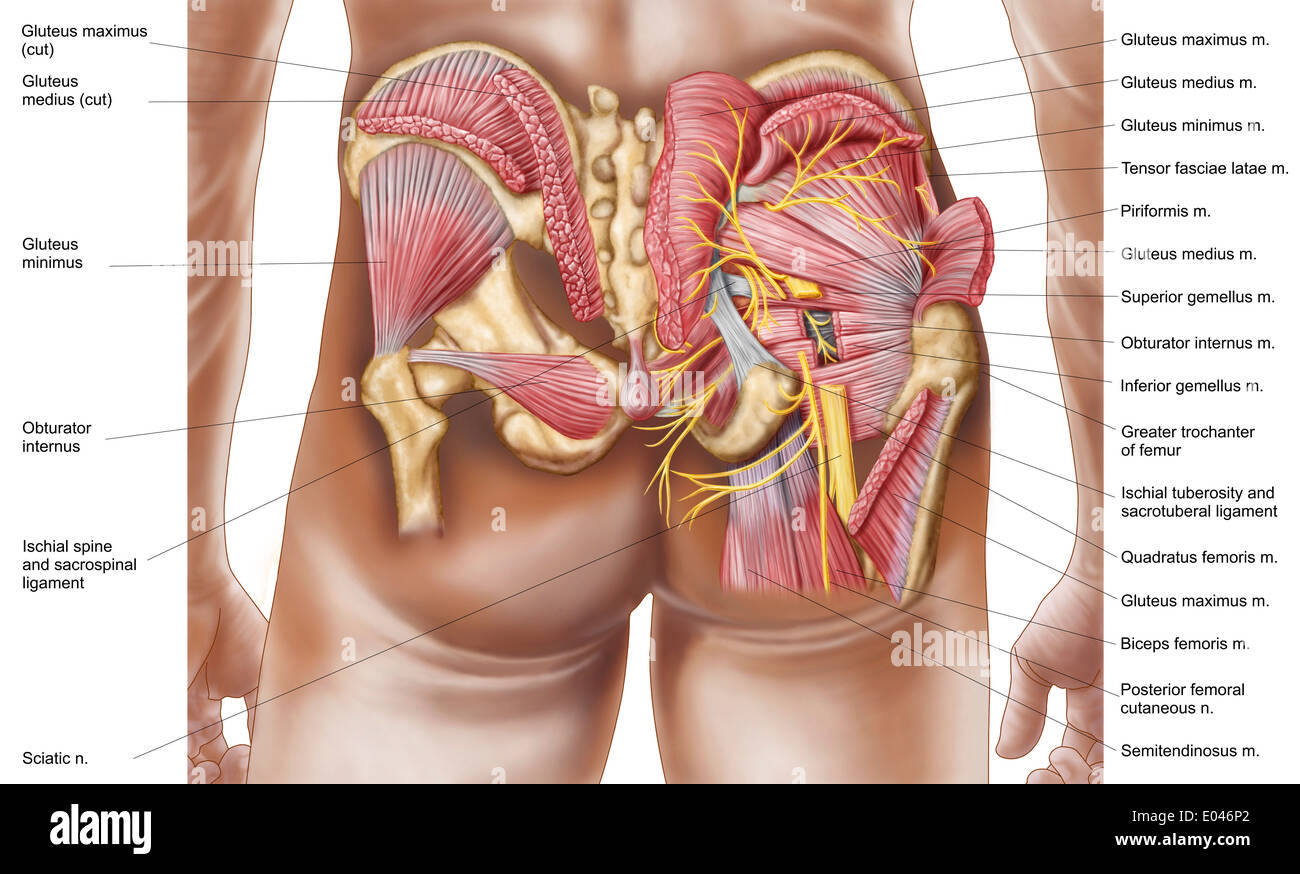 Anatomy of the gluteal muscles in the human buttocks. Stock Photohttps://www.alamy.com/image-license-details/?v=1https://www.alamy.com/anatomy-of-the-gluteal-muscles-in-the-human-buttocks-image68934602.html
RFE046P2–Anatomy of the gluteal muscles in the human buttocks.
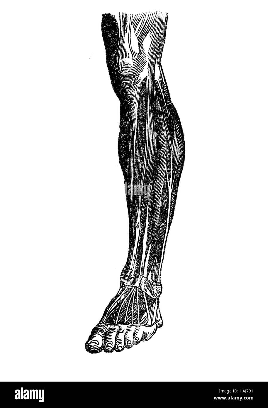 Anatomy, human musculature; foot, shank,knee Stock Photohttps://www.alamy.com/image-license-details/?v=1https://www.alamy.com/stock-photo-anatomy-human-musculature-foot-shankknee-127020013.html
RFHAJ791–Anatomy, human musculature; foot, shank,knee
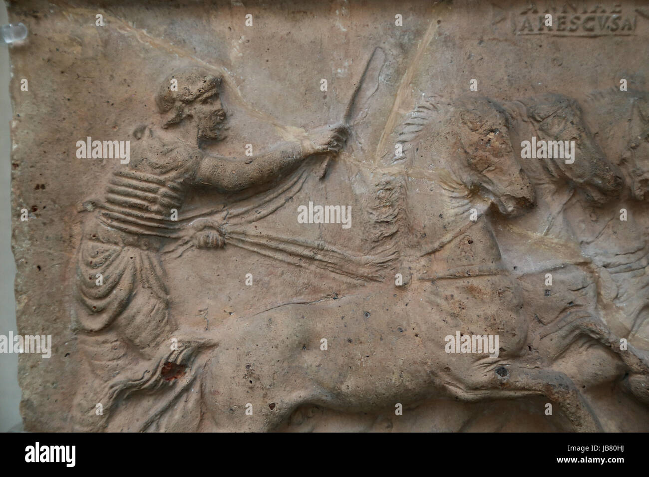 Terracotta decorative relief. Chariot race. c. 70 AD. British Museum. London. UK Stock Photohttps://www.alamy.com/image-license-details/?v=1https://www.alamy.com/stock-photo-terracotta-decorative-relief-chariot-race-c-70-ad-british-museum-london-144620270.html
RMJB80HJ–Terracotta decorative relief. Chariot race. c. 70 AD. British Museum. London. UK
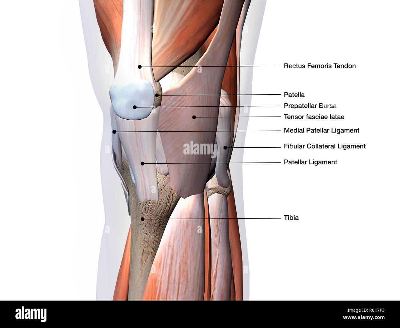 Knee joint showing muscles and ligaments with labels. Stock Photohttps://www.alamy.com/image-license-details/?v=1https://www.alamy.com/knee-joint-showing-muscles-and-ligaments-with-labels-image224157979.html
RFR0K7P3–Knee joint showing muscles and ligaments with labels.
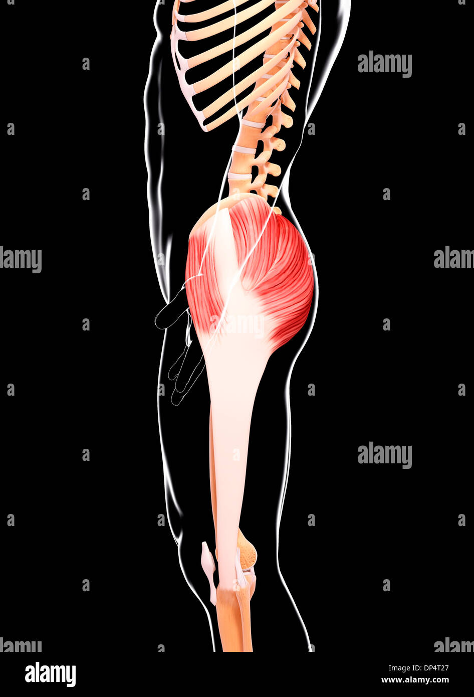 Human musculature, artwork Stock Photohttps://www.alamy.com/image-license-details/?v=1https://www.alamy.com/human-musculature-artwork-image65260223.html
RFDP4T27–Human musculature, artwork
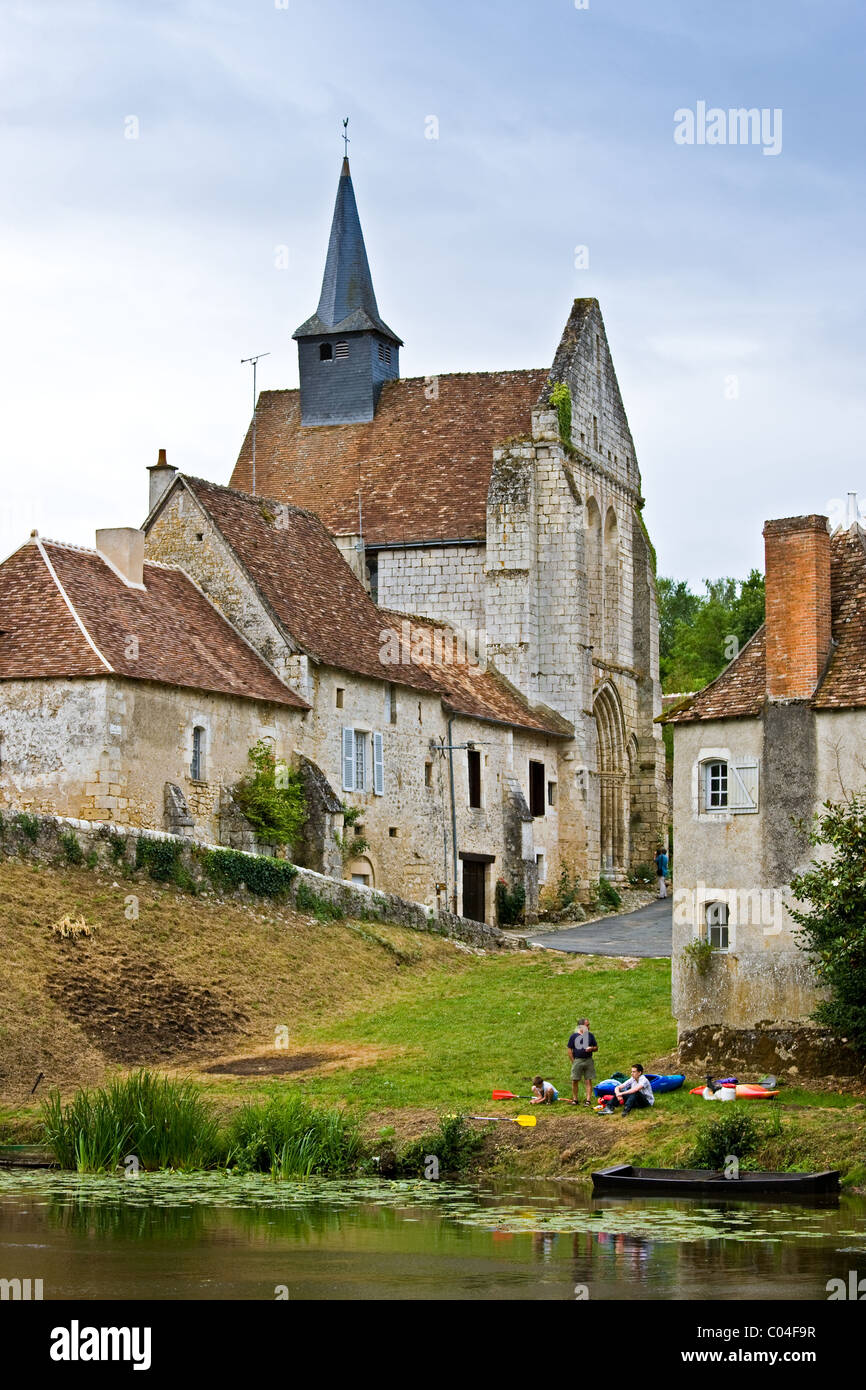 People with kayaks in traditional medieval village of Angles Sur L'Anglin, Vienne, near Poitiers, France Stock Photohttps://www.alamy.com/image-license-details/?v=1https://www.alamy.com/stock-photo-people-with-kayaks-in-traditional-medieval-village-of-angles-sur-langlin-34520579.html
RMC04F9R–People with kayaks in traditional medieval village of Angles Sur L'Anglin, Vienne, near Poitiers, France
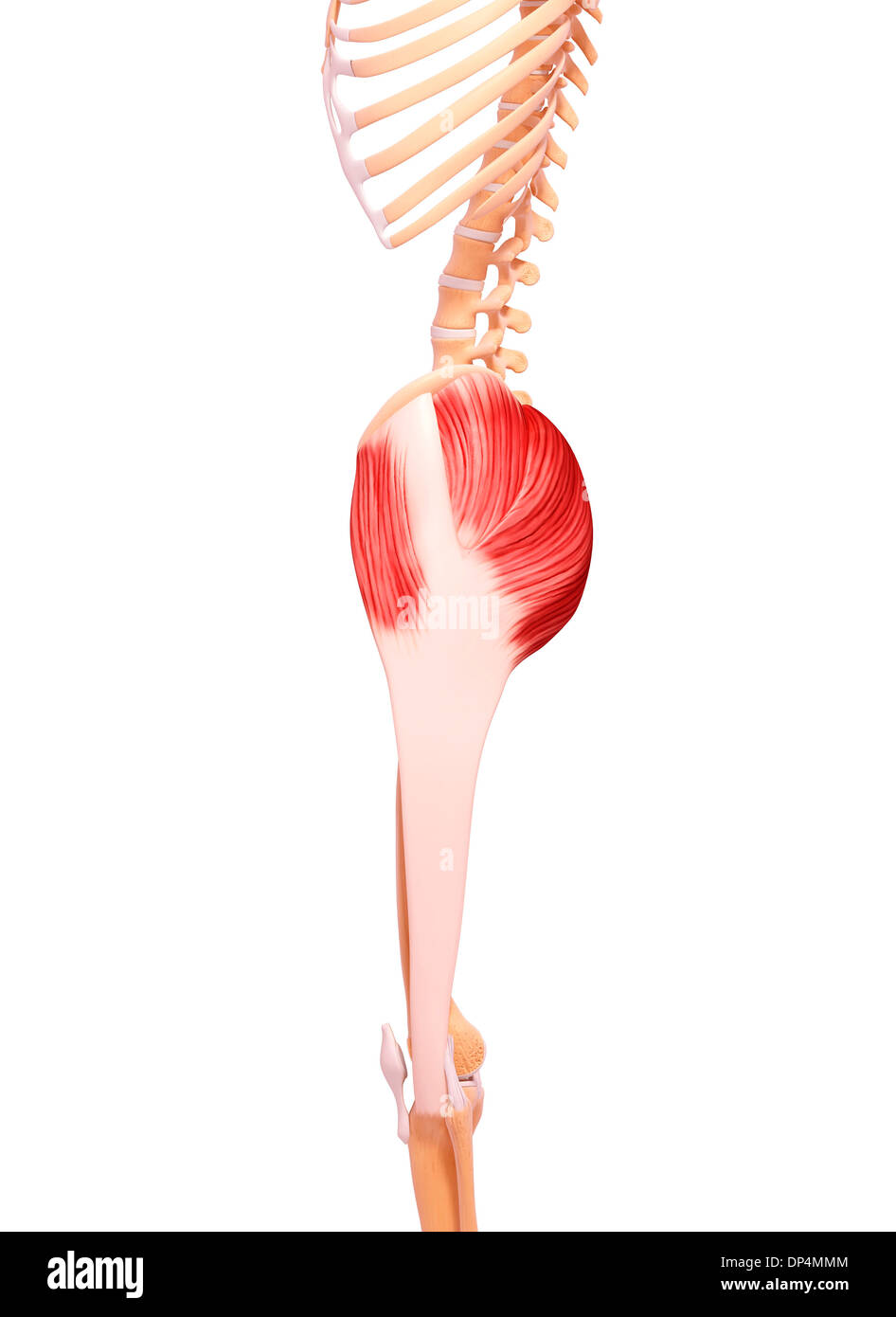 Human musculature, artwork Stock Photohttps://www.alamy.com/image-license-details/?v=1https://www.alamy.com/human-musculature-artwork-image65257604.html
RFDP4MMM–Human musculature, artwork
 Yellow-shouldered Stout Hover Fly (Simosyrphus grandicornis) feeding on nectar, South Australia Stock Photohttps://www.alamy.com/image-license-details/?v=1https://www.alamy.com/yellow-shouldered-stout-hover-fly-simosyrphus-grandicornis-feeding-on-nectar-south-australia-image630427774.html
RF2YHJD1J–Yellow-shouldered Stout Hover Fly (Simosyrphus grandicornis) feeding on nectar, South Australia
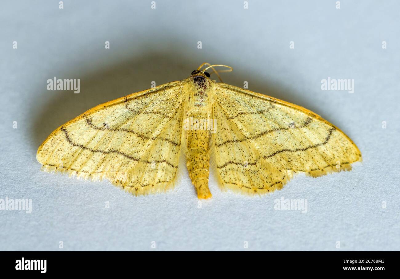 Idaea aversata Riband Wave Moth Stock Photohttps://www.alamy.com/image-license-details/?v=1https://www.alamy.com/idaea-aversata-riband-wave-moth-image365858867.html
RF2C768M3–Idaea aversata Riband Wave Moth
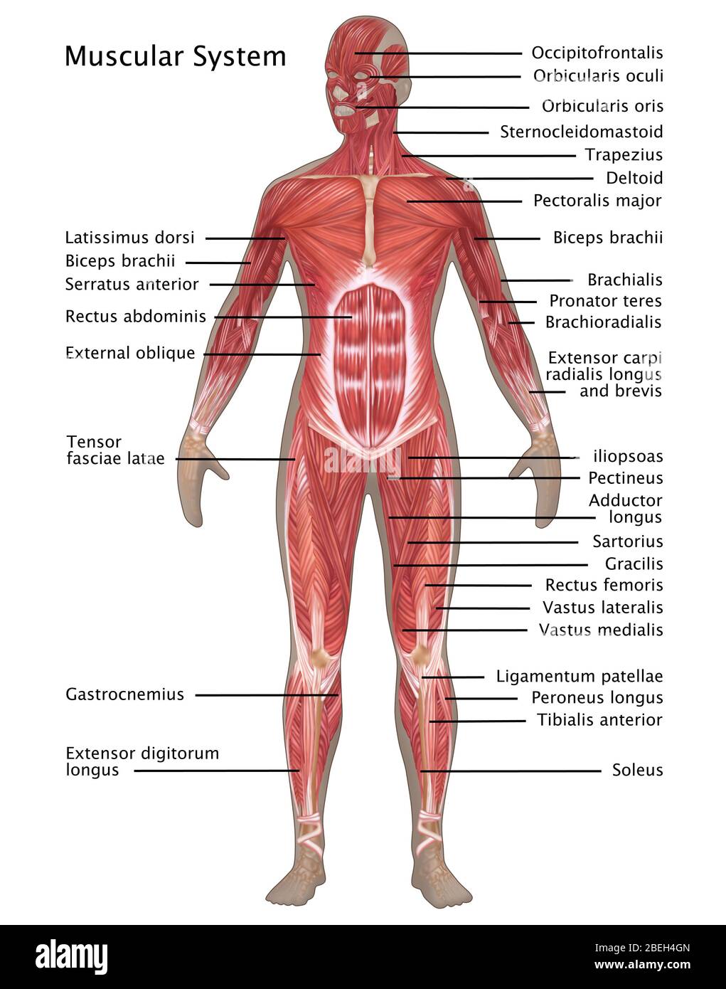 Muscular System in Female Anatomy Stock Photohttps://www.alamy.com/image-license-details/?v=1https://www.alamy.com/muscular-system-in-female-anatomy-image353189333.html
RF2BEH4GN–Muscular System in Female Anatomy
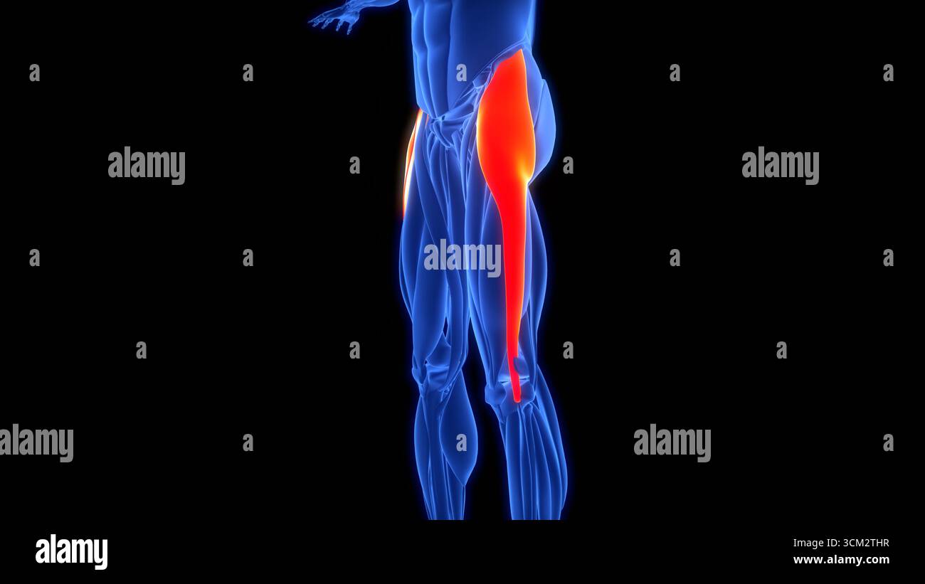 Human Muscular System Leg Muscles Tensor Fasciae Latae Muscles Anatomy Stock Photohttps://www.alamy.com/image-license-details/?v=1https://www.alamy.com/human-muscular-system-leg-muscles-tensor-fasciae-latae-muscles-anatomy-image700771059.html
RF3CM2THR–Human Muscular System Leg Muscles Tensor Fasciae Latae Muscles Anatomy
 Roman mosaic depicting a Gladiator fight. In the lower part, the murmillones Symmanchus and Maternus are fighting in the arena cheered by the lanistae. At the top, Maternus lies slain by Symmachus. 3rd century AD. Limestone and vitreous paste. From Rome (Italy). National Archaeological Museum. Madrid. Spain. Stock Photohttps://www.alamy.com/image-license-details/?v=1https://www.alamy.com/roman-mosaic-depicting-a-gladiator-fight-in-the-lower-part-the-murmillones-symmanchus-and-maternus-are-fighting-in-the-arena-cheered-by-the-lanistae-at-the-top-maternus-lies-slain-by-symmachus-3rd-century-ad-limestone-and-vitreous-paste-from-rome-italy-national-archaeological-museum-madrid-spain-image245310856.html
RMT72TE0–Roman mosaic depicting a Gladiator fight. In the lower part, the murmillones Symmanchus and Maternus are fighting in the arena cheered by the lanistae. At the top, Maternus lies slain by Symmachus. 3rd century AD. Limestone and vitreous paste. From Rome (Italy). National Archaeological Museum. Madrid. Spain.
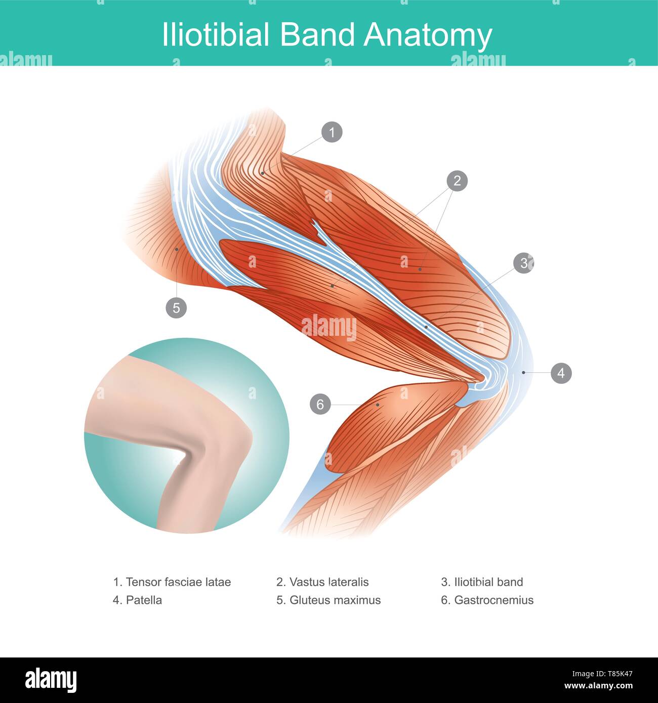 The Iliotibial Band is a longitudinal fibrous reinforcement of the fascia lata in a knee muscle. Part of anatomy human body. Illustration. Stock Vectorhttps://www.alamy.com/image-license-details/?v=1https://www.alamy.com/the-iliotibial-band-is-a-longitudinal-fibrous-reinforcement-of-the-fascia-lata-in-a-knee-muscle-part-of-anatomy-human-body-illustration-image245987175.html
RFT85K47–The Iliotibial Band is a longitudinal fibrous reinforcement of the fascia lata in a knee muscle. Part of anatomy human body. Illustration.
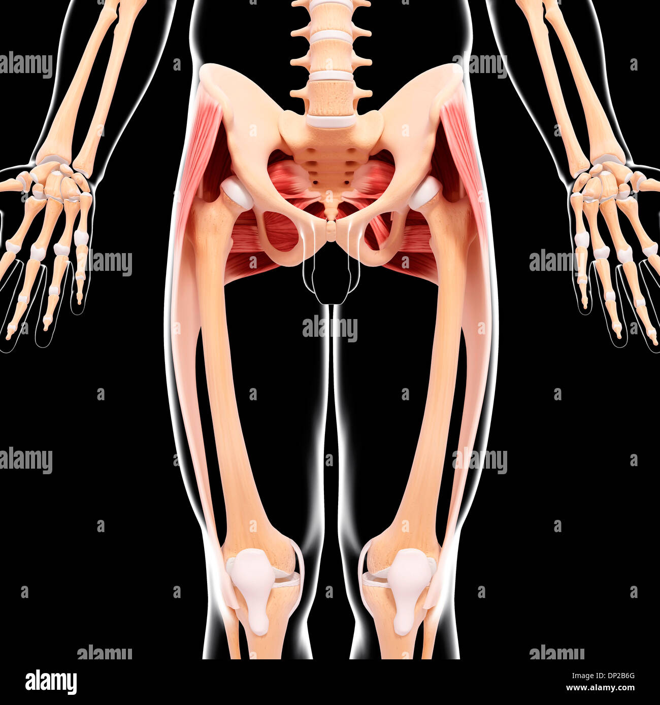 Human hip musculature, artwork Stock Photohttps://www.alamy.com/image-license-details/?v=1https://www.alamy.com/human-hip-musculature-artwork-image65206248.html
RFDP2B6G–Human hip musculature, artwork
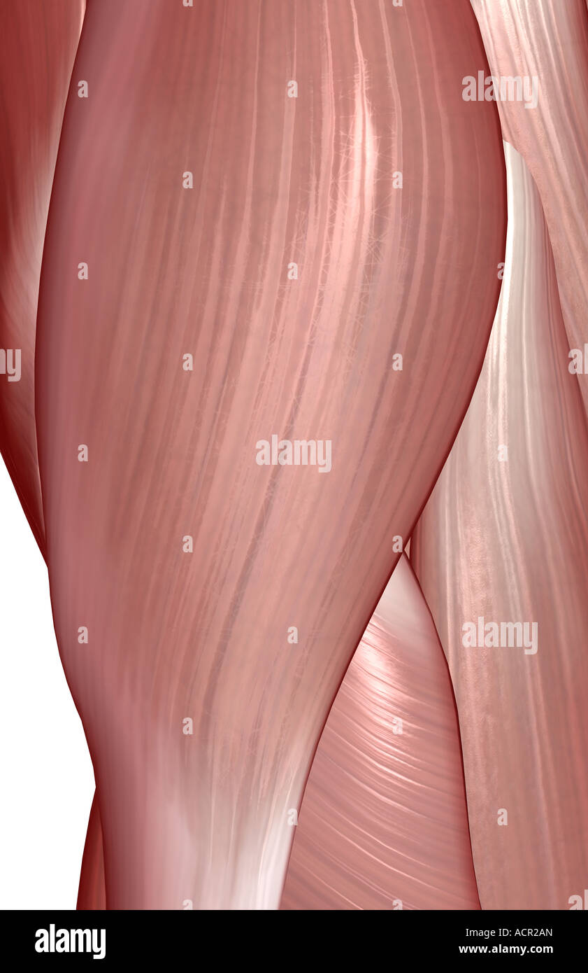 The muscles of the hip Stock Photohttps://www.alamy.com/image-license-details/?v=1https://www.alamy.com/stock-photo-the-muscles-of-the-hip-13212764.html
RFACR2AN–The muscles of the hip
 Black Roll training in the fitness room Stock Photohttps://www.alamy.com/image-license-details/?v=1https://www.alamy.com/stock-photo-black-roll-training-in-the-fitness-room-114567778.html
RMGJB0AA–Black Roll training in the fitness room
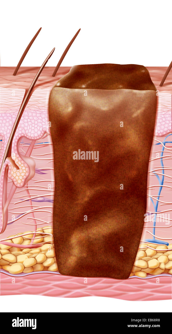 WOUND HEALING, ILLUSTRATION Stock Photohttps://www.alamy.com/image-license-details/?v=1https://www.alamy.com/stock-photo-wound-healing-illustration-75741324.html
RMEB68R8–WOUND HEALING, ILLUSTRATION
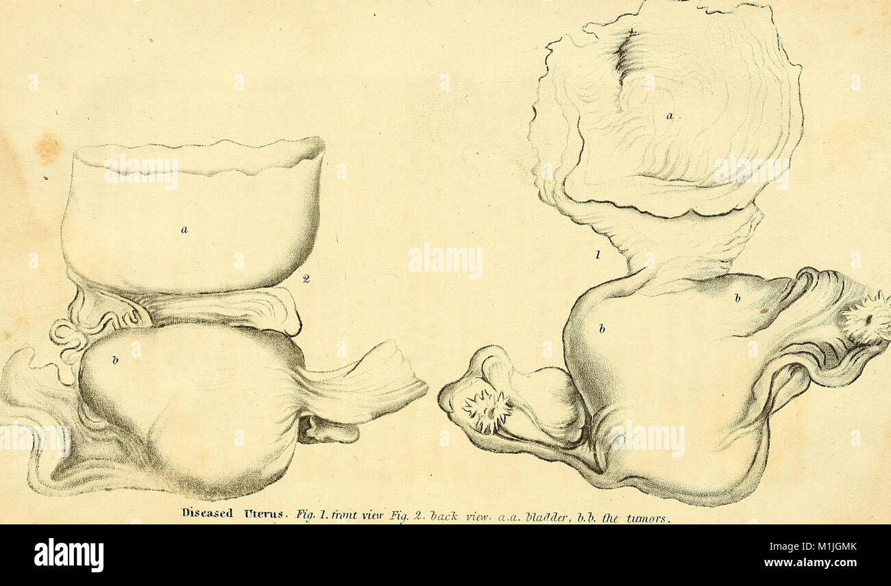 Anatomical investigations, comprising descriptions of various fasciae of the human body to which is added an account of some irregularities of structure and morbid anatomy; with a description of a new (17549108733) Stock Photohttps://www.alamy.com/image-license-details/?v=1https://www.alamy.com/stock-photo-anatomical-investigations-comprising-descriptions-of-various-fasciae-173126595.html
RMM1JGMK–Anatomical investigations, comprising descriptions of various fasciae of the human body to which is added an account of some irregularities of structure and morbid anatomy; with a description of a new (17549108733)
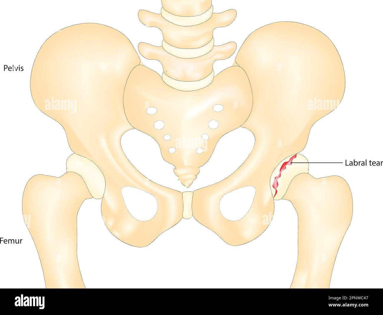 hip labral tear Stock Vectorhttps://www.alamy.com/image-license-details/?v=1https://www.alamy.com/hip-labral-tear-image546987511.html
RF2PNWC47–hip labral tear
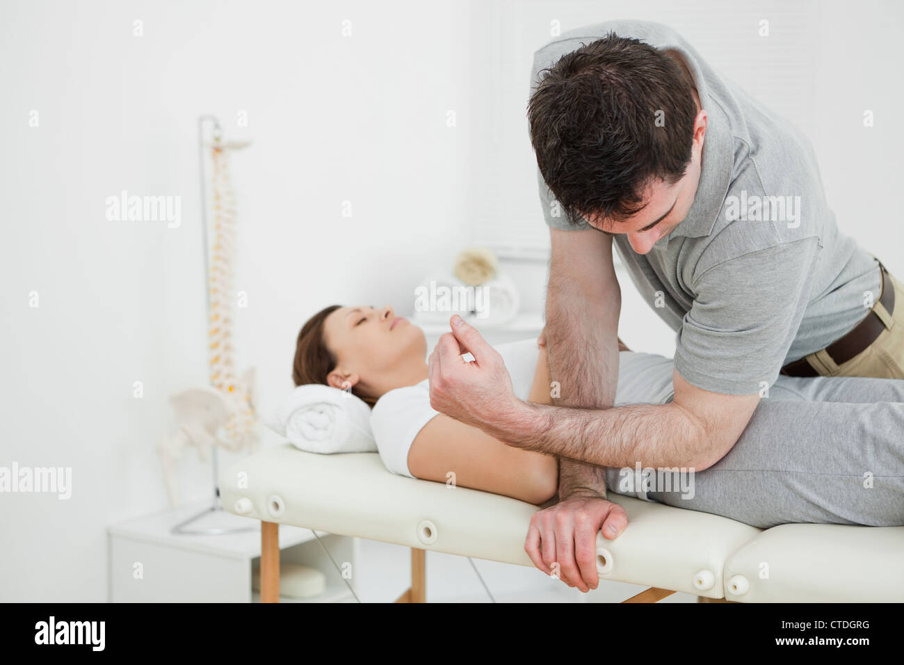 Physiotherapist massaging the pelvis of a woman Stock Photohttps://www.alamy.com/image-license-details/?v=1https://www.alamy.com/stock-photo-physiotherapist-massaging-the-pelvis-of-a-woman-49471060.html
RFCTDGRG–Physiotherapist massaging the pelvis of a woman
 Pressure ulcers NHS information booklet England UK Stock Photohttps://www.alamy.com/image-license-details/?v=1https://www.alamy.com/stock-photo-pressure-ulcers-nhs-information-booklet-england-uk-71171021.html
RME3P3A5–Pressure ulcers NHS information booklet England UK
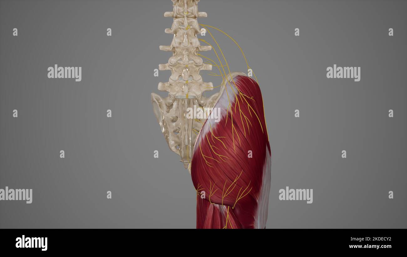 Superficial Nerves of Gluteal Region Stock Photohttps://www.alamy.com/image-license-details/?v=1https://www.alamy.com/superficial-nerves-of-gluteal-region-image490198326.html
RF2KDECY2–Superficial Nerves of Gluteal Region
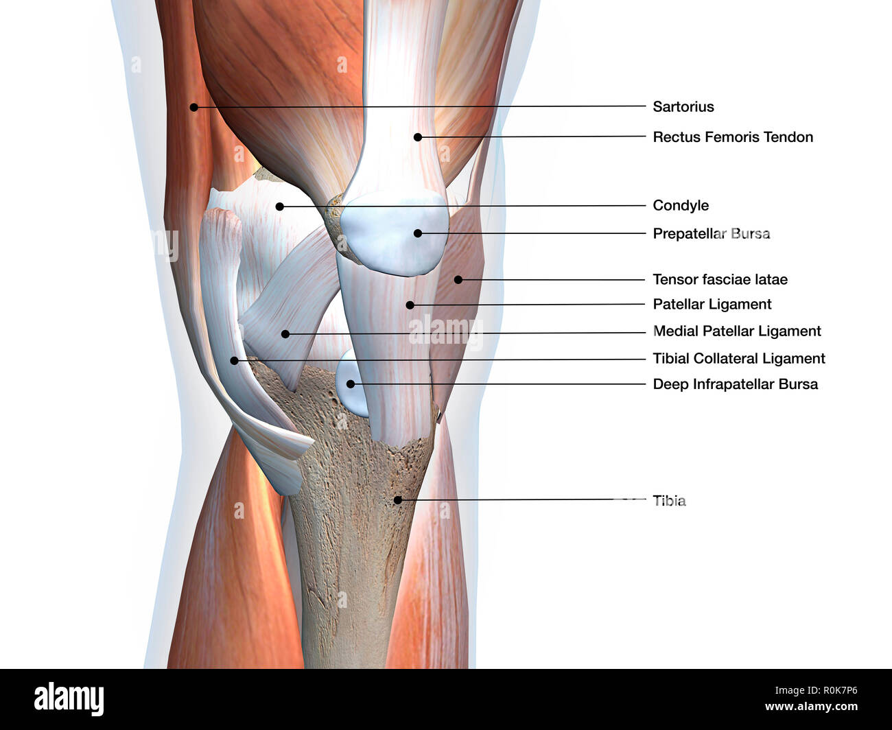 Knee joint showing muscles and ligaments with labels. Stock Photohttps://www.alamy.com/image-license-details/?v=1https://www.alamy.com/knee-joint-showing-muscles-and-ligaments-with-labels-image224157982.html
RFR0K7P6–Knee joint showing muscles and ligaments with labels.
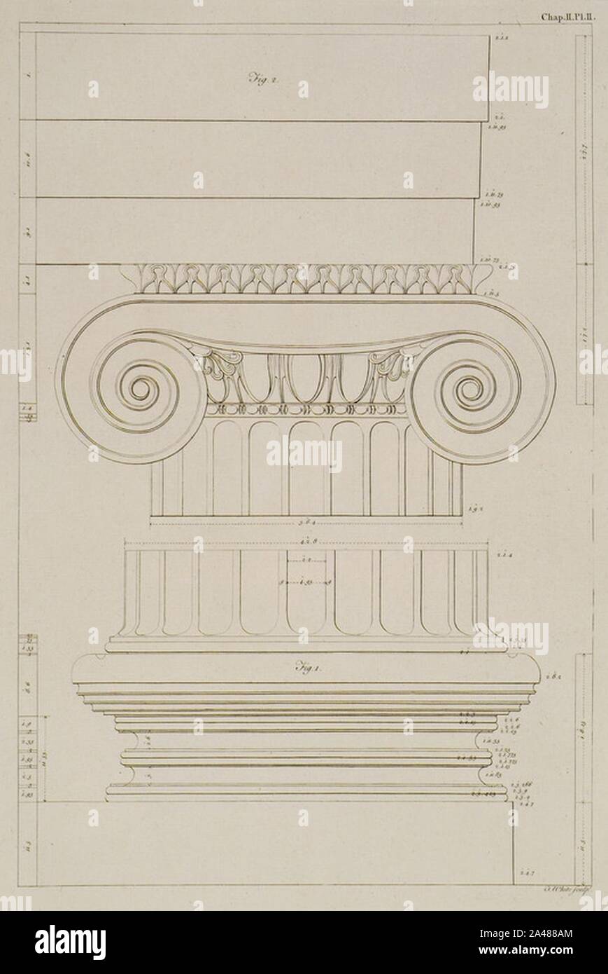 Fig I- The uppermost Step and Base, with the lower part of the Shaft of the Column,Fig II- The Capital and Fasciae of th - Society Of Dilettanti - 1769. Stock Photohttps://www.alamy.com/image-license-details/?v=1https://www.alamy.com/fig-i-the-uppermost-step-and-base-with-the-lower-part-of-the-shaft-of-the-columnfig-ii-the-capital-and-fasciae-of-th-society-of-dilettanti-1769-image329637804.html
RM2A488AM–Fig I- The uppermost Step and Base, with the lower part of the Shaft of the Column,Fig II- The Capital and Fasciae of th - Society Of Dilettanti - 1769.
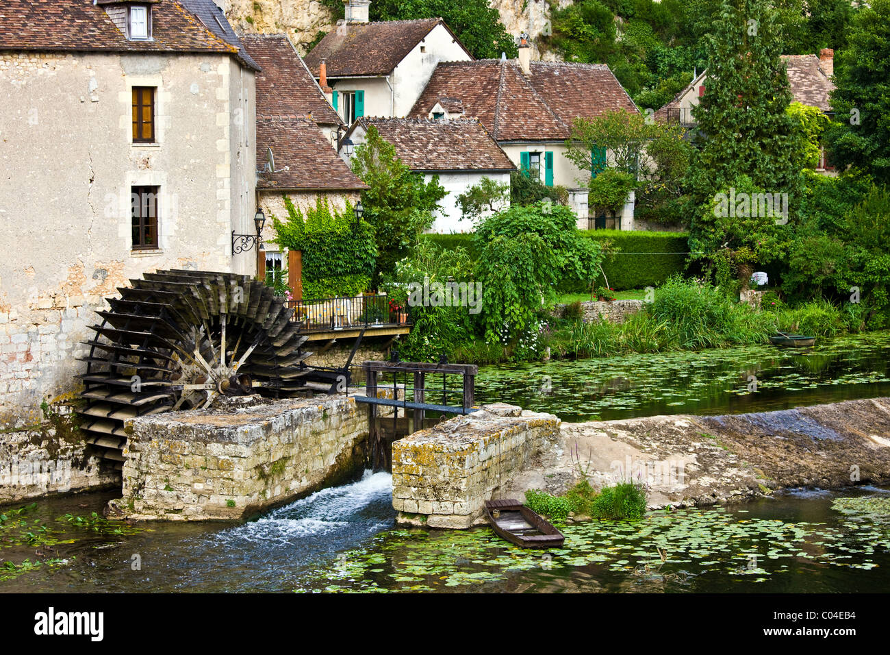 Traditional French houses, water wheel and millrace at Angles Sur L'Anglin medieval village, Vienne, near Poitiers, France Stock Photohttps://www.alamy.com/image-license-details/?v=1https://www.alamy.com/stock-photo-traditional-french-houses-water-wheel-and-millrace-at-angles-sur-langlin-34519832.html
RMC04EB4–Traditional French houses, water wheel and millrace at Angles Sur L'Anglin medieval village, Vienne, near Poitiers, France
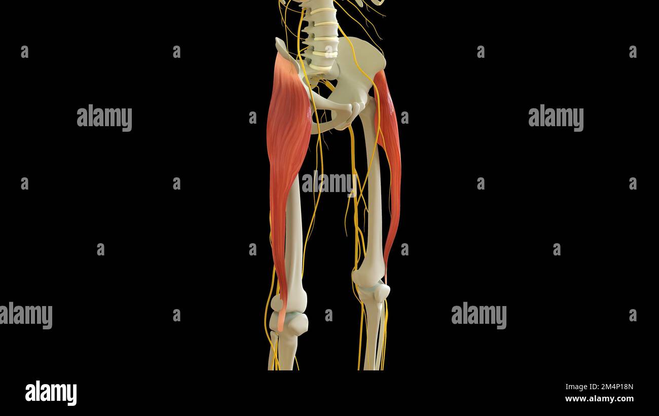 Tensor Fasciae Latae Muscle anatomy for medical concept 3D illustration Stock Photohttps://www.alamy.com/image-license-details/?v=1https://www.alamy.com/tensor-fasciae-latae-muscle-anatomy-for-medical-concept-3d-illustration-image502043269.html
RF2M4P18N–Tensor Fasciae Latae Muscle anatomy for medical concept 3D illustration
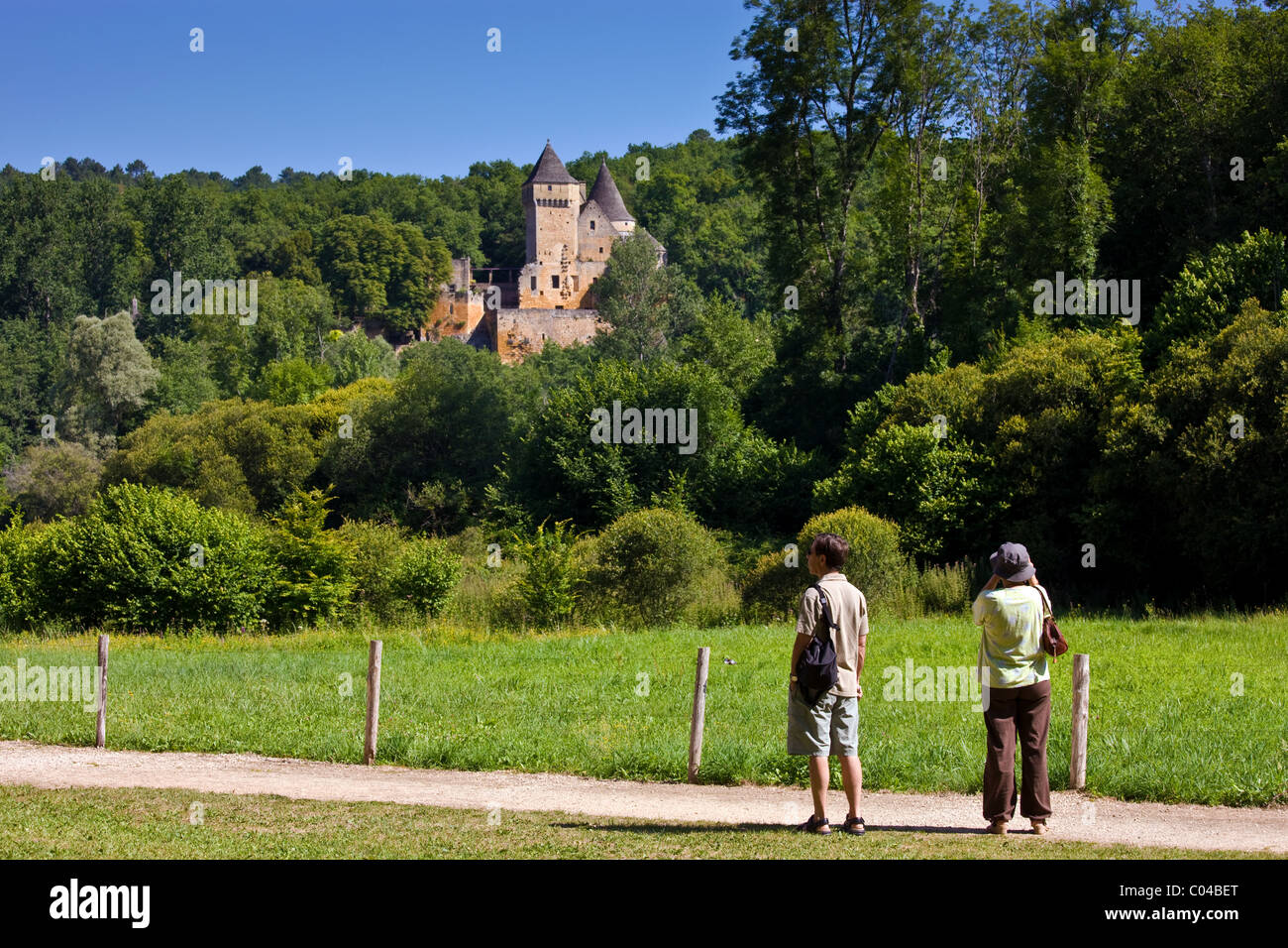 Historic chateau near Les Eyzies, Dordogne, France Stock Photohttps://www.alamy.com/image-license-details/?v=1https://www.alamy.com/stock-photo-historic-chateau-near-les-eyzies-dordogne-france-34517584.html
RMC04BET–Historic chateau near Les Eyzies, Dordogne, France
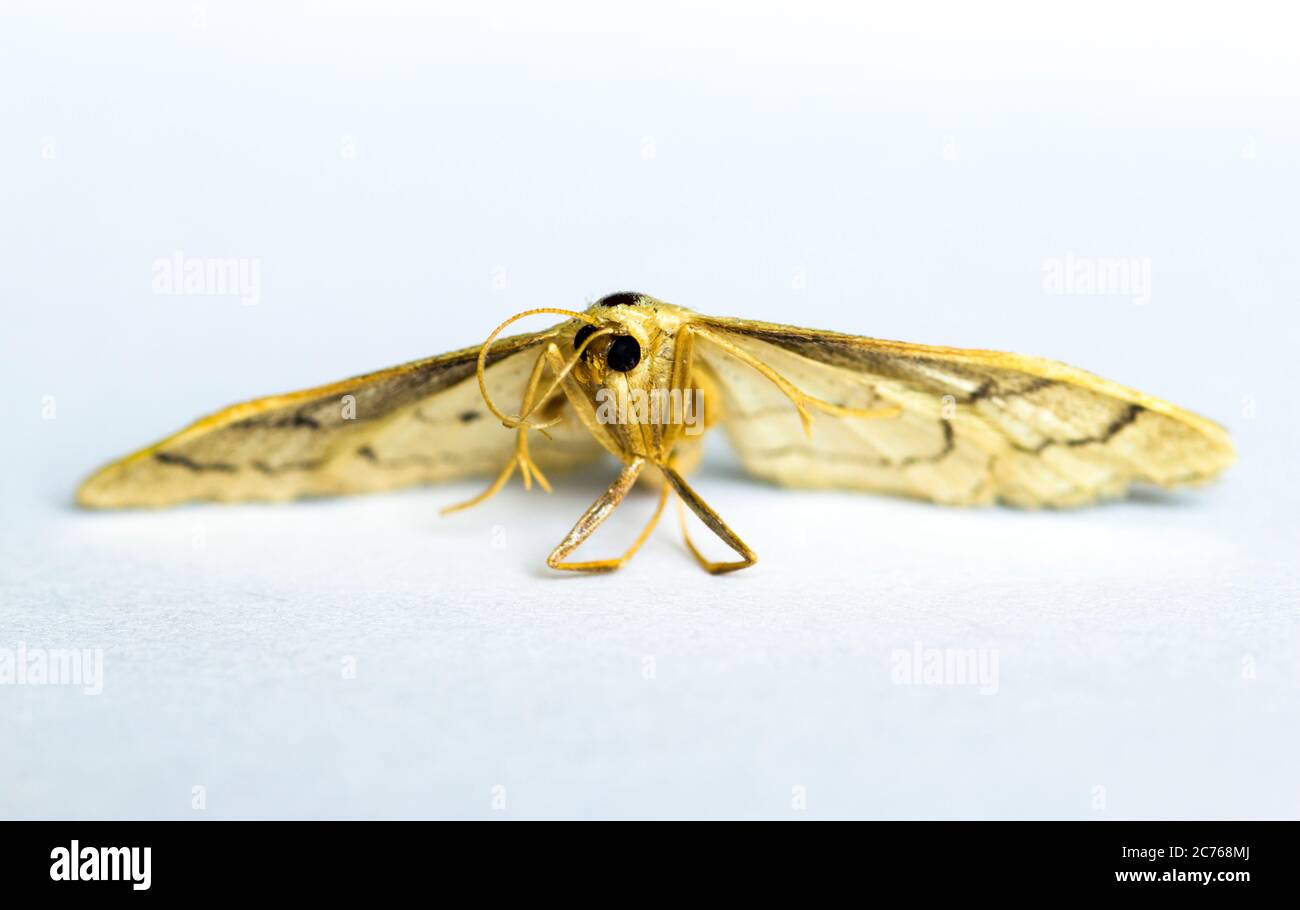 Idaea aversata Riband Wave Moth Stock Photohttps://www.alamy.com/image-license-details/?v=1https://www.alamy.com/idaea-aversata-riband-wave-moth-image365858882.html
RF2C768MJ–Idaea aversata Riband Wave Moth
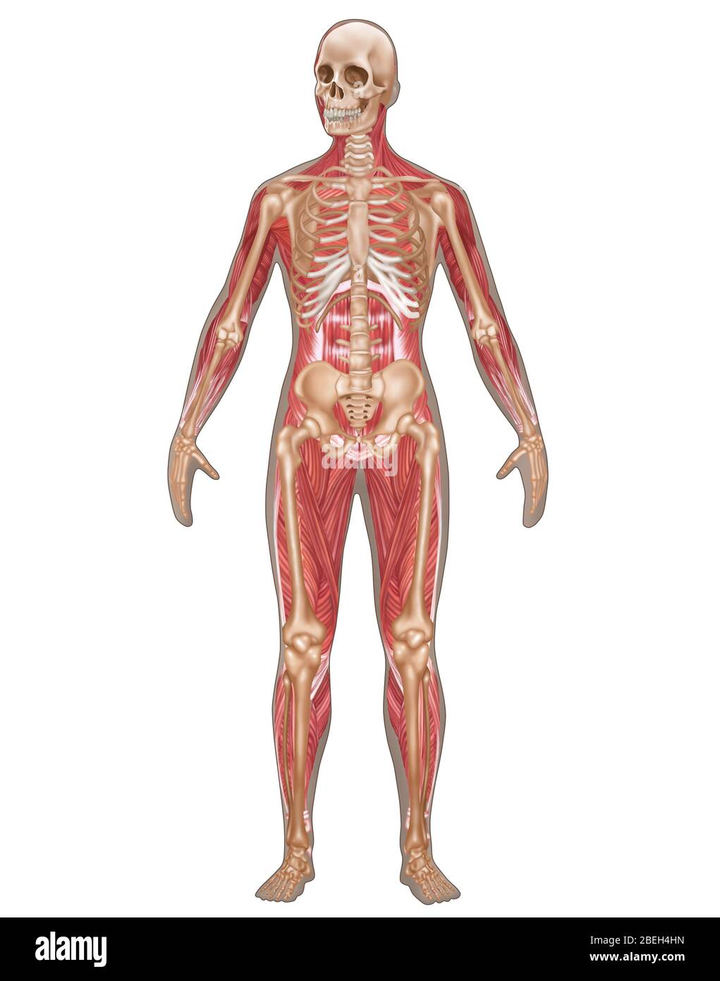 Skeletal & Muscular Systems, Female Anatomy Stock Photohttps://www.alamy.com/image-license-details/?v=1https://www.alamy.com/skeletal-muscular-systems-female-anatomy-image353189361.html
RF2BEH4HN–Skeletal & Muscular Systems, Female Anatomy
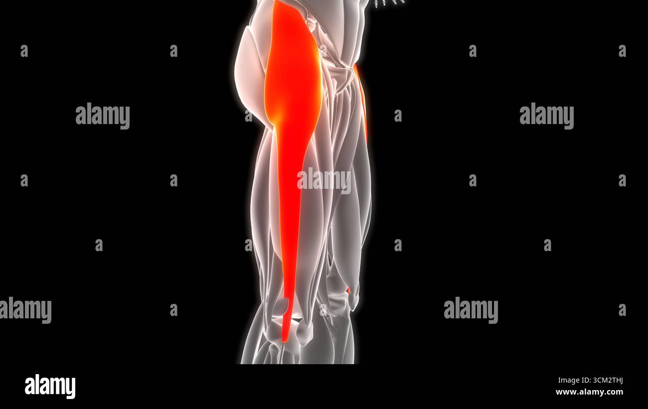 Human Muscular System Leg Muscles Tensor Fasciae Latae Muscles Anatomy Stock Photohttps://www.alamy.com/image-license-details/?v=1https://www.alamy.com/human-muscular-system-leg-muscles-tensor-fasciae-latae-muscles-anatomy-image700771054.html
RF3CM2THJ–Human Muscular System Leg Muscles Tensor Fasciae Latae Muscles Anatomy
 French houses and Chateau Guichard ruins at Angles Sur L'Anglin medieval village, Vienne, nr Poitiers, France Stock Photohttps://www.alamy.com/image-license-details/?v=1https://www.alamy.com/stock-photo-french-houses-and-chateau-guichard-ruins-at-angles-sur-langlin-medieval-34521277.html
RMC04G6N–French houses and Chateau Guichard ruins at Angles Sur L'Anglin medieval village, Vienne, nr Poitiers, France
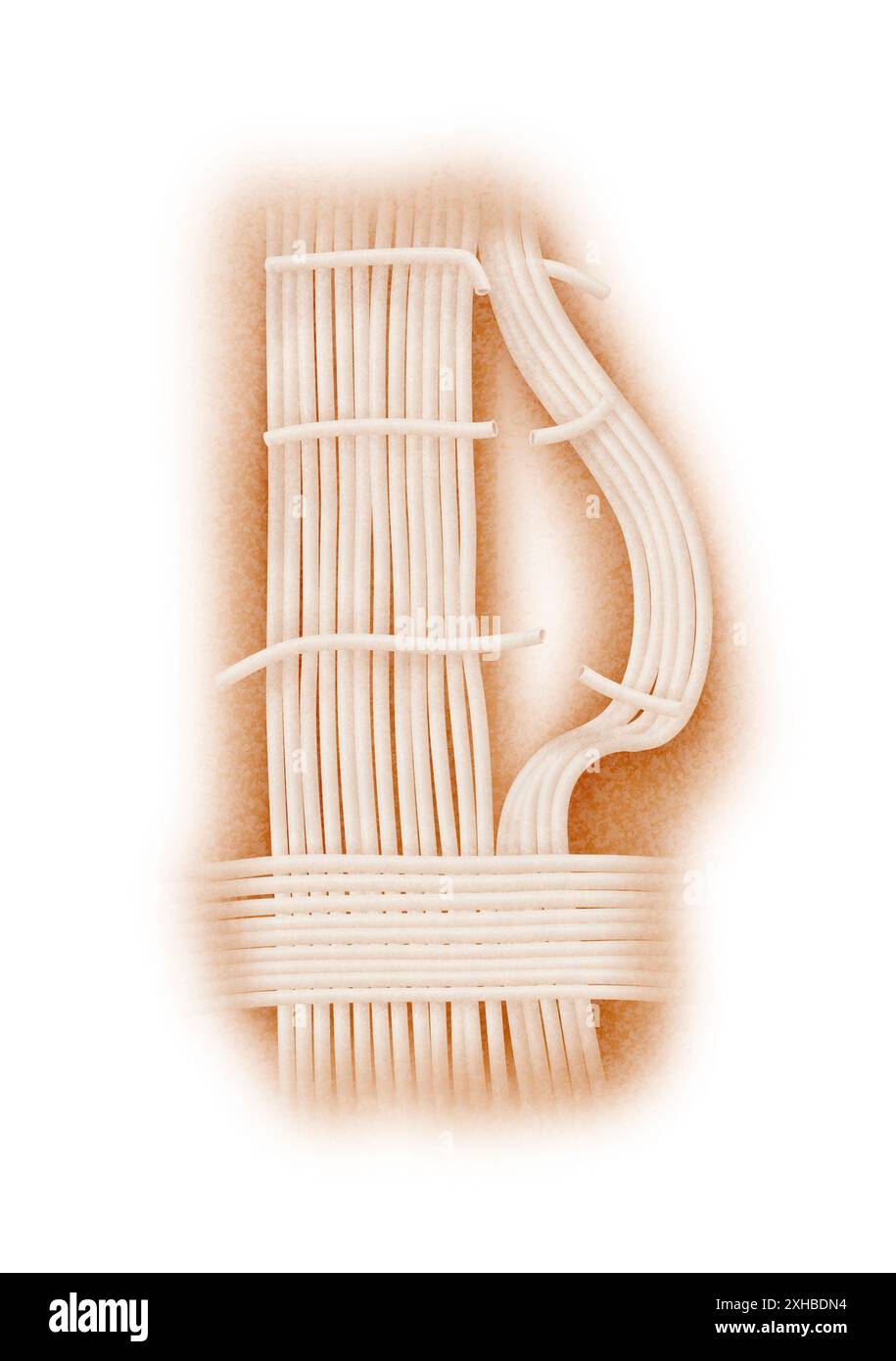 This is a thorough anatomical representation of arm muscles with a detailed focus fasciae Stock Photohttps://www.alamy.com/image-license-details/?v=1https://www.alamy.com/this-is-a-thorough-anatomical-representation-of-arm-muscles-with-a-detailed-focus-fasciae-image613064288.html
RF2XHBDN4–This is a thorough anatomical representation of arm muscles with a detailed focus fasciae
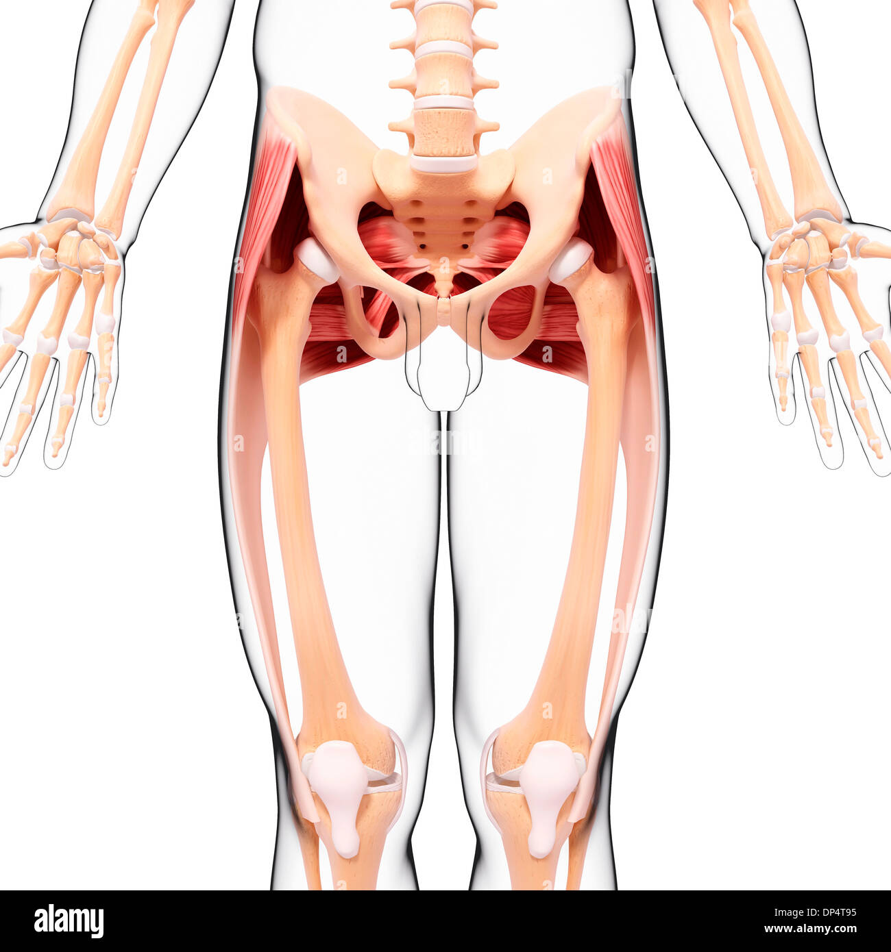 Human musculature, artwork Stock Photohttps://www.alamy.com/image-license-details/?v=1https://www.alamy.com/human-musculature-artwork-image65260417.html
RFDP4T95–Human musculature, artwork
 Chateau de Bourdeilles fortress in town of Bourdeilles popular tourist destination near Brantome in Dordogne, France Stock Photohttps://www.alamy.com/image-license-details/?v=1https://www.alamy.com/stock-photo-chateau-de-bourdeilles-fortress-in-town-of-bourdeilles-popular-tourist-34513028.html
RMC045M4–Chateau de Bourdeilles fortress in town of Bourdeilles popular tourist destination near Brantome in Dordogne, France
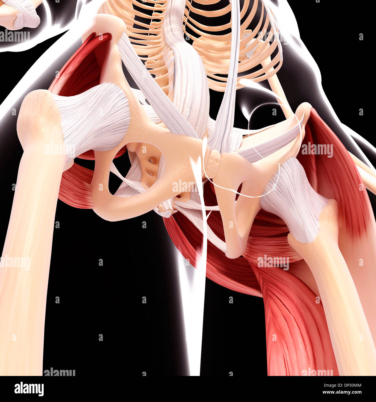 Human hip musculature, artwork Stock Photohttps://www.alamy.com/image-license-details/?v=1https://www.alamy.com/human-hip-musculature-artwork-image65219972.html
RFDP30MM–Human hip musculature, artwork
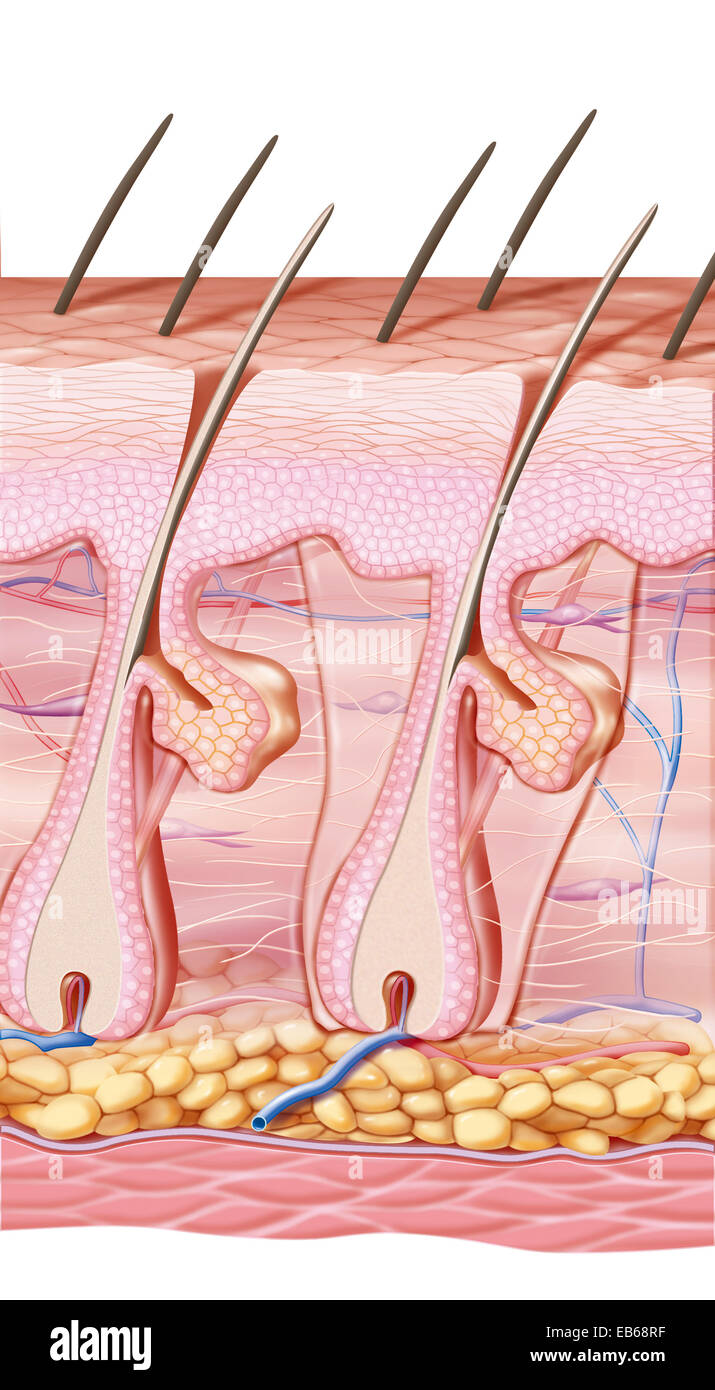 SCAR DRAWING Stock Photohttps://www.alamy.com/image-license-details/?v=1https://www.alamy.com/stock-photo-scar-drawing-75741331.html
RMEB68RF–SCAR DRAWING
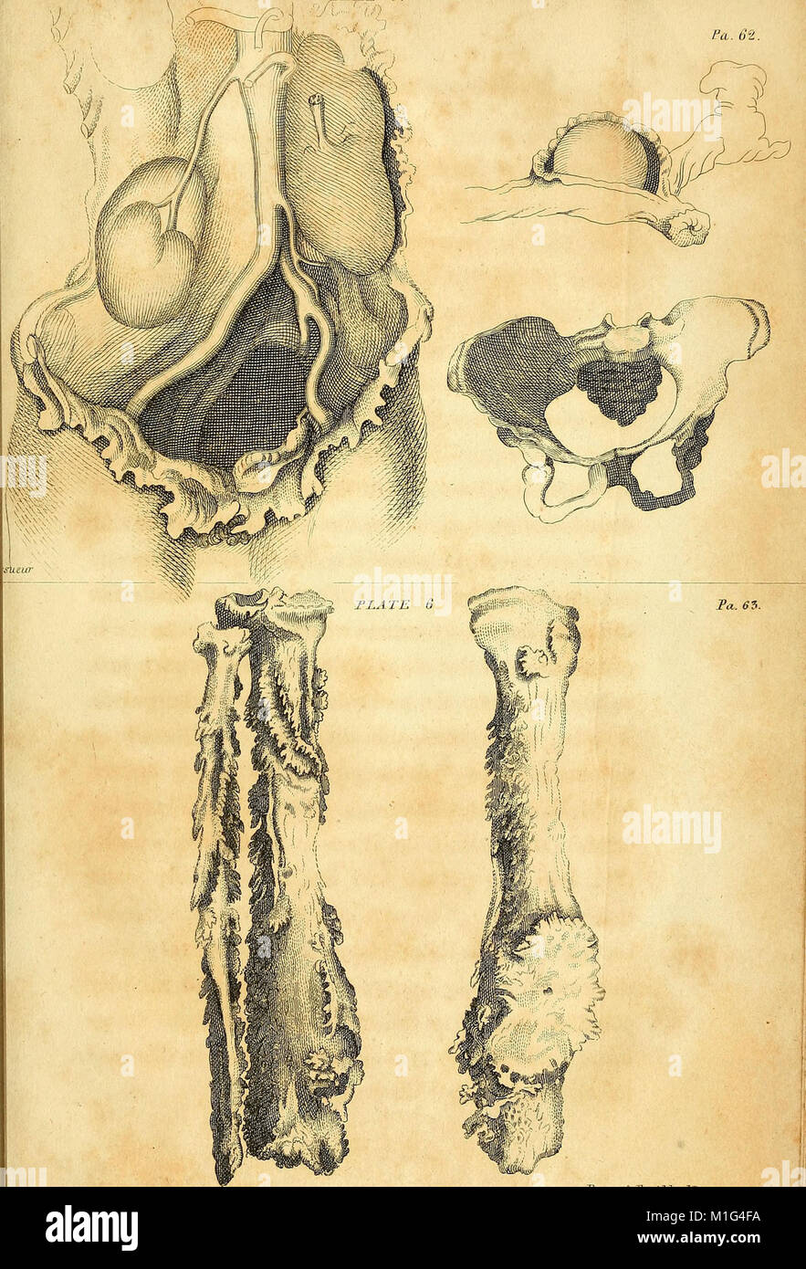 'Anatomical Investigations' (1798) offers detailed descriptions of human fasciae, irregularities in body structure, and cases in morbid anatomy. It also introduces a new anatomical discovery, contributing to medical understanding of human anatomy. Stock Photohttps://www.alamy.com/image-license-details/?v=1https://www.alamy.com/stock-photo-anatomical-investigations-1798-offers-detailed-descriptions-of-human-173073134.html
RMM1G4FA–'Anatomical Investigations' (1798) offers detailed descriptions of human fasciae, irregularities in body structure, and cases in morbid anatomy. It also introduces a new anatomical discovery, contributing to medical understanding of human anatomy.
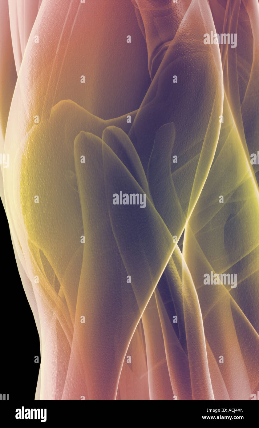 The muscles of the hip Stock Photohttps://www.alamy.com/image-license-details/?v=1https://www.alamy.com/stock-photo-the-muscles-of-the-hip-13166588.html
RFACJ4XN–The muscles of the hip
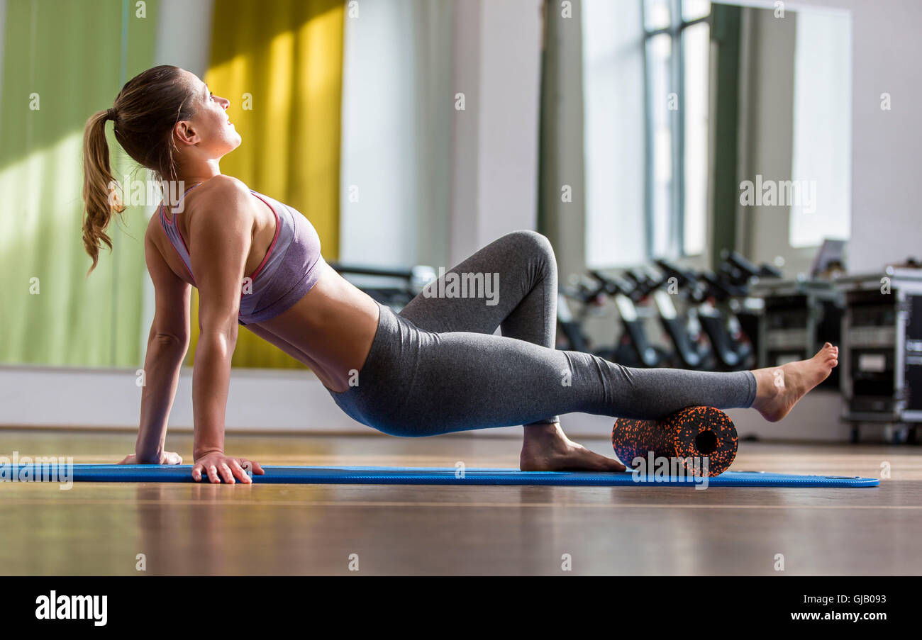 Black Roll training in the fitness room Stock Photohttps://www.alamy.com/image-license-details/?v=1https://www.alamy.com/stock-photo-black-roll-training-in-the-fitness-room-114567743.html
RMGJB093–Black Roll training in the fitness room
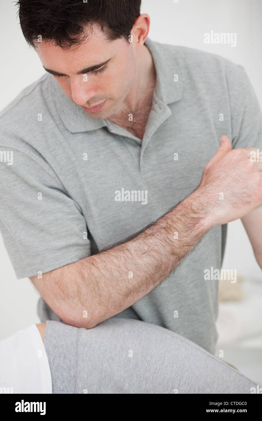 Physiotherapist using his elbow on the hip of a woman Stock Photohttps://www.alamy.com/image-license-details/?v=1https://www.alamy.com/stock-photo-physiotherapist-using-his-elbow-on-the-hip-of-a-woman-49470736.html
RFCTDGC0–Physiotherapist using his elbow on the hip of a woman
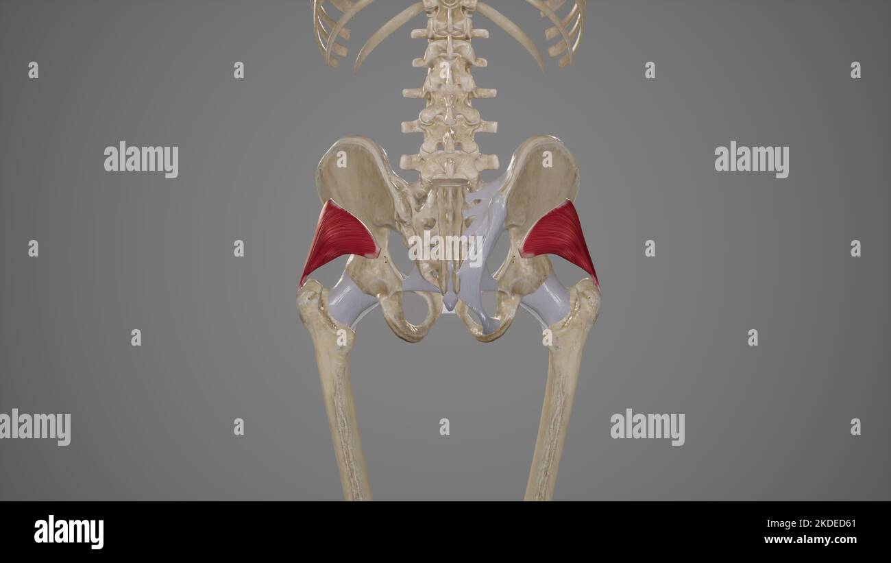 Medical Illustration of Gluteus Minimus Muscle Stock Photohttps://www.alamy.com/image-license-details/?v=1https://www.alamy.com/medical-illustration-of-gluteus-minimus-muscle-image490198521.html
RF2KDED61–Medical Illustration of Gluteus Minimus Muscle
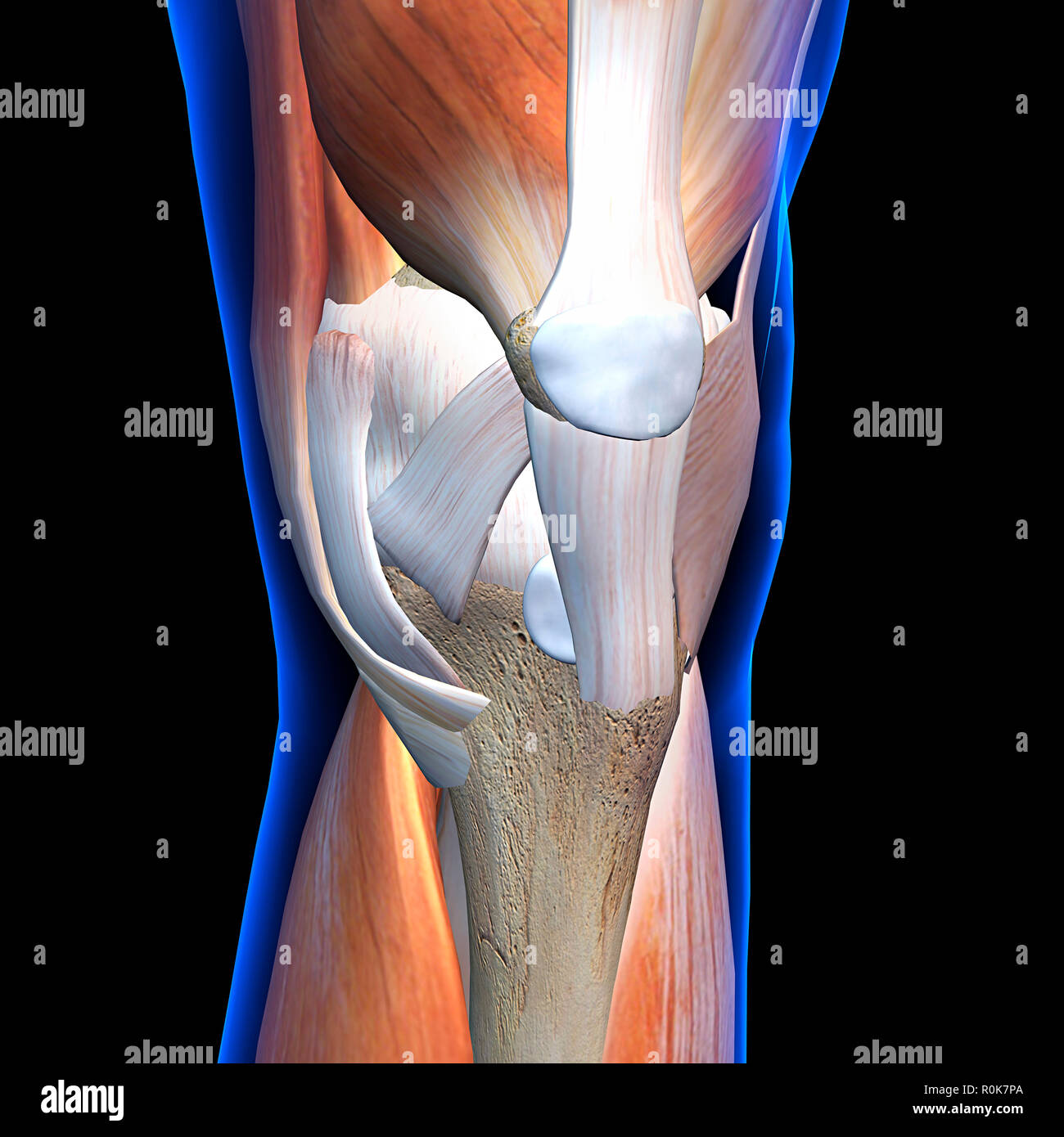 Knee muscles and ligaments, anterior x-ray view. Stock Photohttps://www.alamy.com/image-license-details/?v=1https://www.alamy.com/knee-muscles-and-ligaments-anterior-x-ray-view-image224157986.html
RFR0K7PA–Knee muscles and ligaments, anterior x-ray view.
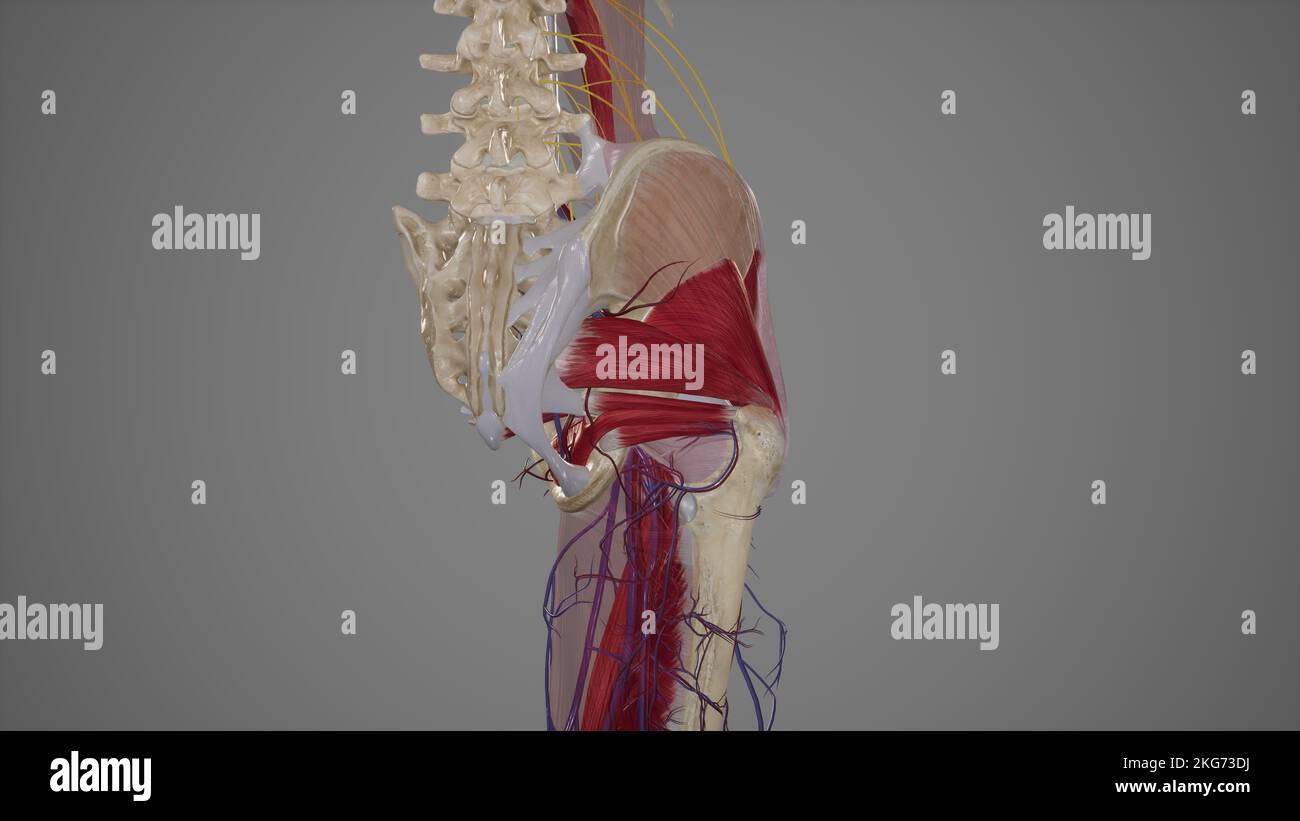 Arteries of Gluteal Region Stock Photohttps://www.alamy.com/image-license-details/?v=1https://www.alamy.com/arteries-of-gluteal-region-image491881198.html
RF2KG73DJ–Arteries of Gluteal Region
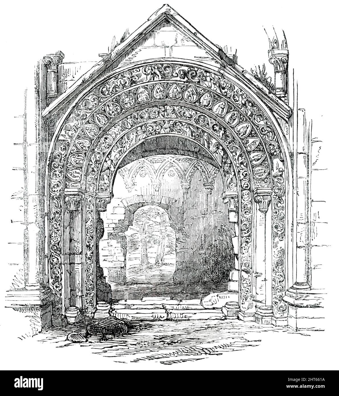 Glastonbury Abbey - North Door of St. Joseph's Chapel, 1850. Ruined medieval abbey in Glastonbury, Somerset. View showing '...the North Portal of St. Joseph's Chapel, than which nothing in freestone masonry can be more highly sculptured. This building is attributed to the abbacy of Hirlewinus, between the years 1102 and 1120. It consists of semicircular arches (four in number), receding gently in succession into the body of the wall, and diminishing in size as they recede, each resting on pillars, and their fasciae thickly covered with a profusion of sculptured representations. We perceive the Stock Photohttps://www.alamy.com/image-license-details/?v=1https://www.alamy.com/glastonbury-abbey-north-door-of-st-josephs-chapel-1850-ruined-medieval-abbey-in-glastonbury-somerset-view-showing-the-north-portal-of-st-josephs-chapel-than-which-nothing-in-freestone-masonry-can-be-more-highly-sculptured-this-building-is-attributed-to-the-abbacy-of-hirlewinus-between-the-years-1102-and-1120-it-consists-of-semicircular-arches-four-in-number-receding-gently-in-succession-into-the-body-of-the-wall-and-diminishing-in-size-as-they-recede-each-resting-on-pillars-and-their-fasciae-thickly-covered-with-a-profusion-of-sculptured-representations-we-perceive-the-image462357766.html
RM2HT661A–Glastonbury Abbey - North Door of St. Joseph's Chapel, 1850. Ruined medieval abbey in Glastonbury, Somerset. View showing '...the North Portal of St. Joseph's Chapel, than which nothing in freestone masonry can be more highly sculptured. This building is attributed to the abbacy of Hirlewinus, between the years 1102 and 1120. It consists of semicircular arches (four in number), receding gently in succession into the body of the wall, and diminishing in size as they recede, each resting on pillars, and their fasciae thickly covered with a profusion of sculptured representations. We perceive the
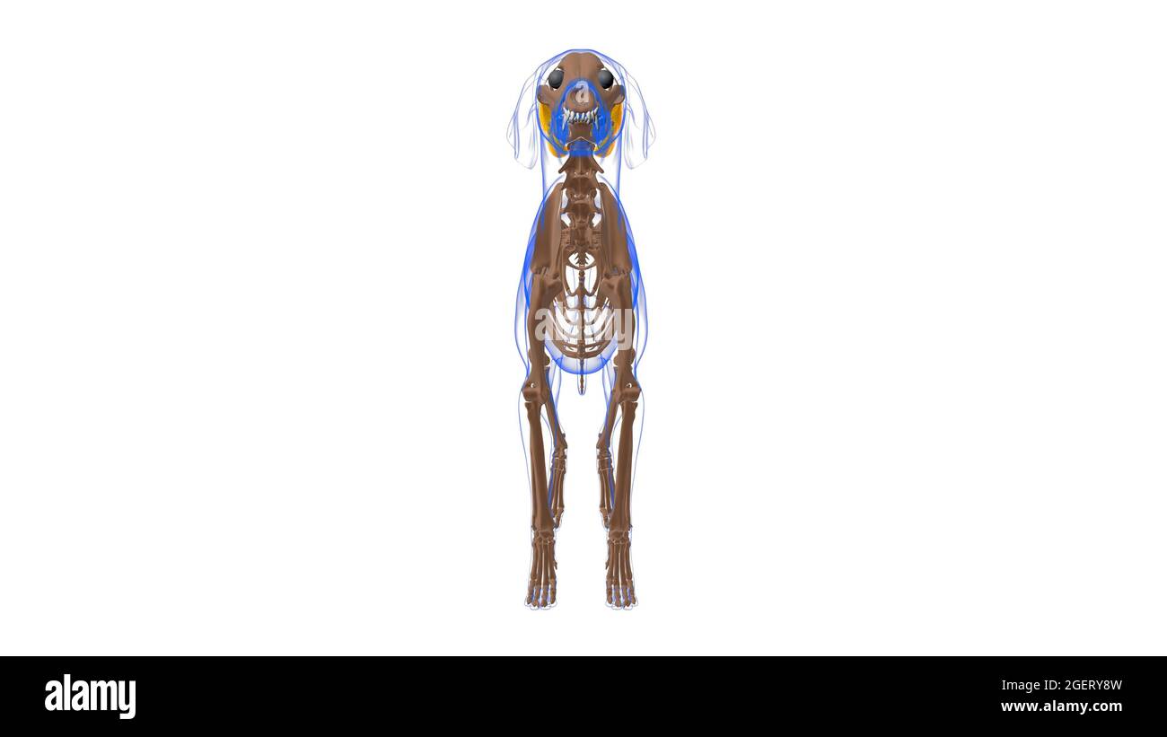 Tensor Fasciae Latae muscle Dog muscle Anatomy For Medical Concept 3D Illustration Stock Photohttps://www.alamy.com/image-license-details/?v=1https://www.alamy.com/tensor-fasciae-latae-muscle-dog-muscle-anatomy-for-medical-concept-3d-illustration-image439390697.html
RF2GERY8W–Tensor Fasciae Latae muscle Dog muscle Anatomy For Medical Concept 3D Illustration
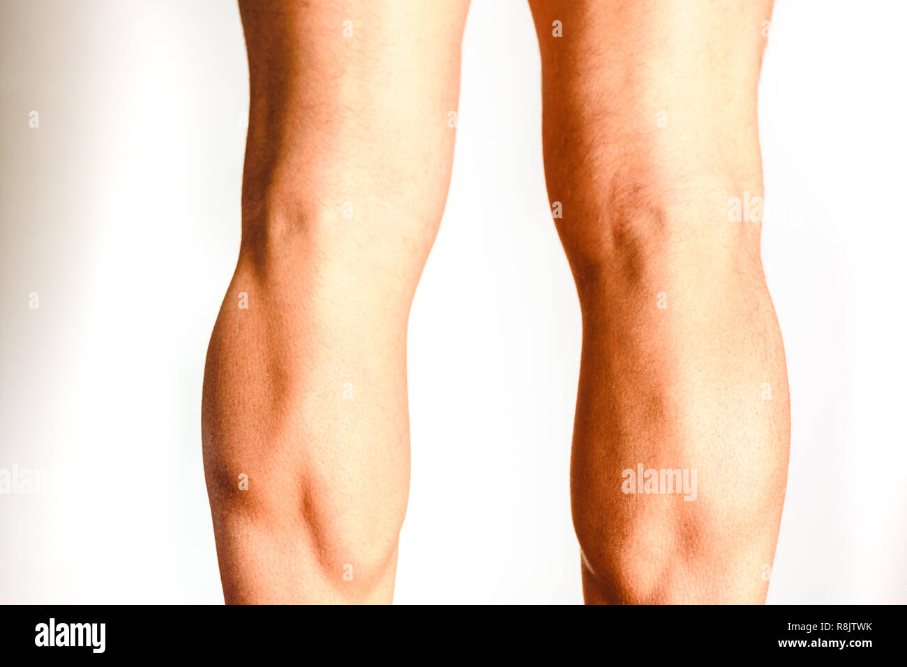 Muscles of the posterior leg, soleus and gastrocnemius muscle, photo of an athlete. Stock Photohttps://www.alamy.com/image-license-details/?v=1https://www.alamy.com/muscles-of-the-posterior-leg-soleus-and-gastrocnemius-muscle-photo-of-an-athlete-image229066703.html
RFR8JTWK–Muscles of the posterior leg, soleus and gastrocnemius muscle, photo of an athlete.
 Idaea aversata Riband Wave Moth Stock Photohttps://www.alamy.com/image-license-details/?v=1https://www.alamy.com/idaea-aversata-riband-wave-moth-image365858878.html
RF2C768ME–Idaea aversata Riband Wave Moth
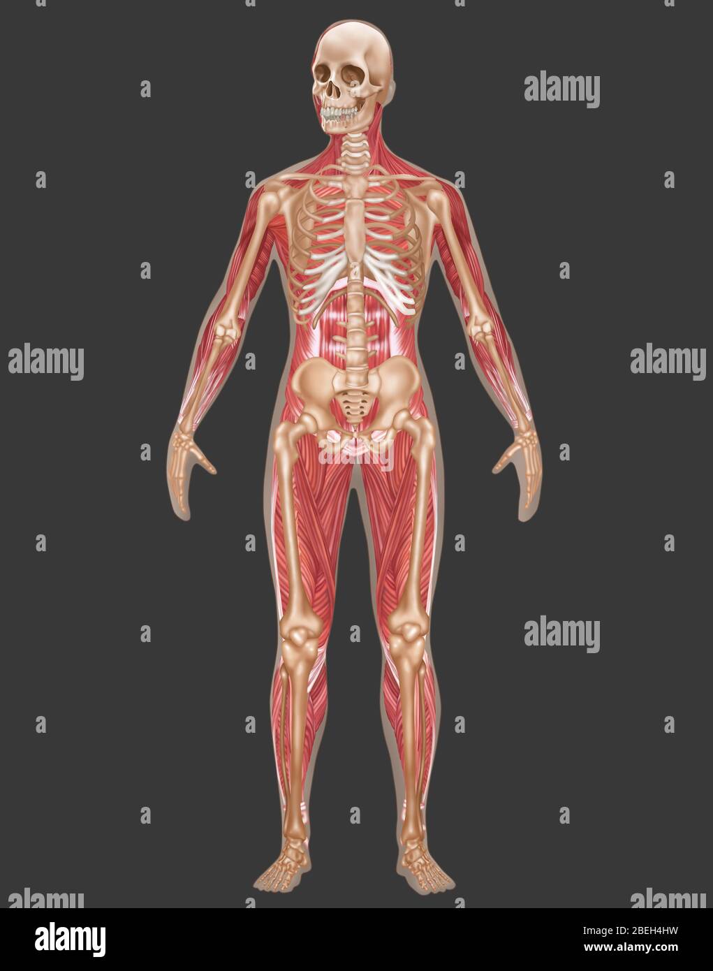 Skeletal & Muscular Systems, Female Anatomy Stock Photohttps://www.alamy.com/image-license-details/?v=1https://www.alamy.com/skeletal-muscular-systems-female-anatomy-image353189365.html
RF2BEH4HW–Skeletal & Muscular Systems, Female Anatomy
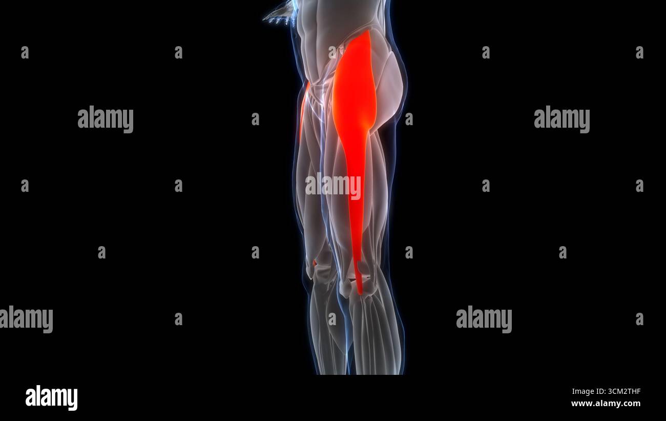 Human Muscular System Leg Muscles Tensor Fasciae Latae Muscles Anatomy Stock Photohttps://www.alamy.com/image-license-details/?v=1https://www.alamy.com/human-muscular-system-leg-muscles-tensor-fasciae-latae-muscles-anatomy-image700771051.html
RF3CM2THF–Human Muscular System Leg Muscles Tensor Fasciae Latae Muscles Anatomy
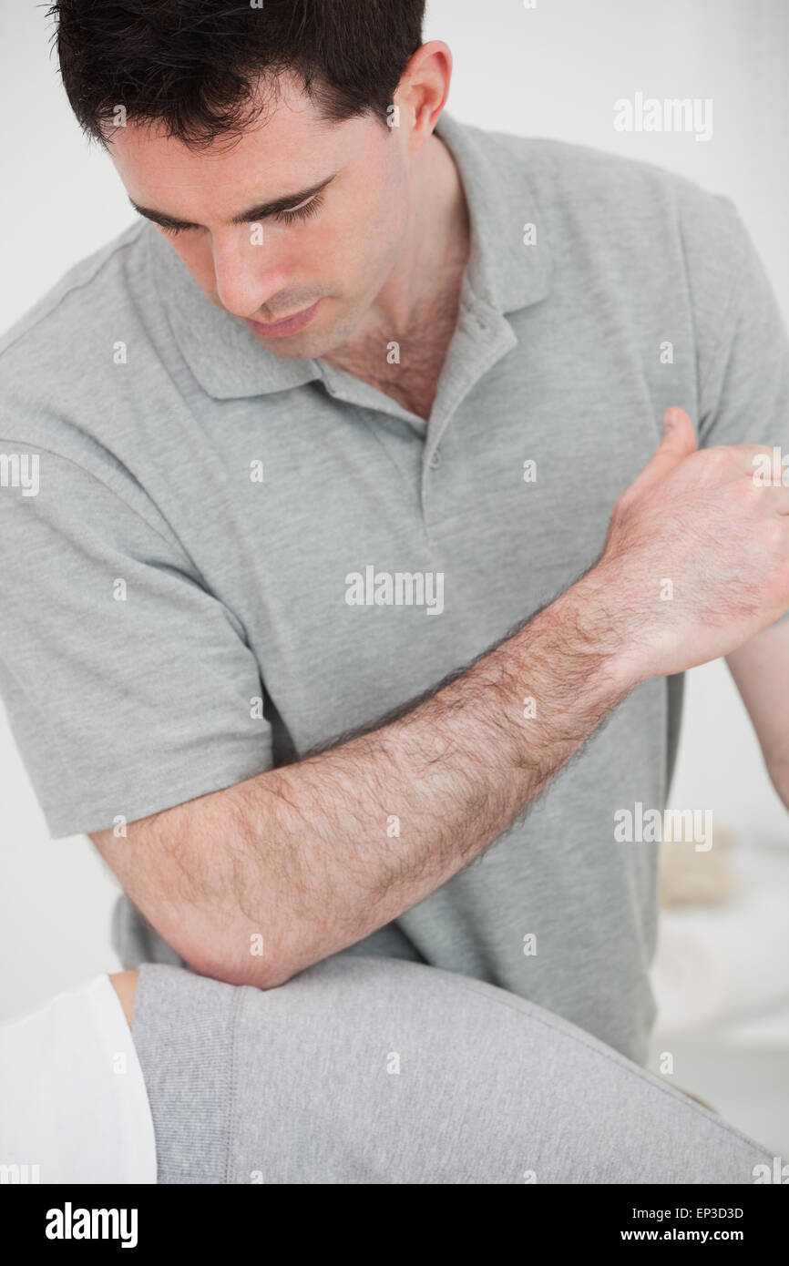 Physiotherapist using his elbow on the hip of a woman Stock Photohttps://www.alamy.com/image-license-details/?v=1https://www.alamy.com/stock-photo-physiotherapist-using-his-elbow-on-the-hip-of-a-woman-82440049.html
RFEP3D3D–Physiotherapist using his elbow on the hip of a woman
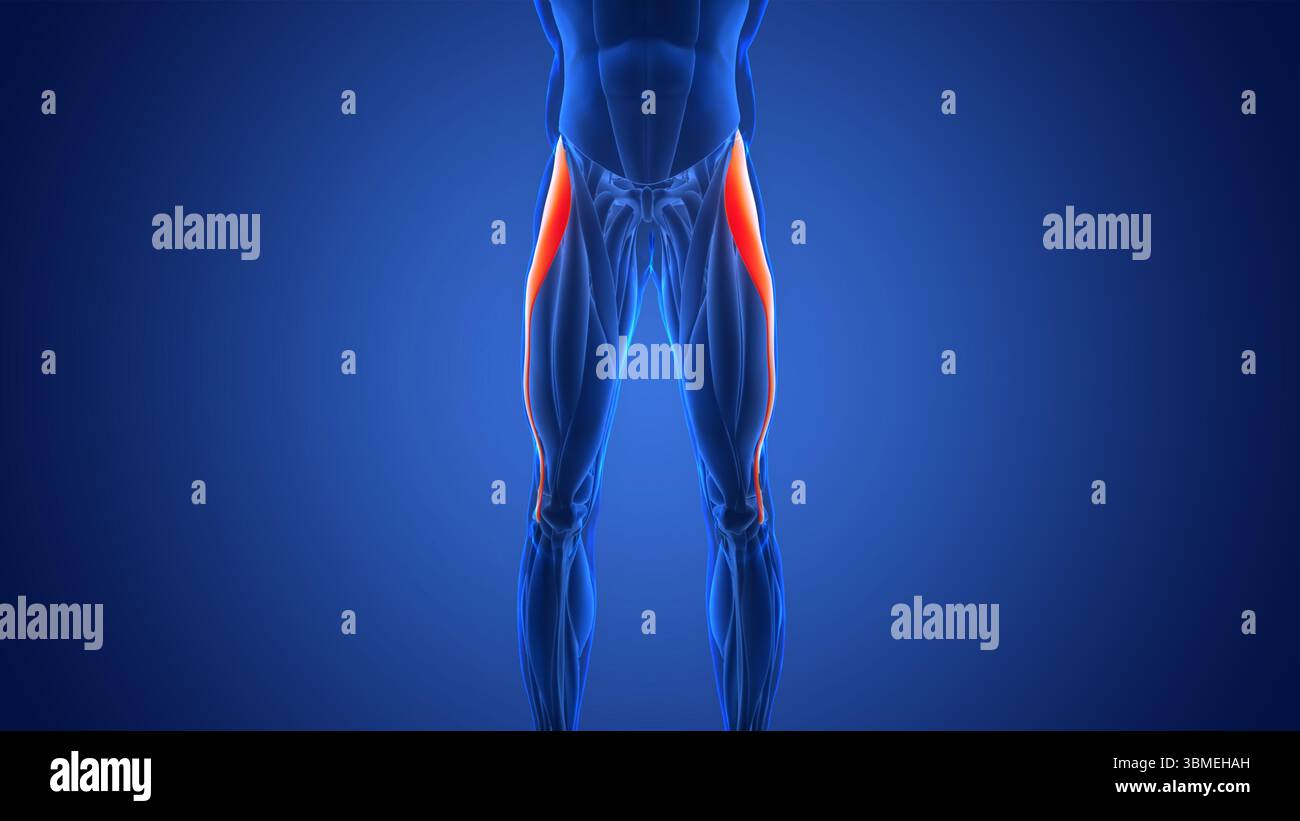 Human Muscular System Leg Muscles Tensor Fasciae Latae Muscles Anatomy Stock Photohttps://www.alamy.com/image-license-details/?v=1https://www.alamy.com/human-muscular-system-leg-muscles-tensor-fasciae-latae-muscles-anatomy-image683818425.html
RF3BMEHAH–Human Muscular System Leg Muscles Tensor Fasciae Latae Muscles Anatomy
 Archive image from page 615 of A handbook of British lepidoptera. A handbook of British lepidoptera CUbiodiversity1126142 Year: 1895 ( [brachmia 1. Termen of fore wings subconcave, stigmata black . 2. i, „ straight, stigmata not black 2. ru/escens. '2. Forewings with dark fasciae . .1. gerronella. „ without transverse markings . 3. inornatella. 1. B. gerronella, Z. 11-12 mm. Forewings with termen subconcave; ochreous, irrorated with brown; an indistinct darker angulated fascia at ; a more distinct oblique dark fuscous fascia beyond middle ; stigmata black, first discal be- yond plioal; an Stock Photohttps://www.alamy.com/image-license-details/?v=1https://www.alamy.com/archive-image-from-page-615-of-a-handbook-of-british-lepidoptera-a-handbook-of-british-lepidoptera-cubiodiversity1126142-year-1895-brachmia-1-termen-of-fore-wings-subconcave-stigmata-black-2-i-straight-stigmata-not-black-2-ruescens-2-forewings-with-dark-fasciae-1-gerronella-without-transverse-markings-3-inornatella-1-b-gerronella-z-11-12-mm-forewings-with-termen-subconcave-ochreous-irrorated-with-brown-an-indistinct-darker-angulated-fascia-at-a-more-distinct-oblique-dark-fuscous-fascia-beyond-middle-stigmata-black-first-discal-be-yond-plioal-an-image264064631.html
RMW9H53K–Archive image from page 615 of A handbook of British lepidoptera. A handbook of British lepidoptera CUbiodiversity1126142 Year: 1895 ( [brachmia 1. Termen of fore wings subconcave, stigmata black . 2. i, „ straight, stigmata not black 2. ru/escens. '2. Forewings with dark fasciae . .1. gerronella. „ without transverse markings . 3. inornatella. 1. B. gerronella, Z. 11-12 mm. Forewings with termen subconcave; ochreous, irrorated with brown; an indistinct darker angulated fascia at ; a more distinct oblique dark fuscous fascia beyond middle ; stigmata black, first discal be- yond plioal; an
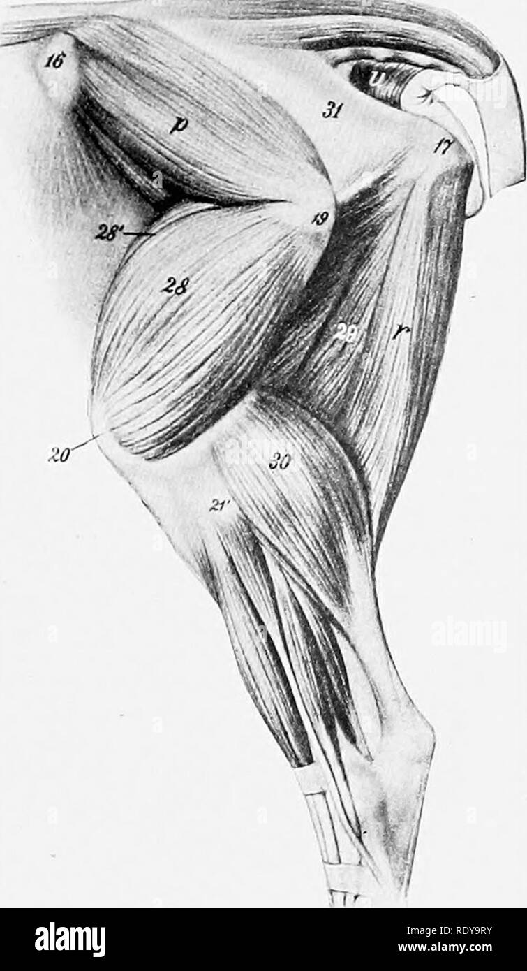 . The anatomy of the domestic animals . Veterinary anatomy. LATERAL MUSCLES OF THE HIP AND THIGH 355 n. LATERAL MUSCLES OF THE HIP AND THIGH (Figs. 303, 309) The tensor fasciae latae is large, and the fleshy part extends further down than in the horse. The gluteus superficialis is not present as such; apparently its anterior part has fused with the tensor fasciae latae and its posterior part with the biceps femoris. The gluteus medius is small, the lumbar part being insignificant and extend- ing forward only to the fourth lumbar vertebra. Its deep portion (gluteus ac- cessorius) is easily sepa Stock Photohttps://www.alamy.com/image-license-details/?v=1https://www.alamy.com/the-anatomy-of-the-domestic-animals-veterinary-anatomy-lateral-muscles-of-the-hip-and-thigh-355-n-lateral-muscles-of-the-hip-and-thigh-figs-303-309-the-tensor-fasciae-latae-is-large-and-the-fleshy-part-extends-further-down-than-in-the-horse-the-gluteus-superficialis-is-not-present-as-such-apparently-its-anterior-part-has-fused-with-the-tensor-fasciae-latae-and-its-posterior-part-with-the-biceps-femoris-the-gluteus-medius-is-small-the-lumbar-part-being-insignificant-and-extend-ing-forward-only-to-the-fourth-lumbar-vertebra-its-deep-portion-gluteus-ac-cessorius-is-easily-sepa-image232325743.html
RMRDY9RY–. The anatomy of the domestic animals . Veterinary anatomy. LATERAL MUSCLES OF THE HIP AND THIGH 355 n. LATERAL MUSCLES OF THE HIP AND THIGH (Figs. 303, 309) The tensor fasciae latae is large, and the fleshy part extends further down than in the horse. The gluteus superficialis is not present as such; apparently its anterior part has fused with the tensor fasciae latae and its posterior part with the biceps femoris. The gluteus medius is small, the lumbar part being insignificant and extend- ing forward only to the fourth lumbar vertebra. Its deep portion (gluteus ac- cessorius) is easily sepa
 Human musculature, artwork Stock Photohttps://www.alamy.com/image-license-details/?v=1https://www.alamy.com/human-musculature-artwork-image65225630.html
RFDP37XP–Human musculature, artwork
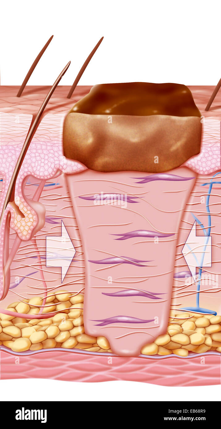 WOUND HEALING, ILLUSTRATION Stock Photohttps://www.alamy.com/image-license-details/?v=1https://www.alamy.com/stock-photo-wound-healing-illustration-75741325.html
RMEB68R9–WOUND HEALING, ILLUSTRATION
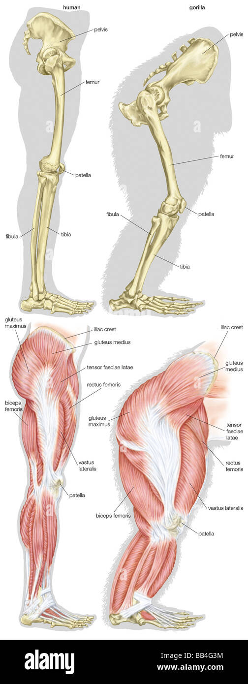 Skeletal and muscular structures of a human's leg (left) and a gorilla's leg (right). Stock Photohttps://www.alamy.com/image-license-details/?v=1https://www.alamy.com/stock-photo-skeletal-and-muscular-structures-of-a-humans-leg-left-and-a-gorillas-24072040.html
RMBB4G3M–Skeletal and muscular structures of a human's leg (left) and a gorilla's leg (right).
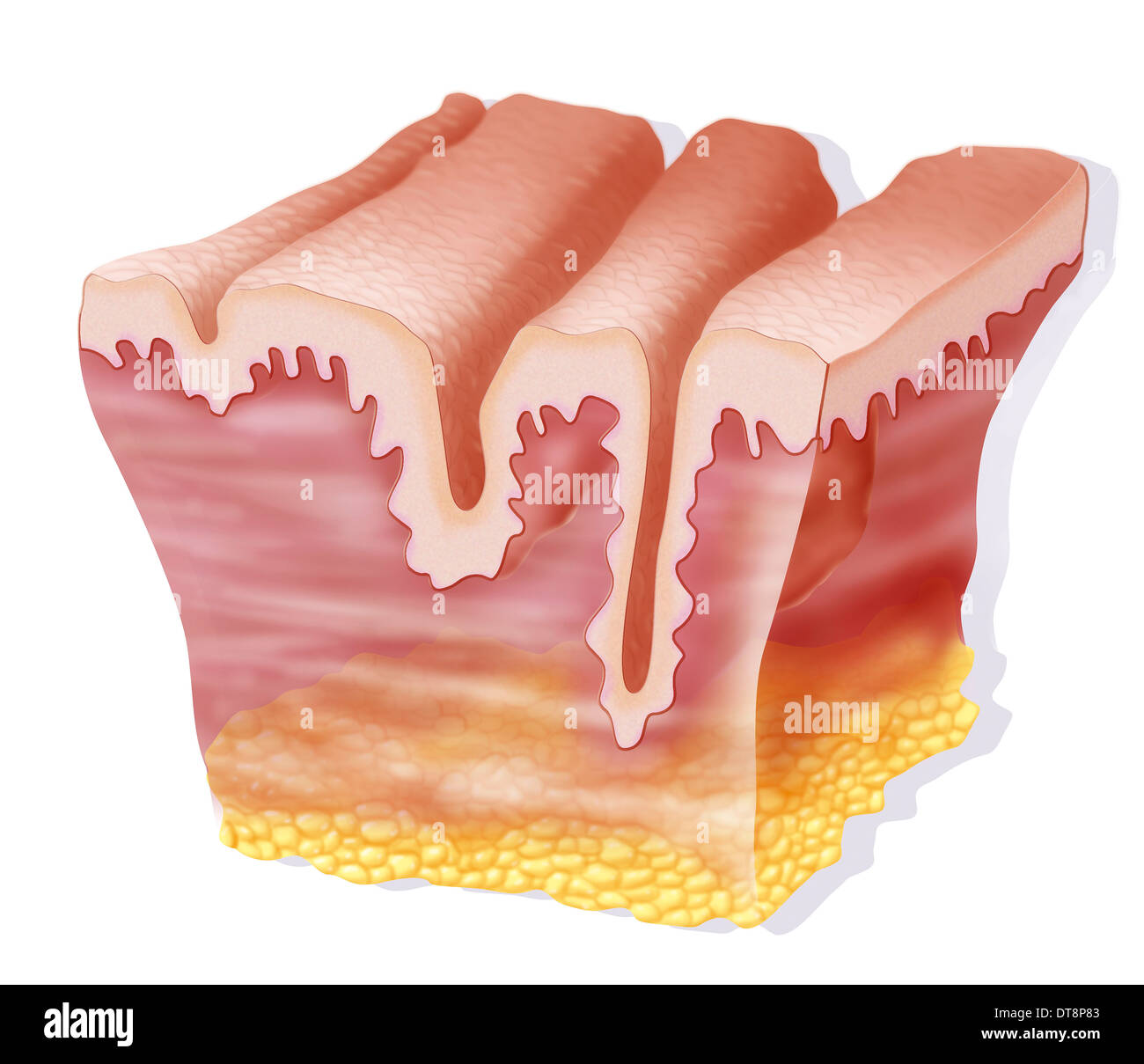 WRINKLE, DRAWING Stock Photohttps://www.alamy.com/image-license-details/?v=1https://www.alamy.com/wrinkle-drawing-image66575939.html
RMDT8P83–WRINKLE, DRAWING
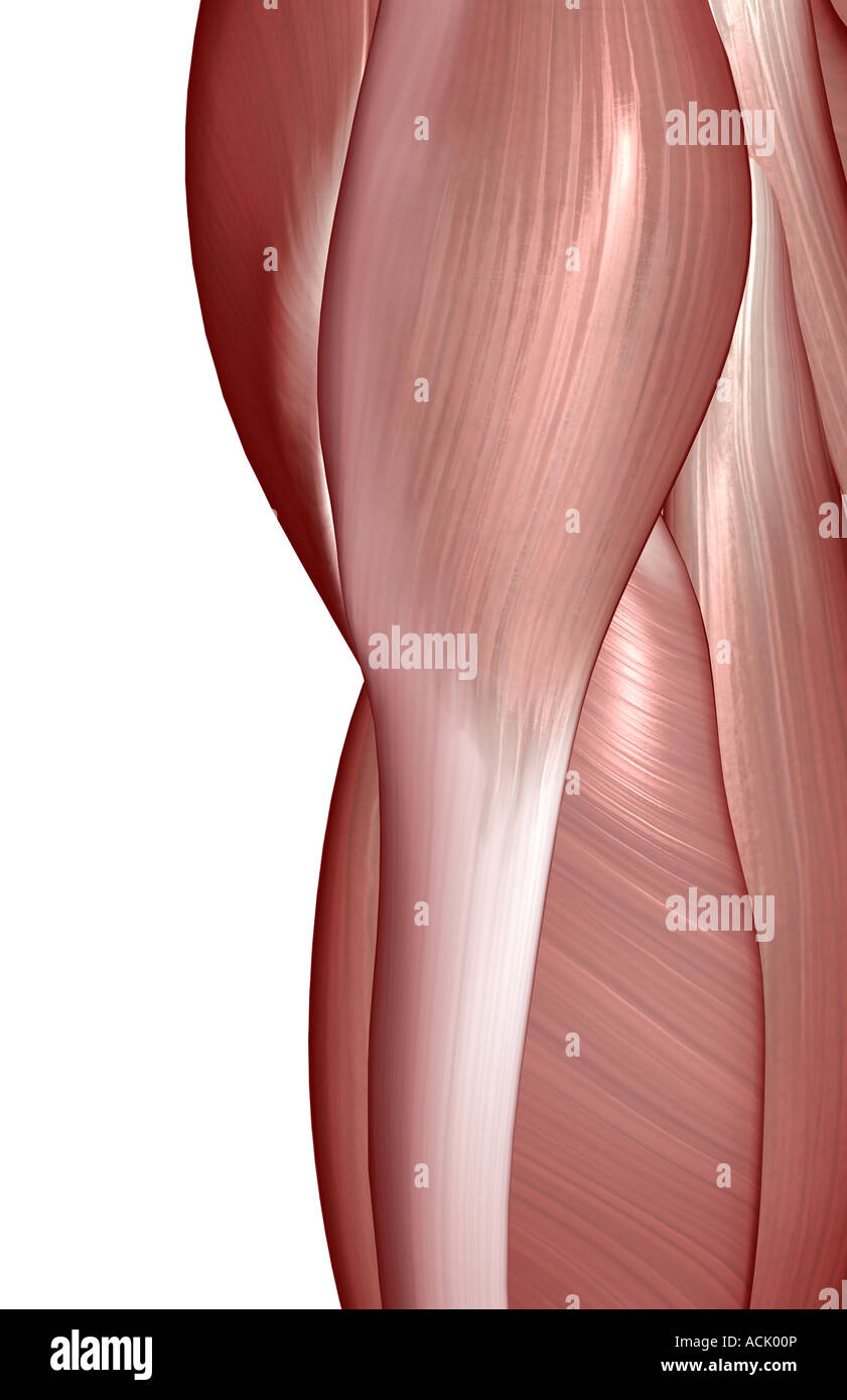 The muscles of the hip Stock Photohttps://www.alamy.com/image-license-details/?v=1https://www.alamy.com/stock-photo-the-muscles-of-the-hip-13174341.html
RFACK00P–The muscles of the hip
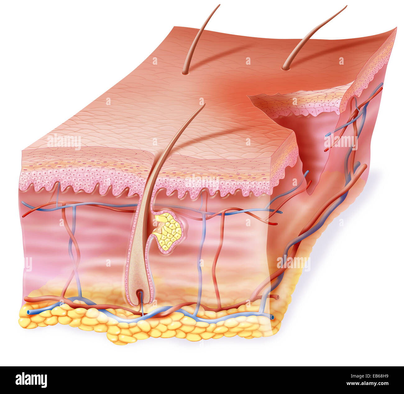 WOUND, ILLUSTRATION Stock Photohttps://www.alamy.com/image-license-details/?v=1https://www.alamy.com/stock-photo-wound-illustration-75741157.html
RMEB68H9–WOUND, ILLUSTRATION
 Black Roll training in the fitness room Stock Photohttps://www.alamy.com/image-license-details/?v=1https://www.alamy.com/stock-photo-black-roll-training-in-the-fitness-room-114567761.html
RMGJB09N–Black Roll training in the fitness room
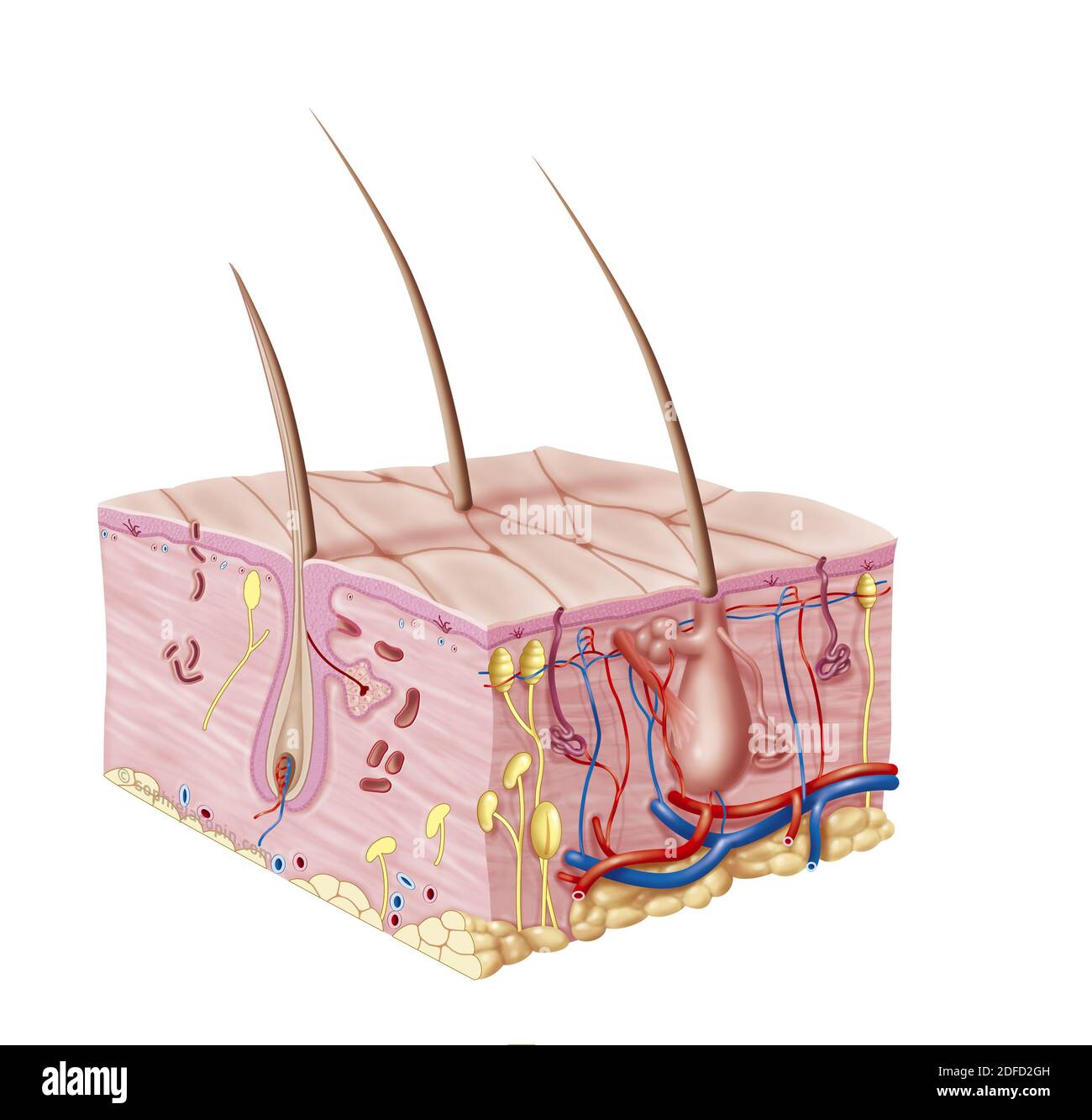 Baby-pediatric skin cut Stock Photohttps://www.alamy.com/image-license-details/?v=1https://www.alamy.com/baby-pediatric-skin-cut-image388135345.html
RM2DFD2GH–Baby-pediatric skin cut
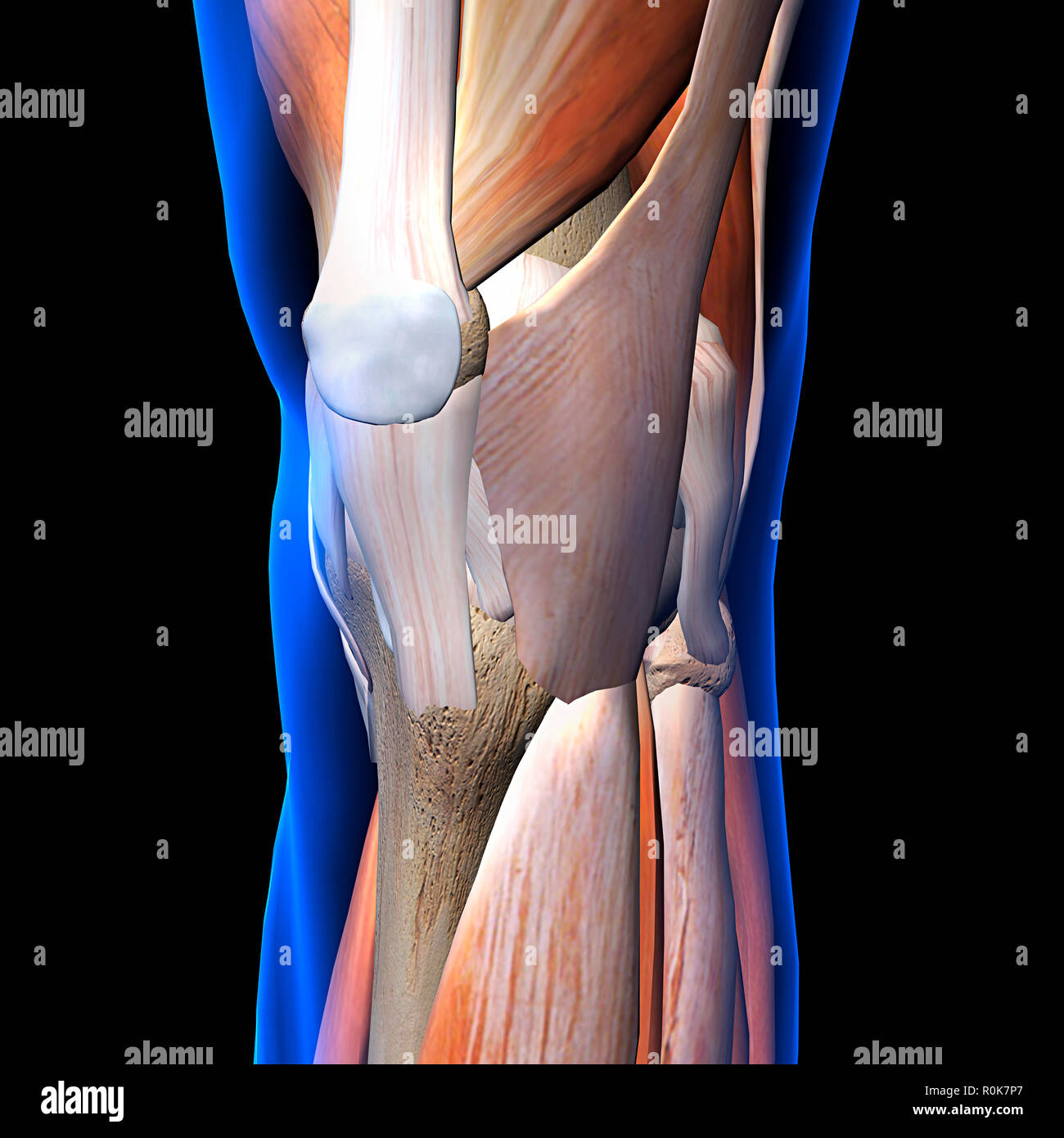 Knee muscles and ligaments, x-ray view on black background. Stock Photohttps://www.alamy.com/image-license-details/?v=1https://www.alamy.com/knee-muscles-and-ligaments-x-ray-view-on-black-background-image224157983.html
RFR0K7P7–Knee muscles and ligaments, x-ray view on black background.
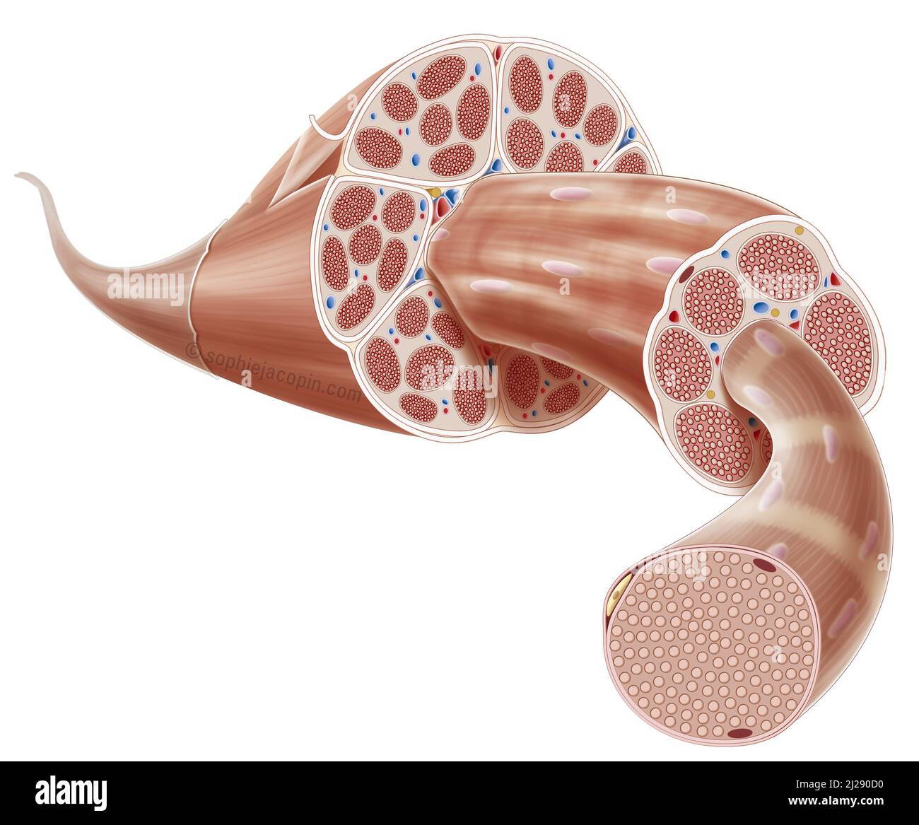 Muscle structure Stock Photohttps://www.alamy.com/image-license-details/?v=1https://www.alamy.com/muscle-structure-image466107180.html
RM2J290D0–Muscle structure
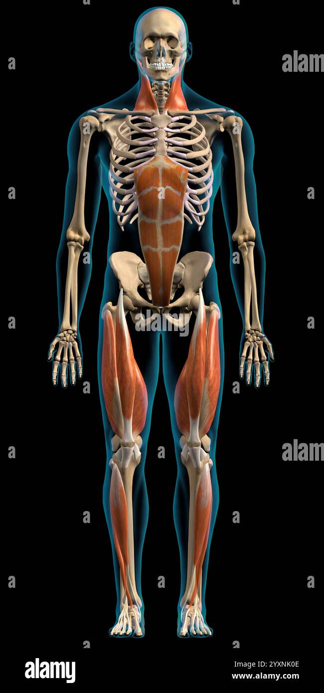 Male superficial network of muscles on white background, frontal view. Stock Photohttps://www.alamy.com/image-license-details/?v=1https://www.alamy.com/male-superficial-network-of-muscles-on-white-background-frontal-view-image636030206.html
RF2YXNK0E–Male superficial network of muscles on white background, frontal view.
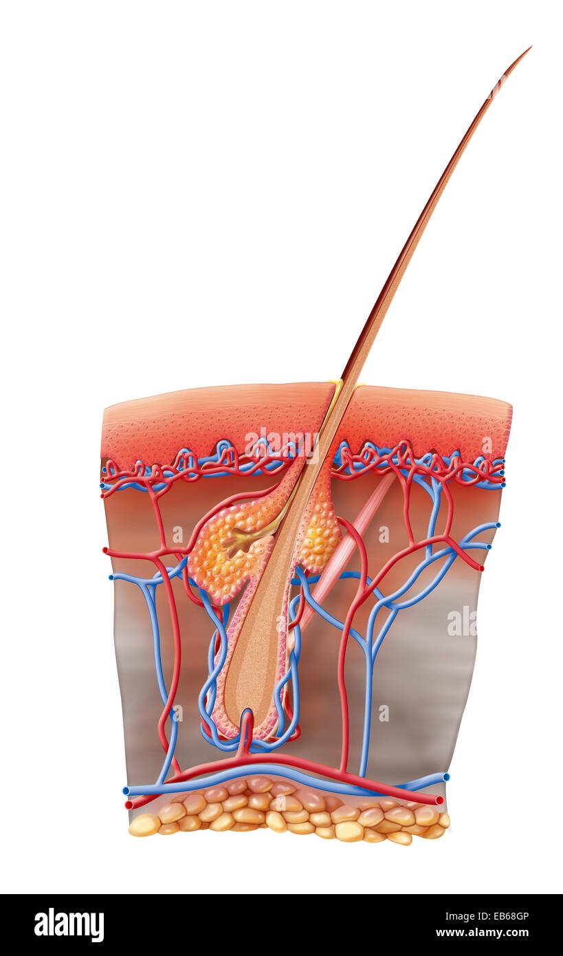 ERYTHEMA ILLUSTRATION Stock Photohttps://www.alamy.com/image-license-details/?v=1https://www.alamy.com/stock-photo-erythema-illustration-75741142.html
RMEB68GP–ERYTHEMA ILLUSTRATION
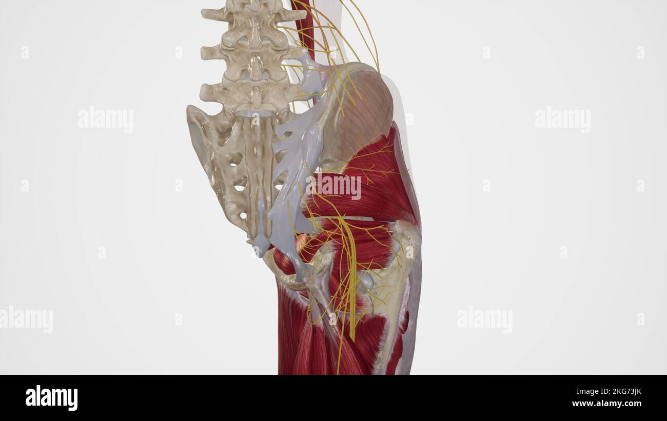 Nerves of Gluteal Region Stock Photohttps://www.alamy.com/image-license-details/?v=1https://www.alamy.com/nerves-of-gluteal-region-image491881339.html
RF2KG73JK–Nerves of Gluteal Region
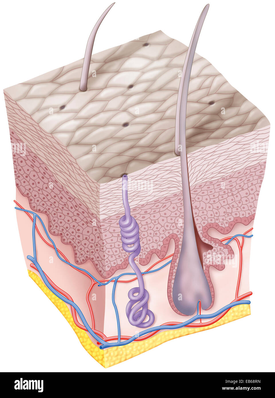 RHINOCEROS SKIN Stock Photohttps://www.alamy.com/image-license-details/?v=1https://www.alamy.com/stock-photo-rhinoceros-skin-75741337.html
RMEB68RN–RHINOCEROS SKIN
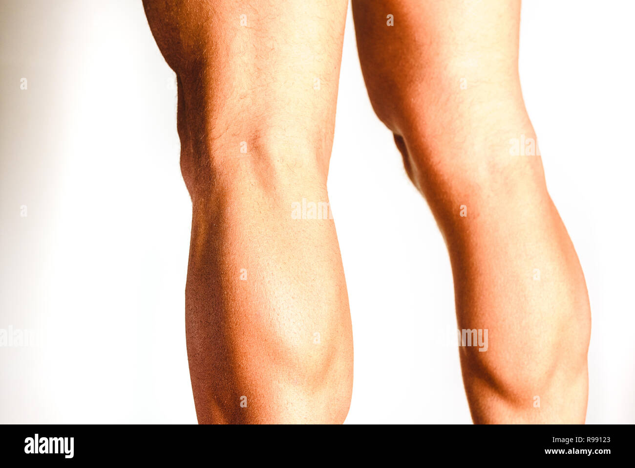 Muscles of the posterior leg, soleus and gastrocnemius muscle, photo of an athlete. Stock Photohttps://www.alamy.com/image-license-details/?v=1https://www.alamy.com/muscles-of-the-posterior-leg-soleus-and-gastrocnemius-muscle-photo-of-an-athlete-image229465099.html
RFR99123–Muscles of the posterior leg, soleus and gastrocnemius muscle, photo of an athlete.