Quick filters:
Articular capsule Stock Photos and Images
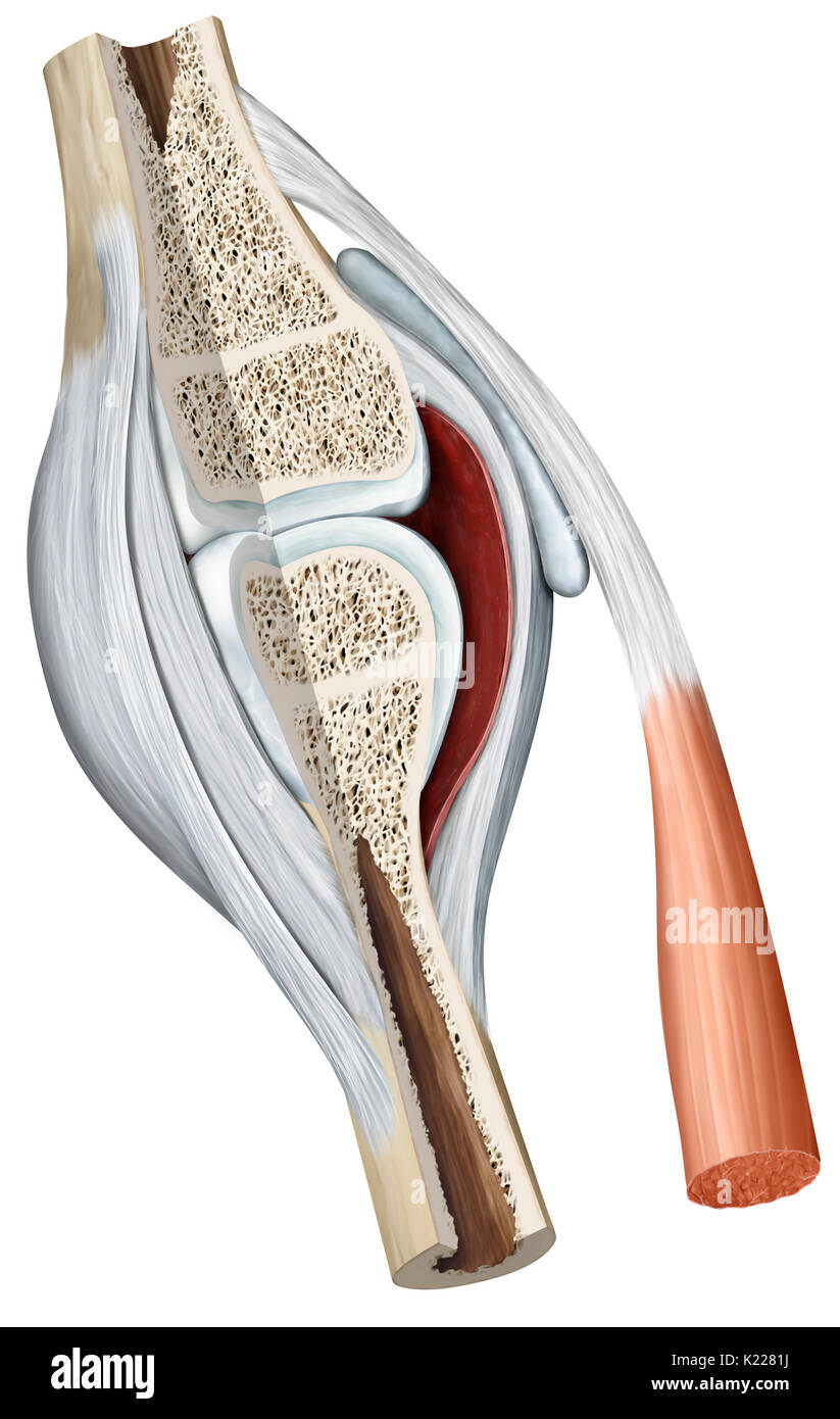 Synovial joint: joint characterized by the presence of an articular capsule filled with a viscous liquid (synovial fluid). This is the most common type of joint. Stock Photohttps://www.alamy.com/image-license-details/?v=1https://www.alamy.com/synovial-joint-joint-characterized-by-the-presence-of-an-articular-image156172846.html
RMK2281J–Synovial joint: joint characterized by the presence of an articular capsule filled with a viscous liquid (synovial fluid). This is the most common type of joint.
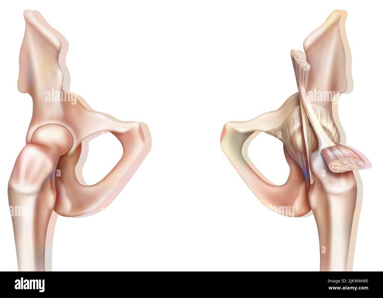 Bone joint of the hip without and with the coxofemoral joint capsule. Stock Photohttps://www.alamy.com/image-license-details/?v=1https://www.alamy.com/bone-joint-of-the-hip-without-and-with-the-coxofemoral-joint-capsule-image476923594.html
RF2JKWMWE–Bone joint of the hip without and with the coxofemoral joint capsule.
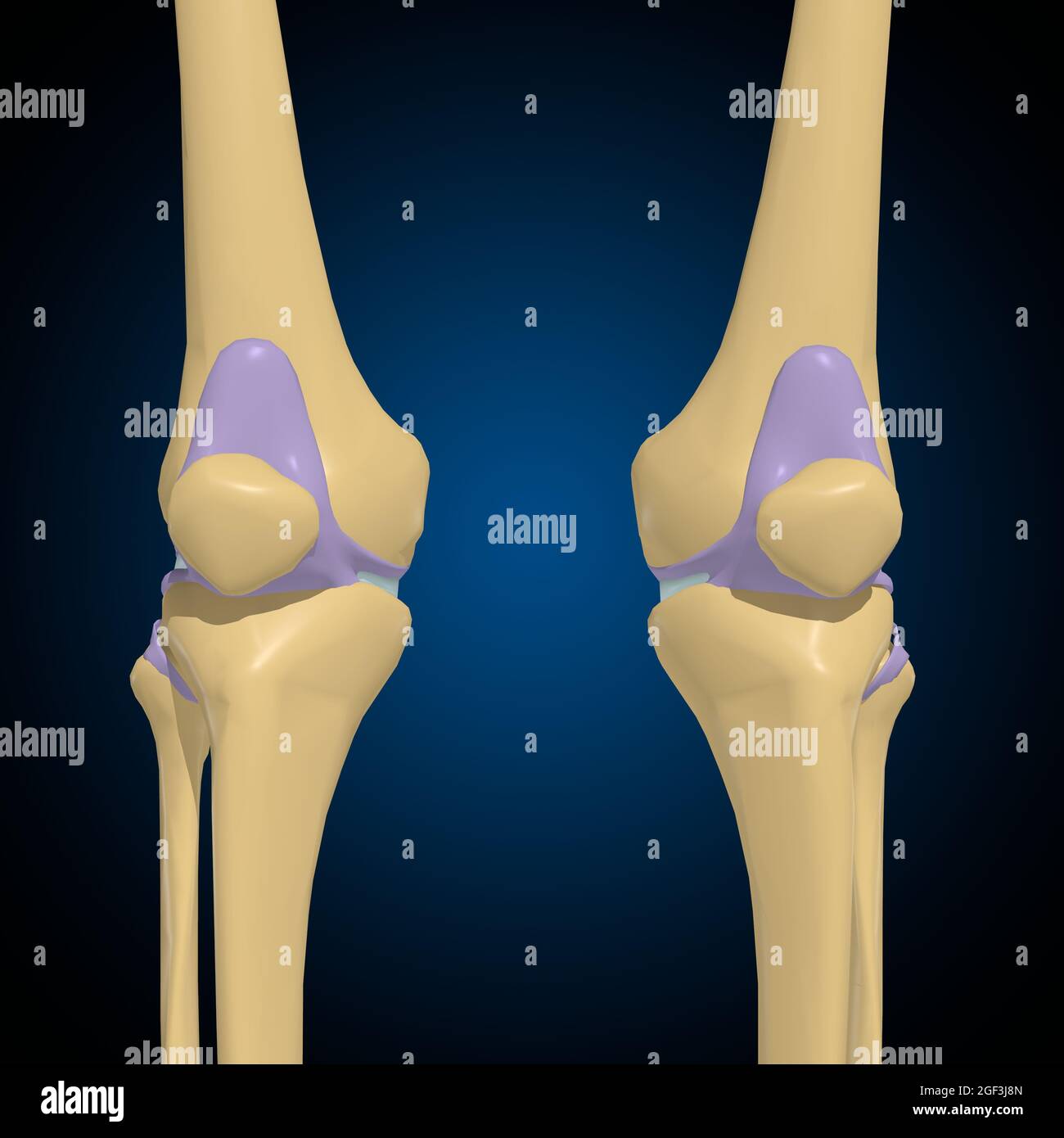 Articular capsule Anatomy For Medical Concept 3D Illustration Stock Photohttps://www.alamy.com/image-license-details/?v=1https://www.alamy.com/articular-capsule-anatomy-for-medical-concept-3d-illustration-image439559253.html
RF2GF3J8N–Articular capsule Anatomy For Medical Concept 3D Illustration
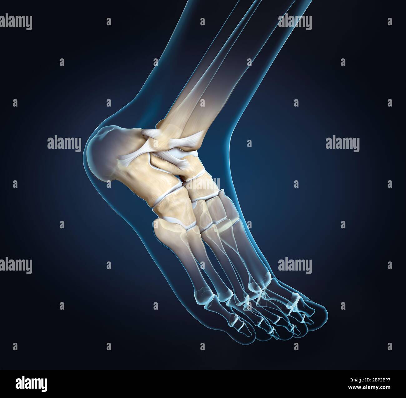 3D illustration showing of a ankle joint with bones, ligaments and articular capsule Stock Photohttps://www.alamy.com/image-license-details/?v=1https://www.alamy.com/3d-illustration-showing-of-a-ankle-joint-with-bones-ligaments-and-articular-capsule-image357782943.html
RF2BP2BP7–3D illustration showing of a ankle joint with bones, ligaments and articular capsule
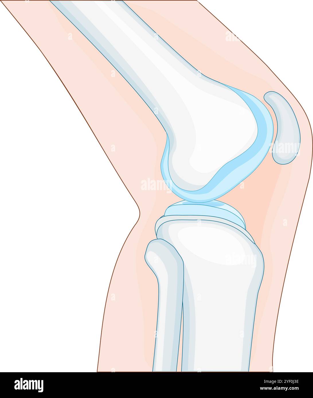 knee anatomy. side view. Cross section of the joint showing the main parts: femur, fibula, articular capsule, menisci, and patella. Vector poster Stock Vectorhttps://www.alamy.com/image-license-details/?v=1https://www.alamy.com/knee-anatomy-side-view-cross-section-of-the-joint-showing-the-main-parts-femur-fibula-articular-capsule-menisci-and-patella-vector-poster-image628807298.html
RF2YF0J3E–knee anatomy. side view. Cross section of the joint showing the main parts: femur, fibula, articular capsule, menisci, and patella. Vector poster
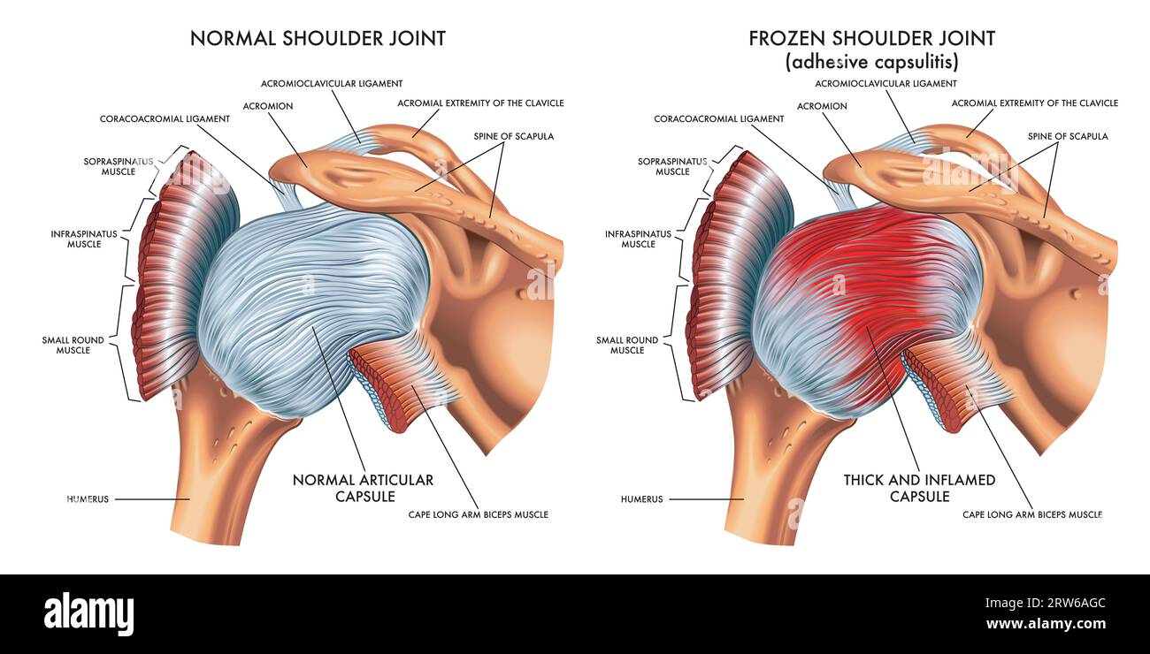 Medical illustration shows the difference between a normal shoulder joint and a frozen shoulder joint, with annotations. Stock Photohttps://www.alamy.com/image-license-details/?v=1https://www.alamy.com/medical-illustration-shows-the-difference-between-a-normal-shoulder-joint-and-a-frozen-shoulder-joint-with-annotations-image566238188.html
RF2RW6AGC–Medical illustration shows the difference between a normal shoulder joint and a frozen shoulder joint, with annotations.
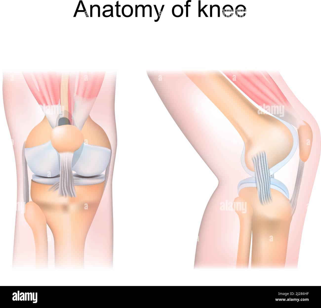 knee anatomy. side and front view. Cross section of the joint showing the main parts: femur, fibula, articular capsule, menisci, muscles and ligaments Stock Vectorhttps://www.alamy.com/image-license-details/?v=1https://www.alamy.com/knee-anatomy-side-and-front-view-cross-section-of-the-joint-showing-the-main-parts-femur-fibula-articular-capsule-menisci-muscles-and-ligaments-image466090059.html
RF2J286HF–knee anatomy. side and front view. Cross section of the joint showing the main parts: femur, fibula, articular capsule, menisci, muscles and ligaments
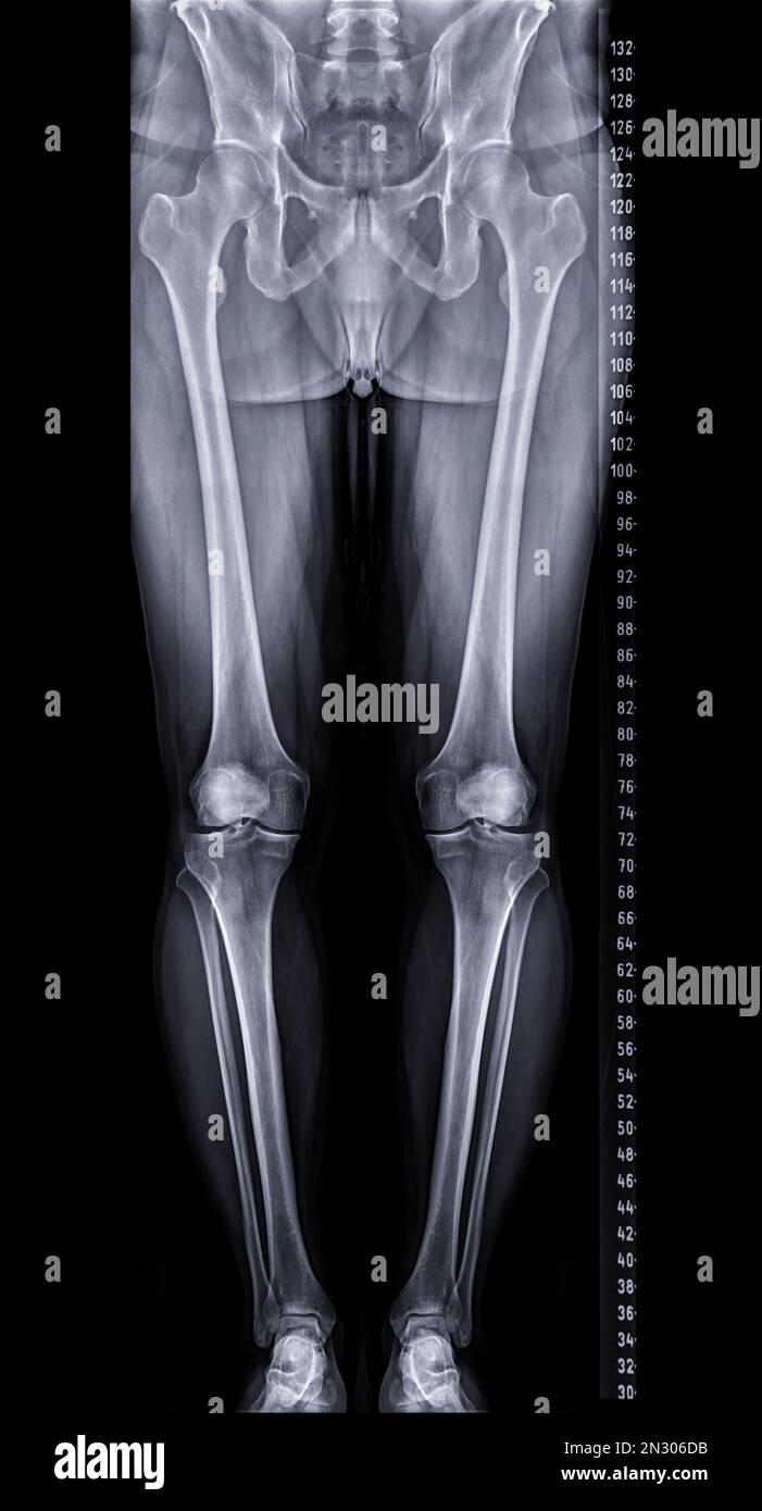 Scanogram is a Full-length standing AP radiograph of both lower extremities including the hip, knee, and ankle. Stock Photohttps://www.alamy.com/image-license-details/?v=1https://www.alamy.com/scanogram-is-a-full-length-standing-ap-radiograph-of-both-lower-extremities-including-the-hip-knee-and-ankle-image518160087.html
RF2N306DB–Scanogram is a Full-length standing AP radiograph of both lower extremities including the hip, knee, and ankle.
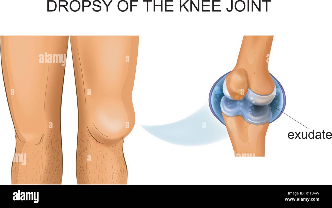 vector illustration of knee hydrarthrosis, articular dropsy Stock Vectorhttps://www.alamy.com/image-license-details/?v=1https://www.alamy.com/vector-illustration-of-knee-hydrarthrosis-articular-dropsy-image224681209.html
RFR1F34W–vector illustration of knee hydrarthrosis, articular dropsy
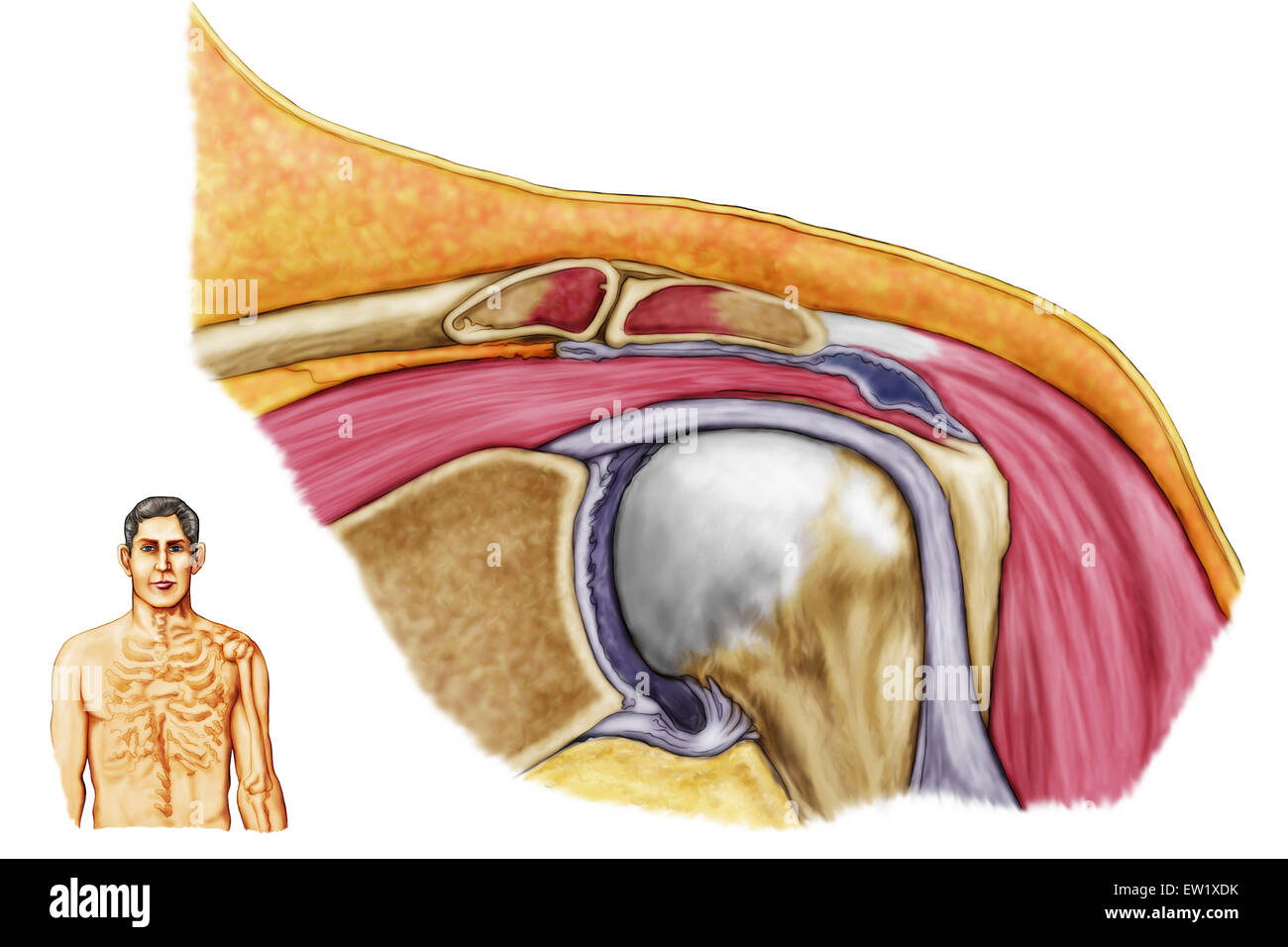 Anatomy of left shoulder, coronal view. Stock Photohttps://www.alamy.com/image-license-details/?v=1https://www.alamy.com/stock-photo-anatomy-of-left-shoulder-coronal-view-84250591.html
RFEW1XDK–Anatomy of left shoulder, coronal view.
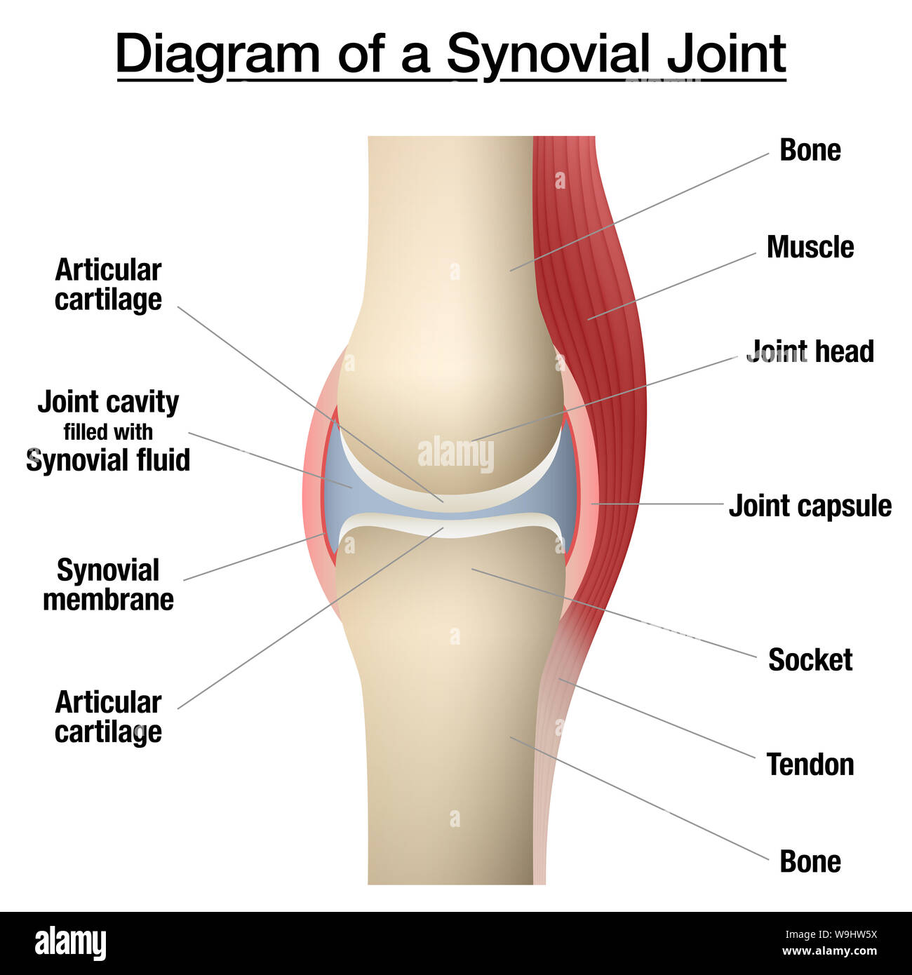 Synovial joint chart. Labeled anatomy infographic with two bones, articular cartilage, joint cavity, synovial fluid, muscle and tendon. Stock Photohttps://www.alamy.com/image-license-details/?v=1https://www.alamy.com/synovial-joint-chart-labeled-anatomy-infographic-with-two-bones-articular-cartilage-joint-cavity-synovial-fluid-muscle-and-tendon-image264080374.html
RFW9HW5X–Synovial joint chart. Labeled anatomy infographic with two bones, articular cartilage, joint cavity, synovial fluid, muscle and tendon.
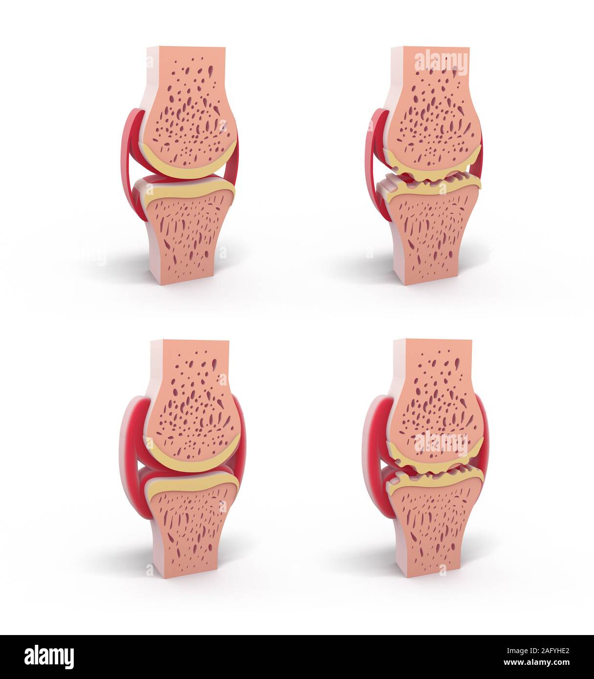 3d illustration of healthy and spherical synovial joint. In four representations standing and isolated on white background. Stock Photohttps://www.alamy.com/image-license-details/?v=1https://www.alamy.com/3d-illustration-of-healthy-and-spherical-synovial-joint-in-four-representations-standing-and-isolated-on-white-background-image336823258.html
RF2AFYHE2–3d illustration of healthy and spherical synovial joint. In four representations standing and isolated on white background.
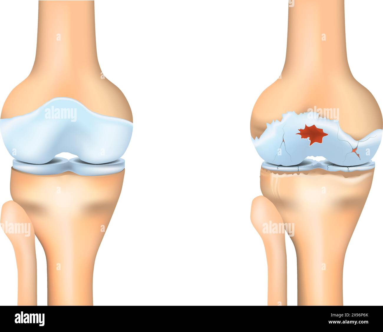 Knee osteoarthritis. Joint degeneration. Vector illustration. Stock Vectorhttps://www.alamy.com/image-license-details/?v=1https://www.alamy.com/knee-osteoarthritis-joint-degeneration-vector-illustration-image608043931.html
RF2X96P6K–Knee osteoarthritis. Joint degeneration. Vector illustration.
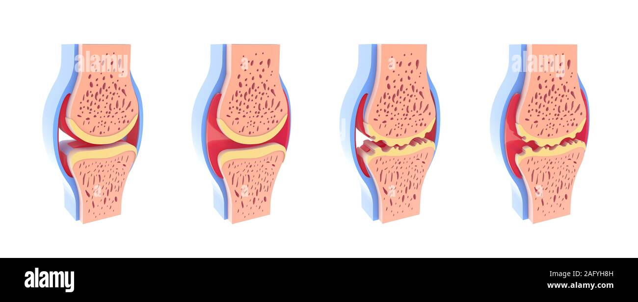 3d illustration of healthy and spherical synovial joint. In four representations standing and isolated on white background. Stock Photohttps://www.alamy.com/image-license-details/?v=1https://www.alamy.com/3d-illustration-of-healthy-and-spherical-synovial-joint-in-four-representations-standing-and-isolated-on-white-background-image336823105.html
RF2AFYH8H–3d illustration of healthy and spherical synovial joint. In four representations standing and isolated on white background.
 Joint knee synovial synovitis pain sinovitis tenosynovitis inflammation cartilage, concept inflamed nodules for seth and team oregon, portland Stock Photohttps://www.alamy.com/image-license-details/?v=1https://www.alamy.com/joint-knee-synovial-synovitis-pain-sinovitis-tenosynovitis-inflammation-cartilage-concept-inflamed-nodules-for-seth-and-team-oregon-portland-image479667437.html
RF2JTAMKW–Joint knee synovial synovitis pain sinovitis tenosynovitis inflammation cartilage, concept inflamed nodules for seth and team oregon, portland
 3d illustration of synovial joint. Next to the graphic representation of a hand placing the joint, front view on white background. Stock Photohttps://www.alamy.com/image-license-details/?v=1https://www.alamy.com/3d-illustration-of-synovial-joint-next-to-the-graphic-representation-of-a-hand-placing-the-joint-front-view-on-white-background-image336823446.html
RF2AFYHMP–3d illustration of synovial joint. Next to the graphic representation of a hand placing the joint, front view on white background.
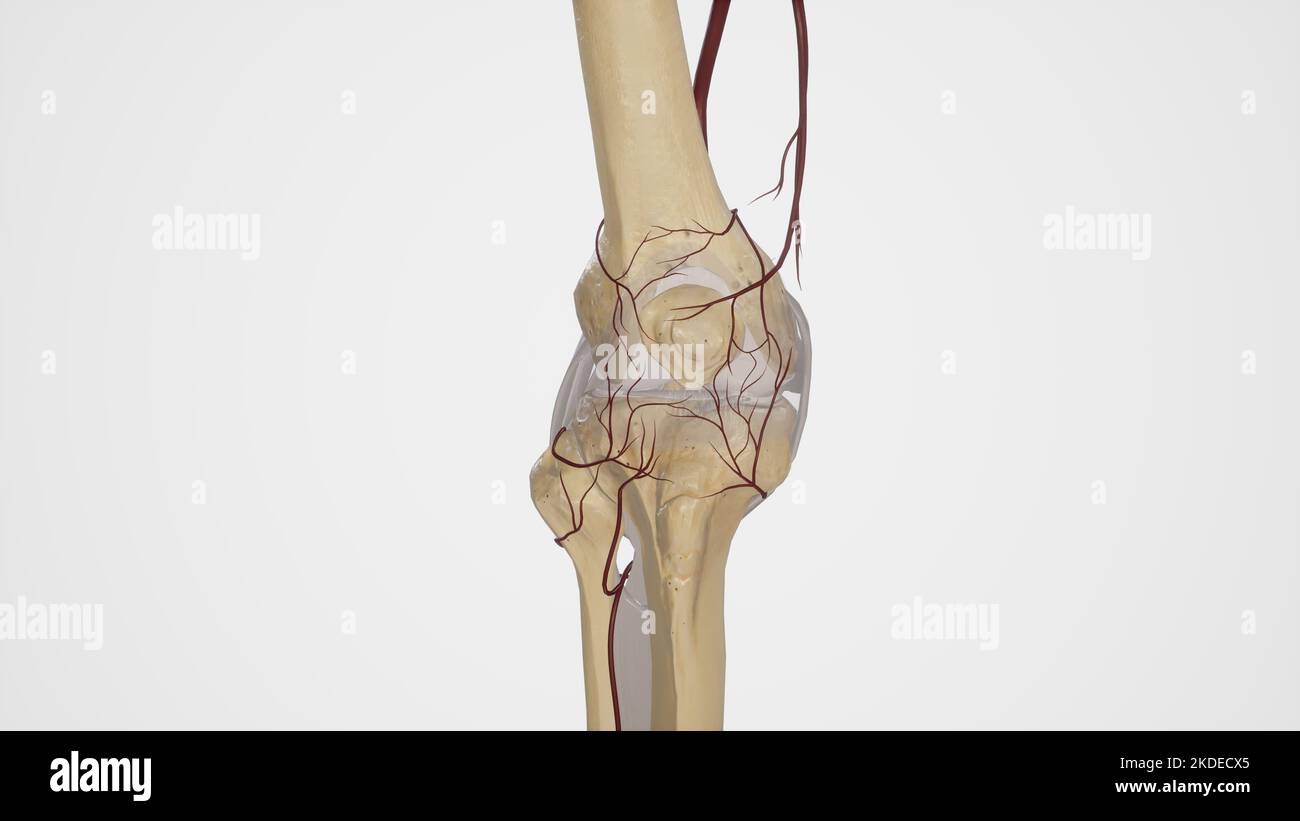 Arterial Anastomosis of Knee Joint Stock Photohttps://www.alamy.com/image-license-details/?v=1https://www.alamy.com/arterial-anastomosis-of-knee-joint-image490198301.html
RF2KDECX5–Arterial Anastomosis of Knee Joint
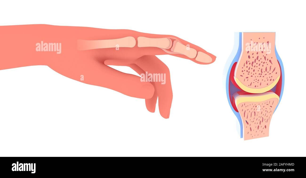 3d illustration of synovial joint. Next to the graphic representation of a hand placing the joint. Stock Photohttps://www.alamy.com/image-license-details/?v=1https://www.alamy.com/3d-illustration-of-synovial-joint-next-to-the-graphic-representation-of-a-hand-placing-the-joint-image336823437.html
RF2AFYHMD–3d illustration of synovial joint. Next to the graphic representation of a hand placing the joint.
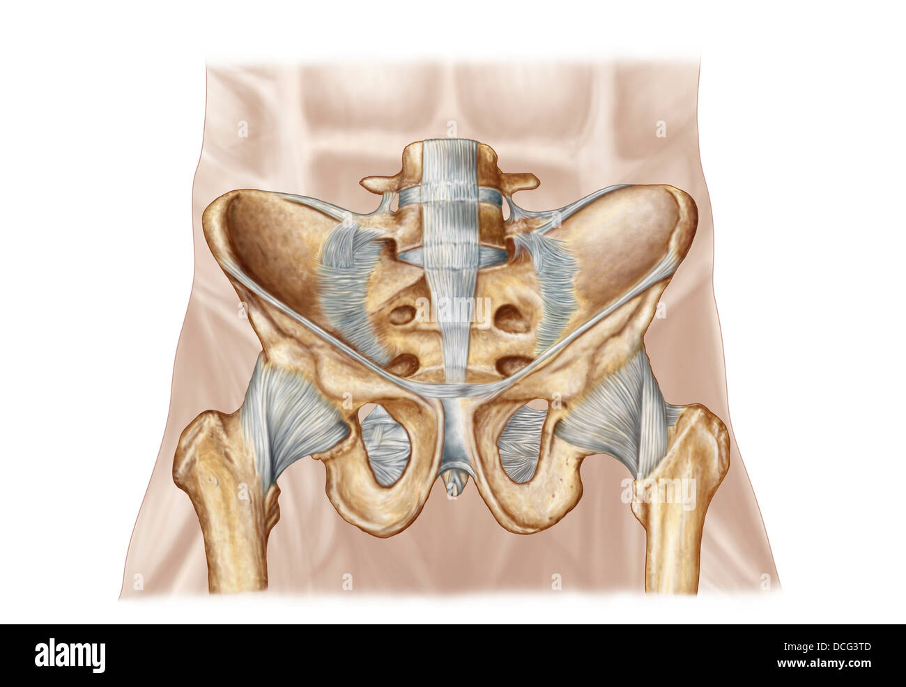 Anatomy of human pelvic bone and ligaments. Stock Photohttps://www.alamy.com/image-license-details/?v=1https://www.alamy.com/stock-photo-anatomy-of-human-pelvic-bone-and-ligaments-59361245.html
RFDCG3TD–Anatomy of human pelvic bone and ligaments.
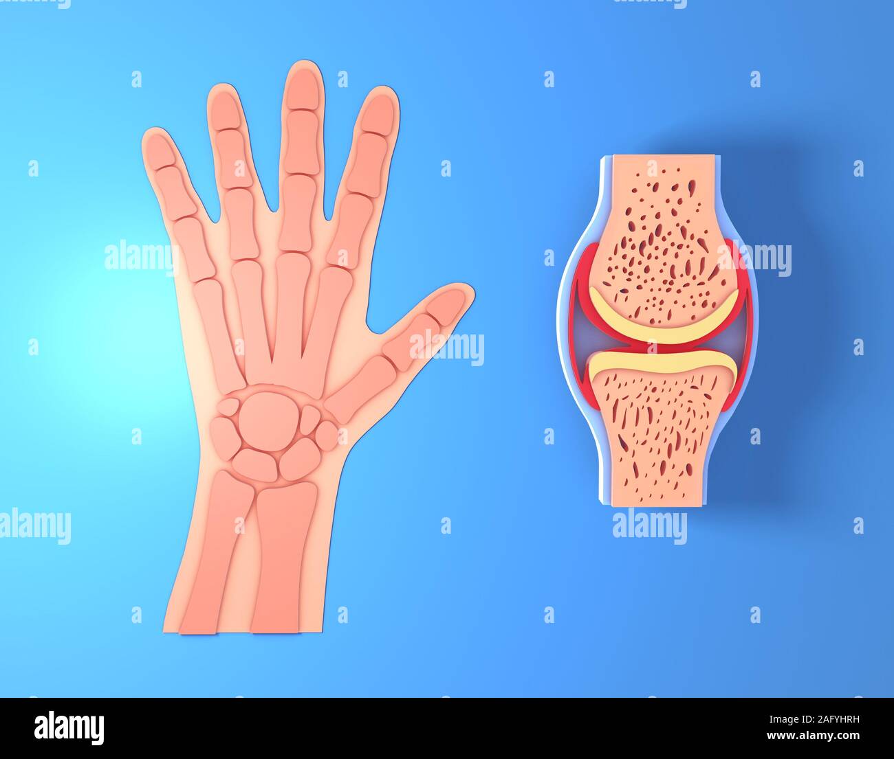 3d illustration of synovial joint. Next to the graphic representation of a hand placing the joint, front view on blue background. Stock Photohttps://www.alamy.com/image-license-details/?v=1https://www.alamy.com/3d-illustration-of-synovial-joint-next-to-the-graphic-representation-of-a-hand-placing-the-joint-front-view-on-blue-background-image336823525.html
RF2AFYHRH–3d illustration of synovial joint. Next to the graphic representation of a hand placing the joint, front view on blue background.
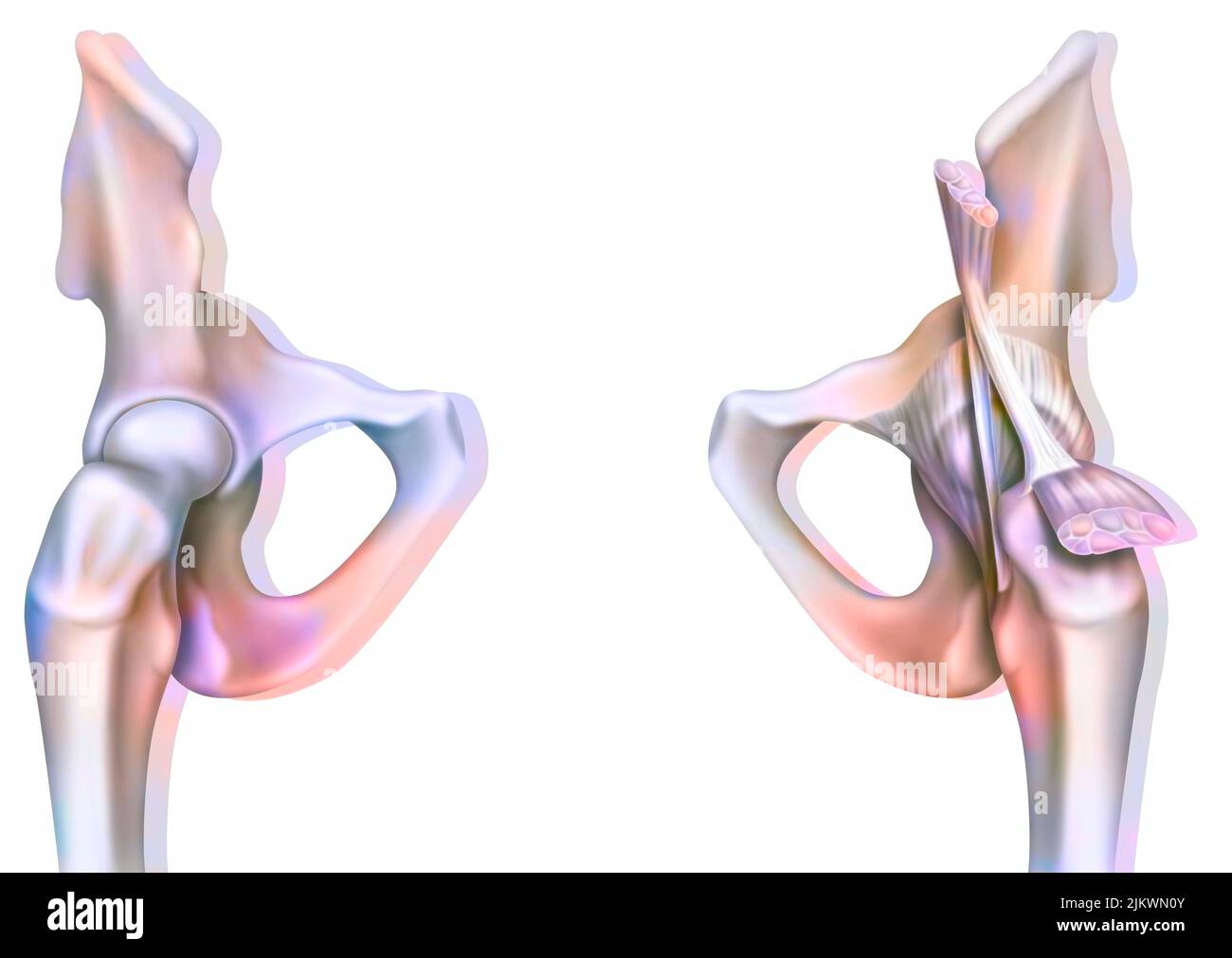 Bone joint of the hip without and with the coxofemoral joint capsule. Stock Photohttps://www.alamy.com/image-license-details/?v=1https://www.alamy.com/bone-joint-of-the-hip-without-and-with-the-coxofemoral-joint-capsule-image476923691.html
RF2JKWN0Y–Bone joint of the hip without and with the coxofemoral joint capsule.
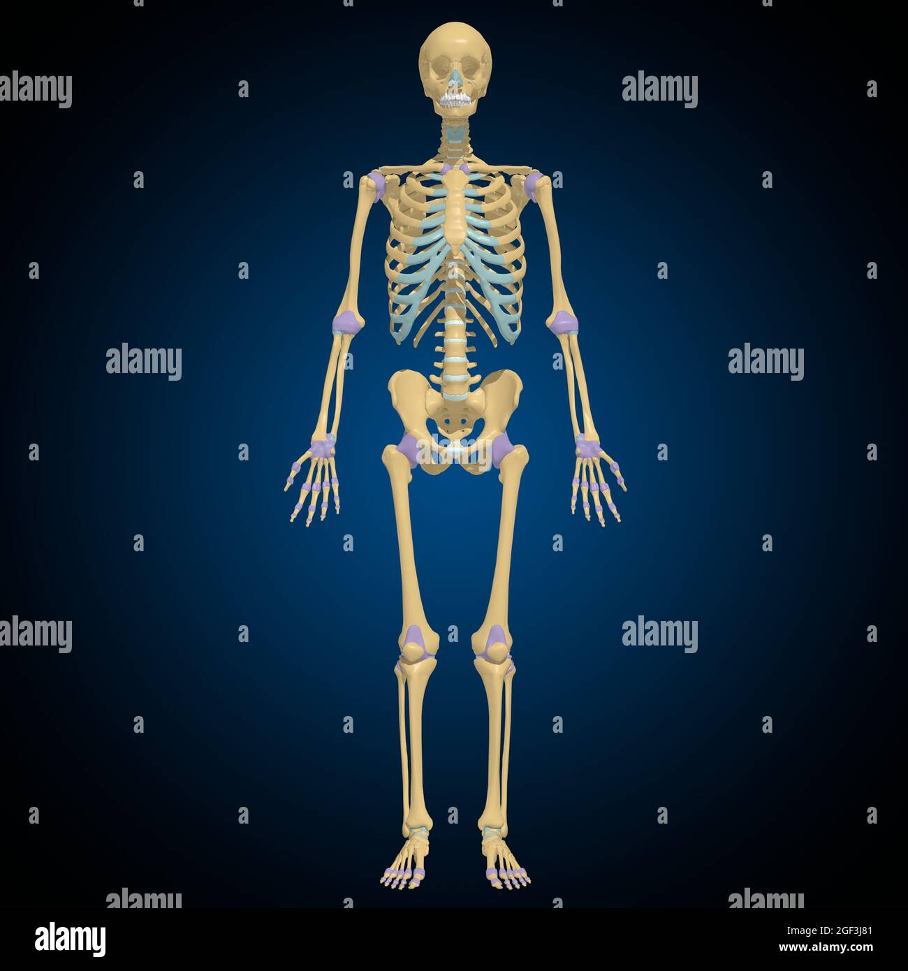 Articular capsule Anatomy For Medical Concept 3D Illustration Stock Photohttps://www.alamy.com/image-license-details/?v=1https://www.alamy.com/articular-capsule-anatomy-for-medical-concept-3d-illustration-image439559233.html
RF2GF3J81–Articular capsule Anatomy For Medical Concept 3D Illustration
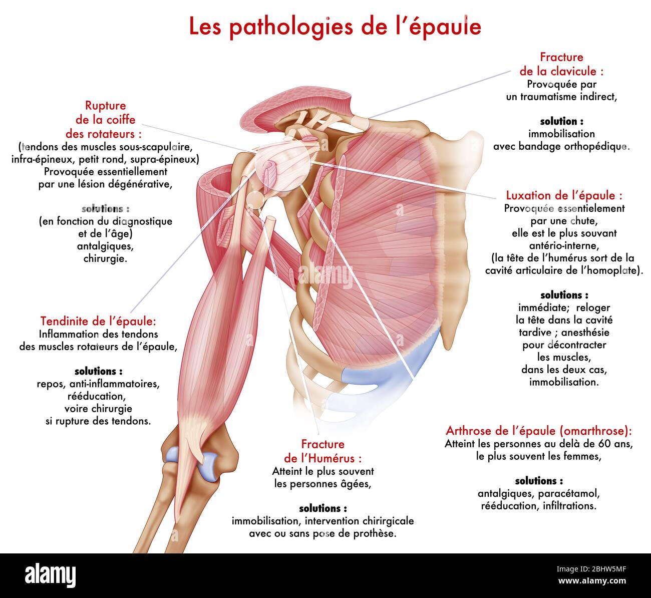 Medical illustration representing pathologies of the shoulder. The articular capsule, the ligaments, the small pectoralis and coraco-brachial tendons Stock Photohttps://www.alamy.com/image-license-details/?v=1https://www.alamy.com/medical-illustration-representing-pathologies-of-the-shoulder-the-articular-capsule-the-ligaments-the-small-pectoralis-and-coraco-brachial-tendons-image355209807.html
RM2BHW5MF–Medical illustration representing pathologies of the shoulder. The articular capsule, the ligaments, the small pectoralis and coraco-brachial tendons
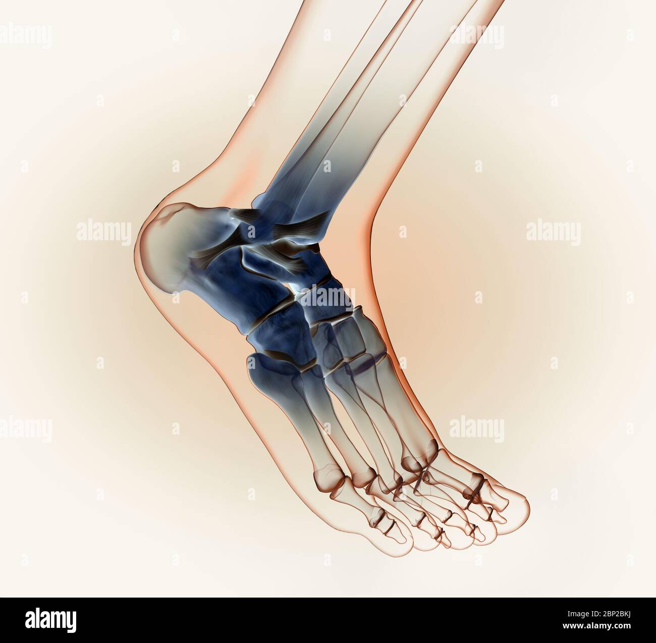 3D illustration showing of a ankle joint with bones, ligaments and articular capsule Stock Photohttps://www.alamy.com/image-license-details/?v=1https://www.alamy.com/3d-illustration-showing-of-a-ankle-joint-with-bones-ligaments-and-articular-capsule-image357782870.html
RF2BP2BKJ–3D illustration showing of a ankle joint with bones, ligaments and articular capsule
 Anatomy of the joint of a healthy knee on the left, and one deformed by osteoarthritis on the right. Stock Photohttps://www.alamy.com/image-license-details/?v=1https://www.alamy.com/anatomy-of-the-joint-of-a-healthy-knee-on-the-left-and-one-deformed-by-osteoarthritis-on-the-right-image476924956.html
RF2JKWPJ4–Anatomy of the joint of a healthy knee on the left, and one deformed by osteoarthritis on the right.
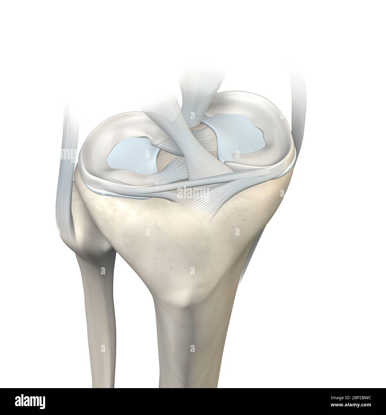 3D illustration showing knee joint with transparent femur and articular capsule, menisci and ligaments Stock Photohttps://www.alamy.com/image-license-details/?v=1https://www.alamy.com/3d-illustration-showing-knee-joint-with-transparent-femur-and-articular-capsule-menisci-and-ligaments-image357783032.html
RF2BP2BWC–3D illustration showing knee joint with transparent femur and articular capsule, menisci and ligaments
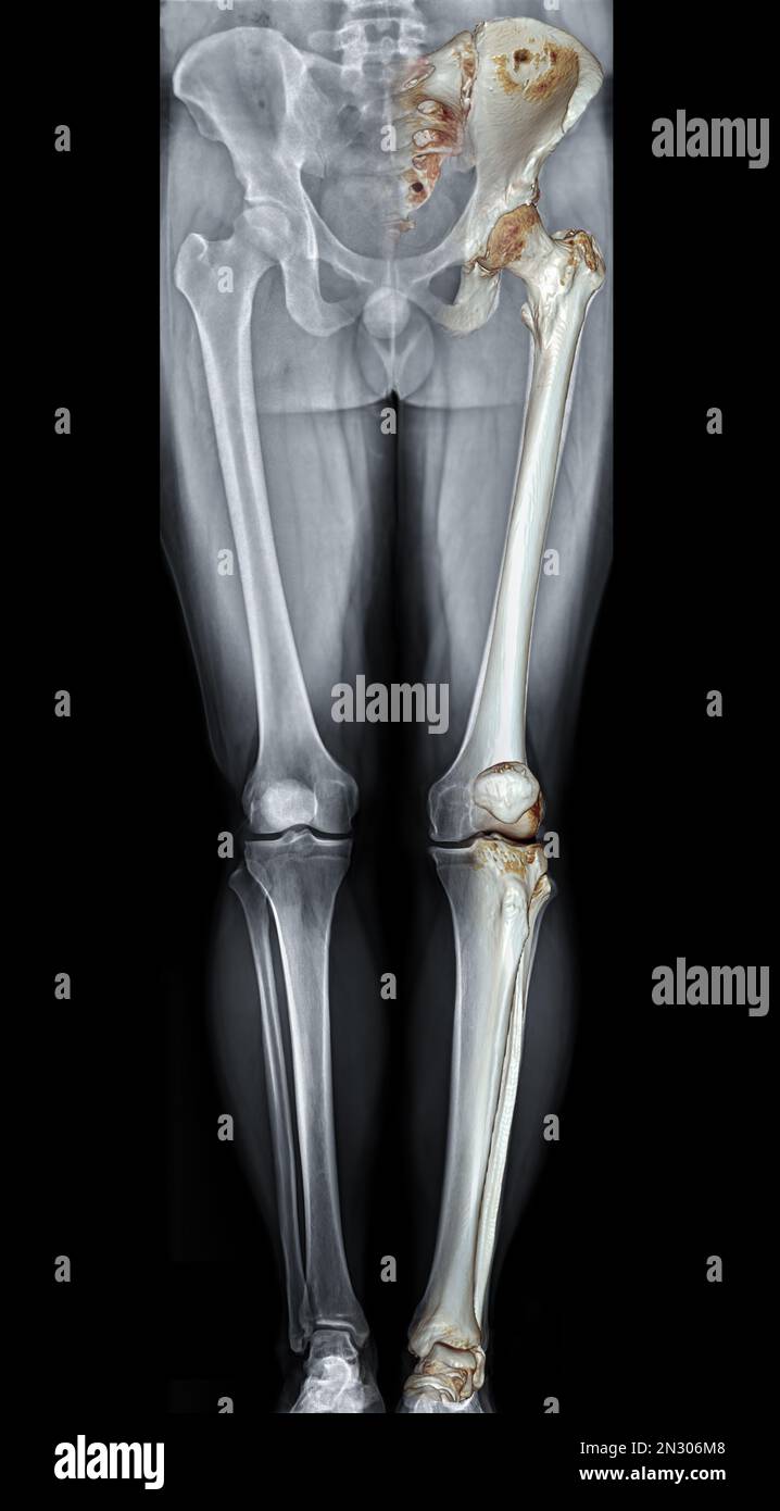 Scanogram is a Full-length standing AP radiograph of both lower extremities including the hip, knee, and ankle with 3D rendering. Stock Photohttps://www.alamy.com/image-license-details/?v=1https://www.alamy.com/scanogram-is-a-full-length-standing-ap-radiograph-of-both-lower-extremities-including-the-hip-knee-and-ankle-with-3d-rendering-image518160280.html
RF2N306M8–Scanogram is a Full-length standing AP radiograph of both lower extremities including the hip, knee, and ankle with 3D rendering.
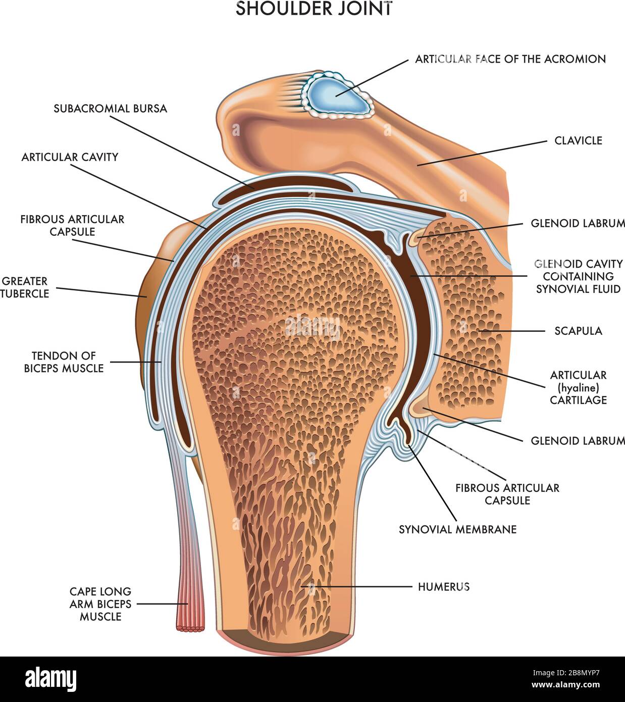 Shoulder joint illustrated and annotated with components on white. Stock Vectorhttps://www.alamy.com/image-license-details/?v=1https://www.alamy.com/shoulder-joint-illustrated-and-annotated-with-components-on-white-image349585439.html
RF2B8MYP7–Shoulder joint illustrated and annotated with components on white.
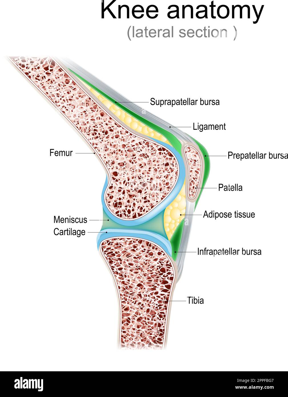 Knee anatomy. Side view. lateral section of joint with ligaments, meniscus, and bursae. knee joint cavity. Cross section of leg bones. detailed vector Stock Vectorhttps://www.alamy.com/image-license-details/?v=1https://www.alamy.com/knee-anatomy-side-view-lateral-section-of-joint-with-ligaments-meniscus-and-bursae-knee-joint-cavity-cross-section-of-leg-bones-detailed-vector-image547382199.html
RF2PPFBG7–Knee anatomy. Side view. lateral section of joint with ligaments, meniscus, and bursae. knee joint cavity. Cross section of leg bones. detailed vector
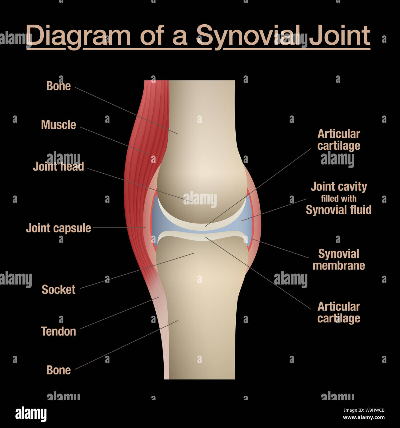 Synovial joint diagram. Labeled anatomy chart with two bones, articular cartilage, joint cavity, synovial fluid, muscle and tendon. Black background. Stock Photohttps://www.alamy.com/image-license-details/?v=1https://www.alamy.com/synovial-joint-diagram-labeled-anatomy-chart-with-two-bones-articular-cartilage-joint-cavity-synovial-fluid-muscle-and-tendon-black-background-image264080555.html
RFW9HWCB–Synovial joint diagram. Labeled anatomy chart with two bones, articular cartilage, joint cavity, synovial fluid, muscle and tendon. Black background.
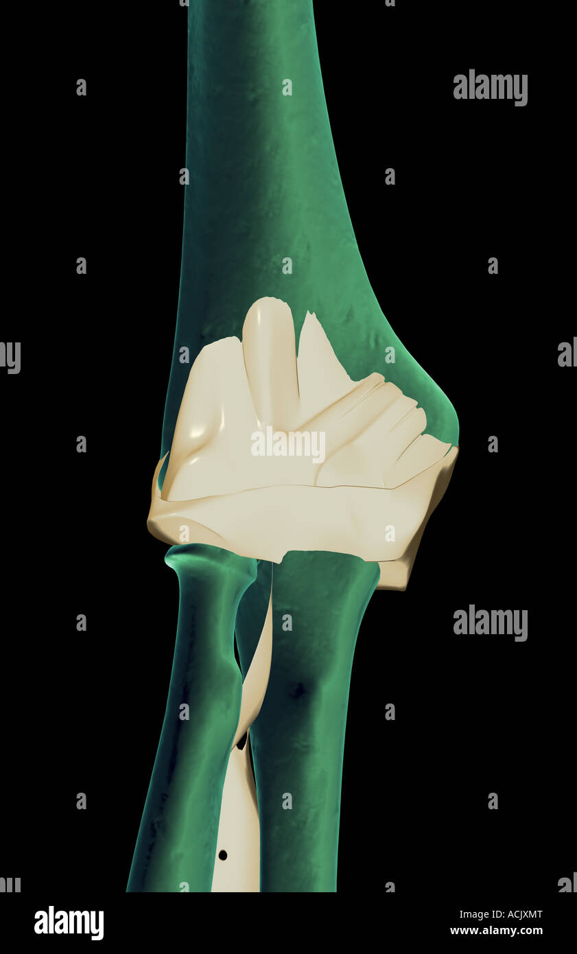 The ligaments of the elbow Stock Photohttps://www.alamy.com/image-license-details/?v=1https://www.alamy.com/stock-photo-the-ligaments-of-the-elbow-13173911.html
RFACJXMT–The ligaments of the elbow
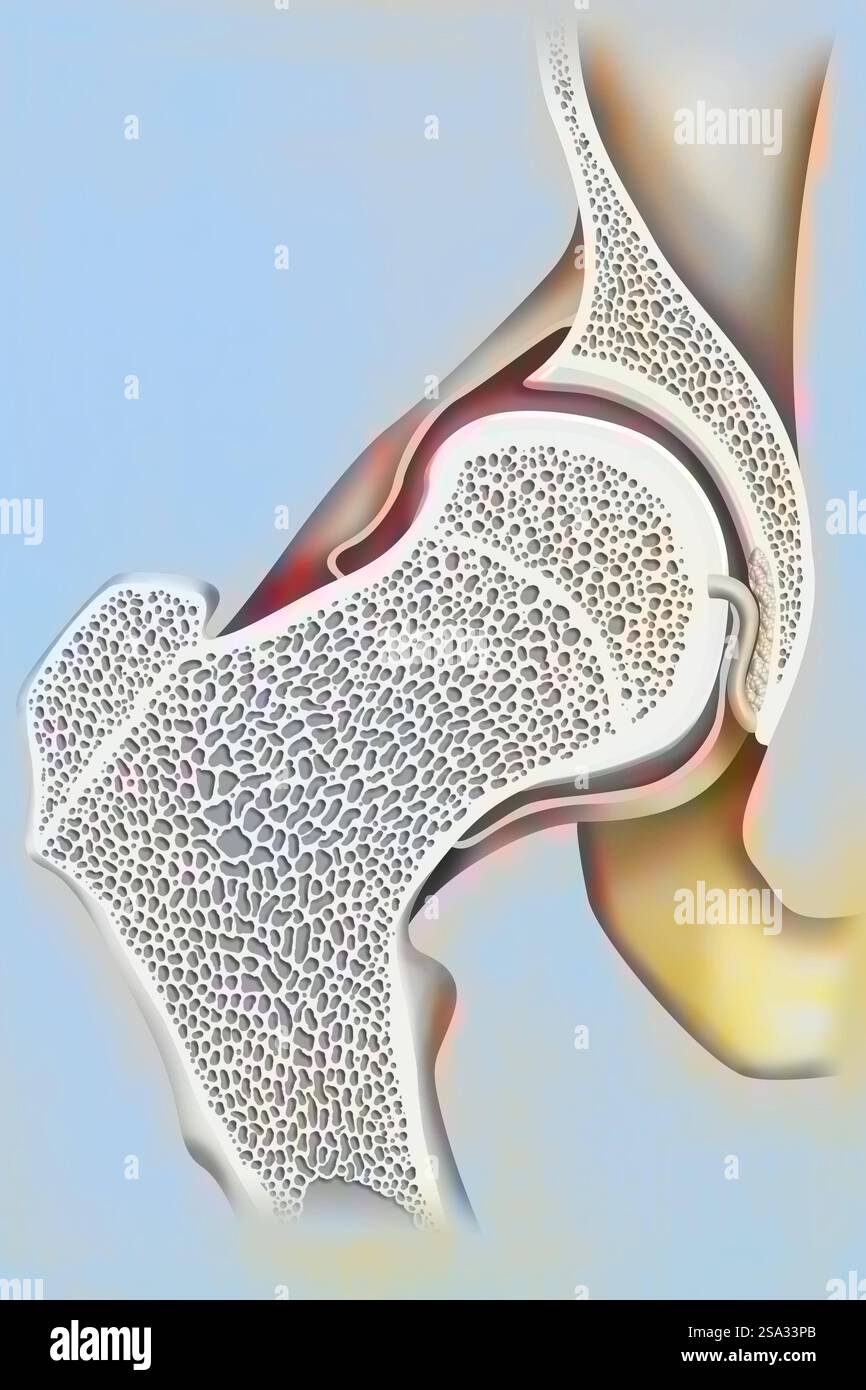 Coxofemoral joint and femoral neck. Hip drawing 016238 052 Stock Photohttps://www.alamy.com/image-license-details/?v=1https://www.alamy.com/coxofemoral-joint-and-femoral-neck-hip-drawing-016238-052-image642999011.html
RM2SA33PB–Coxofemoral joint and femoral neck. Hip drawing 016238 052
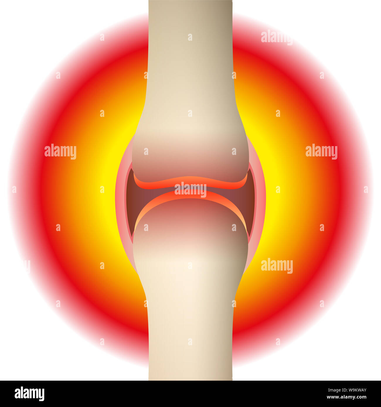 Joint pain - schematic anatomical graphic of a synovial joint with arthritis, rheumatism, gout, osteoarthritis or inflammation. Stock Photohttps://www.alamy.com/image-license-details/?v=1https://www.alamy.com/joint-pain-schematic-anatomical-graphic-of-a-synovial-joint-with-arthritis-rheumatism-gout-osteoarthritis-or-inflammation-image264124419.html
RFW9KWAY–Joint pain - schematic anatomical graphic of a synovial joint with arthritis, rheumatism, gout, osteoarthritis or inflammation.
 Joint synovitis synovial pain knee sinovitis tenosynovitis inflammation fluid, from swelling lupus for soccer and stadium athlete, portland Stock Photohttps://www.alamy.com/image-license-details/?v=1https://www.alamy.com/joint-synovitis-synovial-pain-knee-sinovitis-tenosynovitis-inflammation-fluid-from-swelling-lupus-for-soccer-and-stadium-athlete-portland-image479801989.html
RF2JTGT99–Joint synovitis synovial pain knee sinovitis tenosynovitis inflammation fluid, from swelling lupus for soccer and stadium athlete, portland
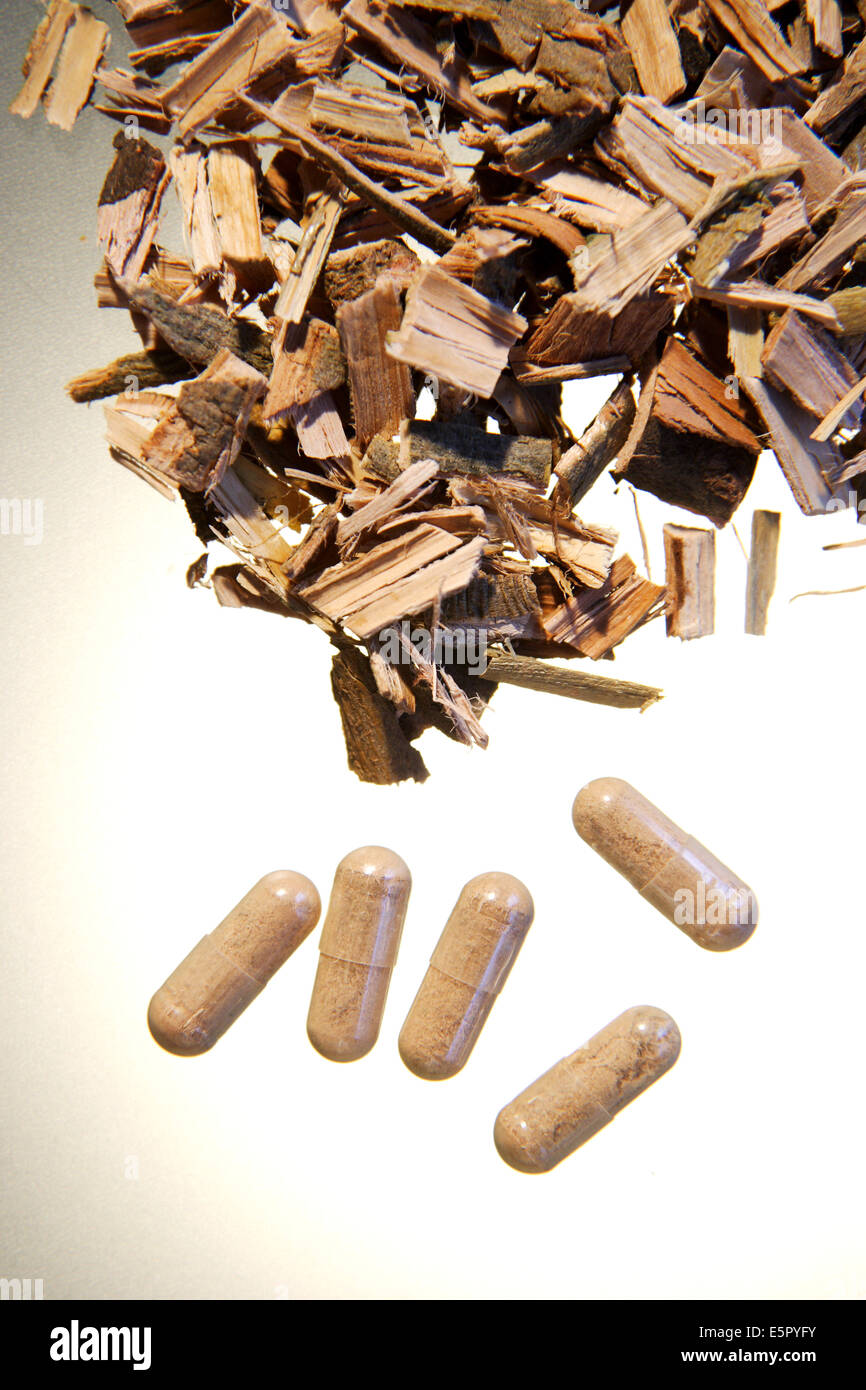 Dryed bark of white willow, used in phytotherapy for its anti-inflammatory properties. Stock Photohttps://www.alamy.com/image-license-details/?v=1https://www.alamy.com/stock-photo-dryed-bark-of-white-willow-used-in-phytotherapy-for-its-anti-inflammatory-72419311.html
RME5PYFY–Dryed bark of white willow, used in phytotherapy for its anti-inflammatory properties.
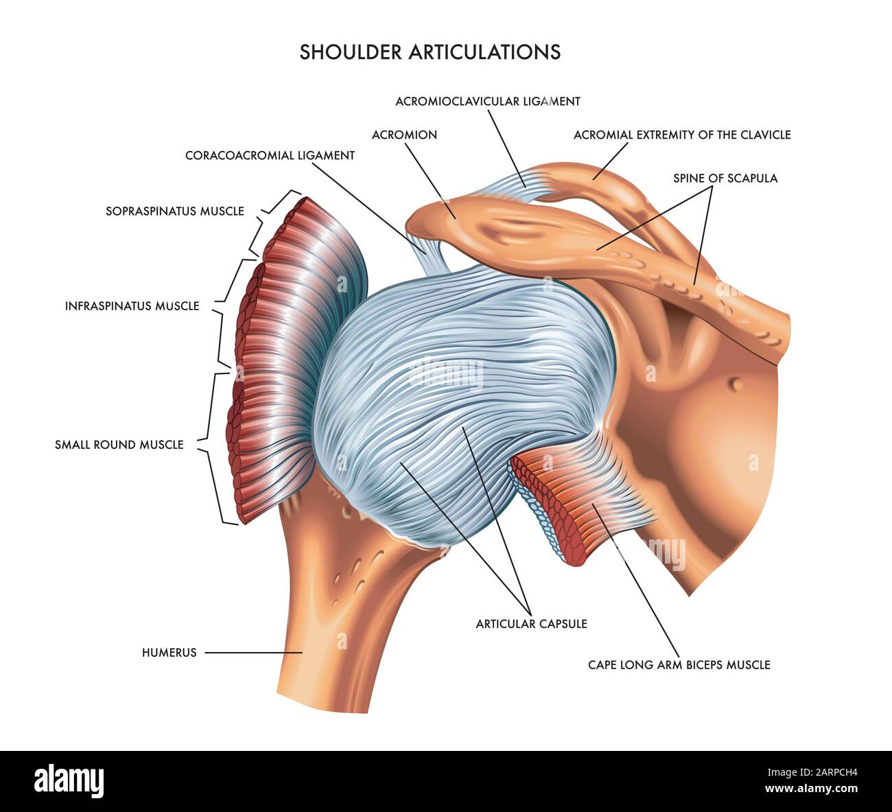 A detailed medical illustration of shoulder articulations. Stock Photohttps://www.alamy.com/image-license-details/?v=1https://www.alamy.com/a-detailed-medical-illustration-of-shoulder-articulations-image341626912.html
RF2ARPCH4–A detailed medical illustration of shoulder articulations.
 Knee and meniscus anatomy medical vector illustration isolated on white background eps 10 Stock Vectorhttps://www.alamy.com/image-license-details/?v=1https://www.alamy.com/knee-and-meniscus-anatomy-medical-vector-illustration-isolated-on-white-background-eps-10-image341381579.html
RF2ARB7K7–Knee and meniscus anatomy medical vector illustration isolated on white background eps 10
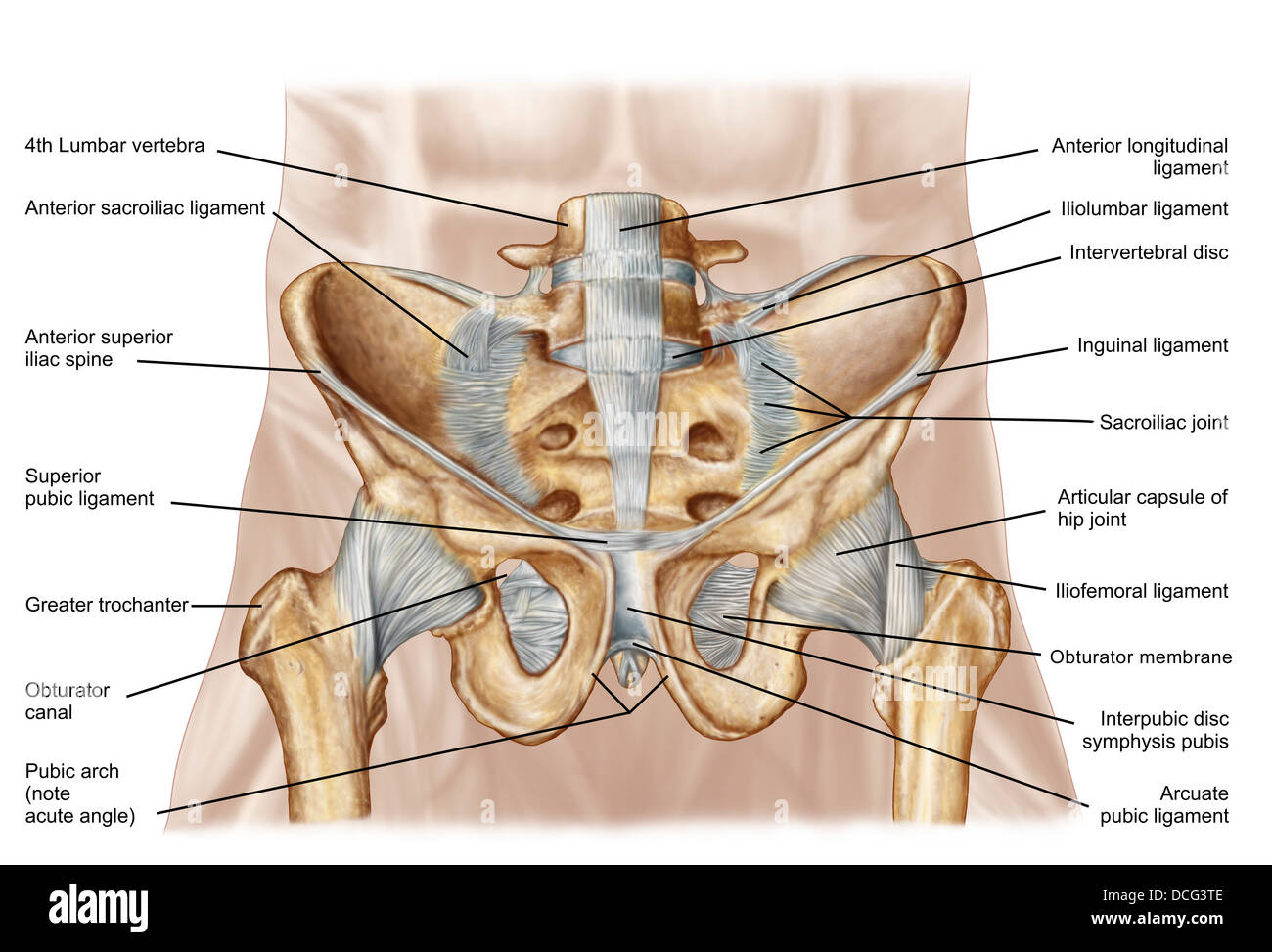 Anatomy of human pelvic bone and ligaments. Stock Photohttps://www.alamy.com/image-license-details/?v=1https://www.alamy.com/stock-photo-anatomy-of-human-pelvic-bone-and-ligaments-59361246.html
RFDCG3TE–Anatomy of human pelvic bone and ligaments.
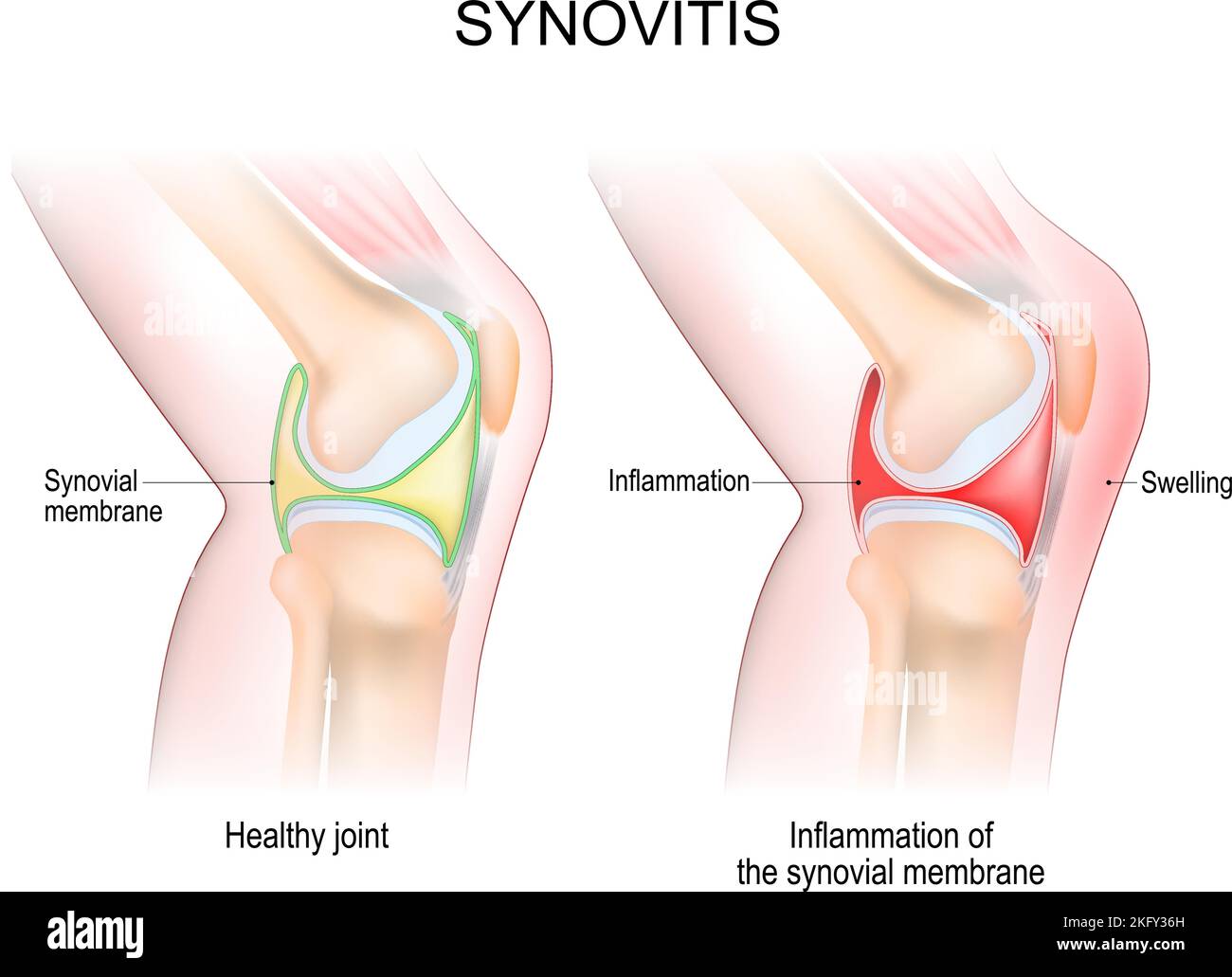 synovitis of a Knee. Close-up of normal joint, and knee with inflammation of the synovial membrane. Signs and symptoms of the disease. side view Stock Vectorhttps://www.alamy.com/image-license-details/?v=1https://www.alamy.com/synovitis-of-a-knee-close-up-of-normal-joint-and-knee-with-inflammation-of-the-synovial-membrane-signs-and-symptoms-of-the-disease-side-view-image491705385.html
RF2KFY36H–synovitis of a Knee. Close-up of normal joint, and knee with inflammation of the synovial membrane. Signs and symptoms of the disease. side view
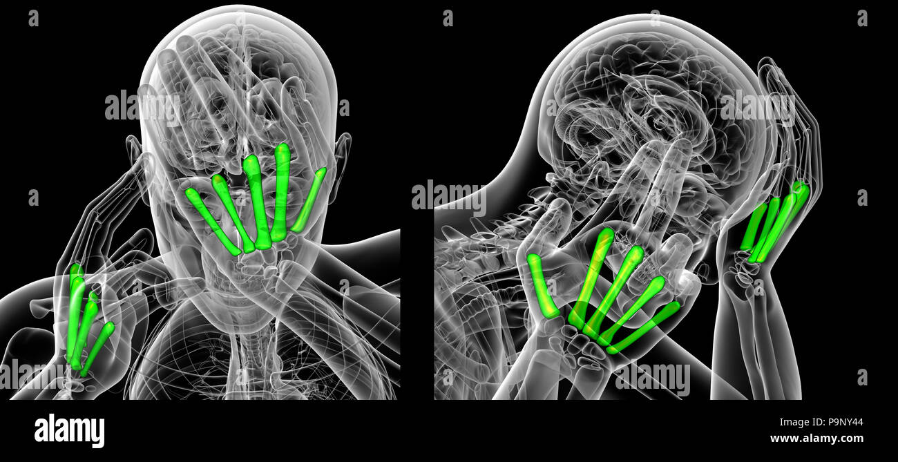 3d rendering of metacarpal Stock Photohttps://www.alamy.com/image-license-details/?v=1https://www.alamy.com/3d-rendering-of-metacarpal-image212538596.html
RFP9NY44–3d rendering of metacarpal
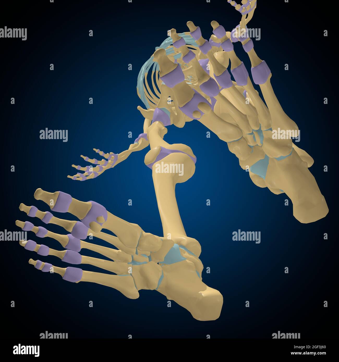 Articular capsule Anatomy For Medical Concept 3D Illustration Stock Photohttps://www.alamy.com/image-license-details/?v=1https://www.alamy.com/articular-capsule-anatomy-for-medical-concept-3d-illustration-image439559176.html
RF2GF3J60–Articular capsule Anatomy For Medical Concept 3D Illustration
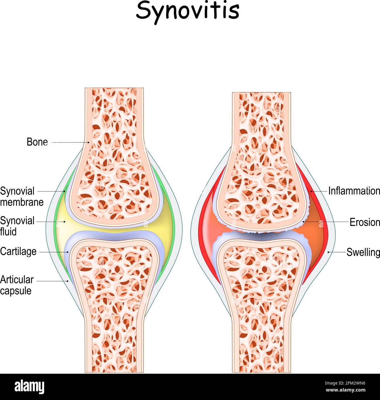 synovitis. Close-up. comparison and difference between a healthy joint and a joint with inflammation of the synovial membrane. Signs and symptoms Stock Vectorhttps://www.alamy.com/image-license-details/?v=1https://www.alamy.com/synovitis-close-up-comparison-and-difference-between-a-healthy-joint-and-a-joint-with-inflammation-of-the-synovial-membrane-signs-and-symptoms-image425406050.html
RF2FM2WN6–synovitis. Close-up. comparison and difference between a healthy joint and a joint with inflammation of the synovial membrane. Signs and symptoms
 Radius and ulna (bones of the forearm) seen by their front face, vintage engraved illustration. Usual Medicine Dictionary by Dr Stock Vectorhttps://www.alamy.com/image-license-details/?v=1https://www.alamy.com/stock-photo-radius-and-ulna-bones-of-the-forearm-seen-by-their-front-face-vintage-84420011.html
RFEW9JGB–Radius and ulna (bones of the forearm) seen by their front face, vintage engraved illustration. Usual Medicine Dictionary by Dr
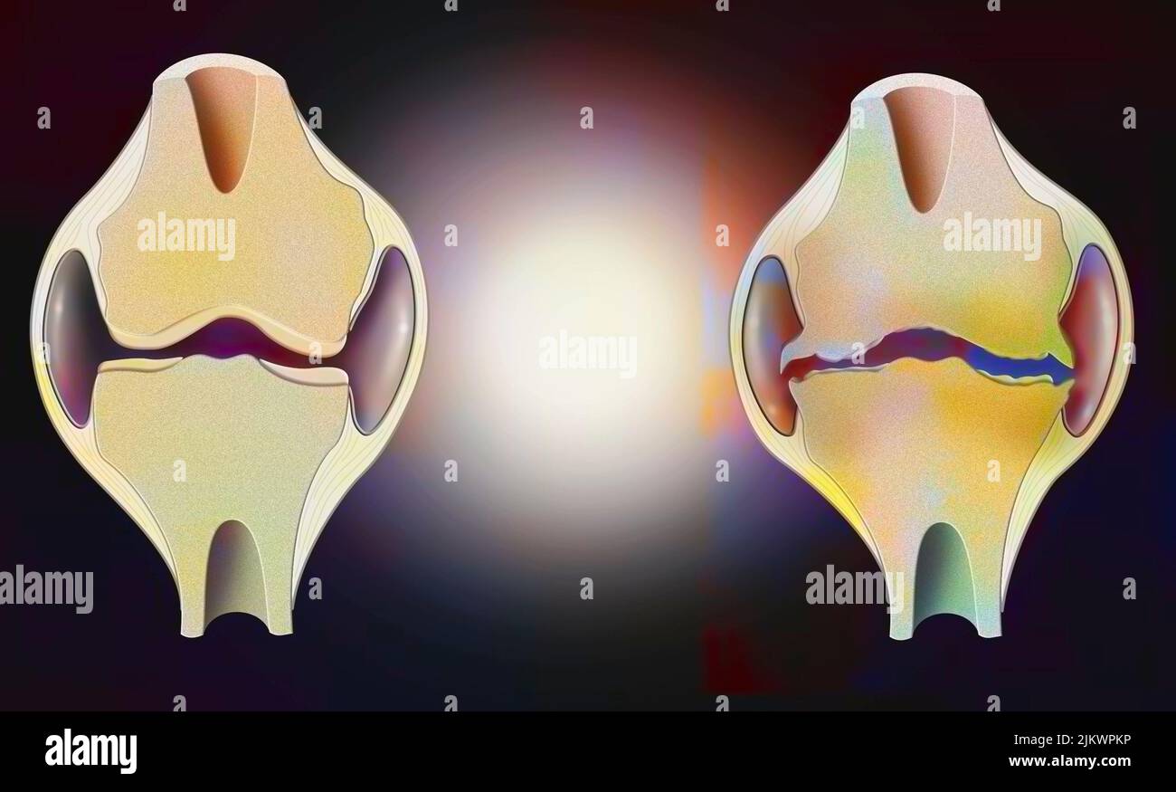 Anatomy of the joint of a healthy knee on the left, and one deformed by osteoarthritis on the right. Stock Photohttps://www.alamy.com/image-license-details/?v=1https://www.alamy.com/anatomy-of-the-joint-of-a-healthy-knee-on-the-left-and-one-deformed-by-osteoarthritis-on-the-right-image476925002.html
RF2JKWPKP–Anatomy of the joint of a healthy knee on the left, and one deformed by osteoarthritis on the right.
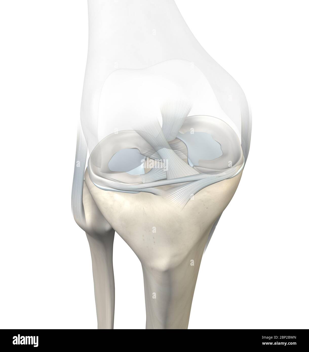 3D illustration showing knee joint with transparent femur and articular capsule, menisci and ligaments Stock Photohttps://www.alamy.com/image-license-details/?v=1https://www.alamy.com/3d-illustration-showing-knee-joint-with-transparent-femur-and-articular-capsule-menisci-and-ligaments-image357783041.html
RF2BP2BWN–3D illustration showing knee joint with transparent femur and articular capsule, menisci and ligaments
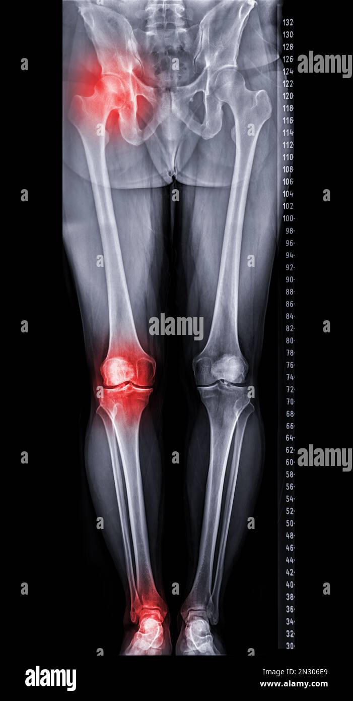 Scanogram is a Full-length standing AP radiograph of both lower extremities including the hip, knee, and ankle. Stock Photohttps://www.alamy.com/image-license-details/?v=1https://www.alamy.com/scanogram-is-a-full-length-standing-ap-radiograph-of-both-lower-extremities-including-the-hip-knee-and-ankle-image518160113.html
RF2N306E9–Scanogram is a Full-length standing AP radiograph of both lower extremities including the hip, knee, and ankle.
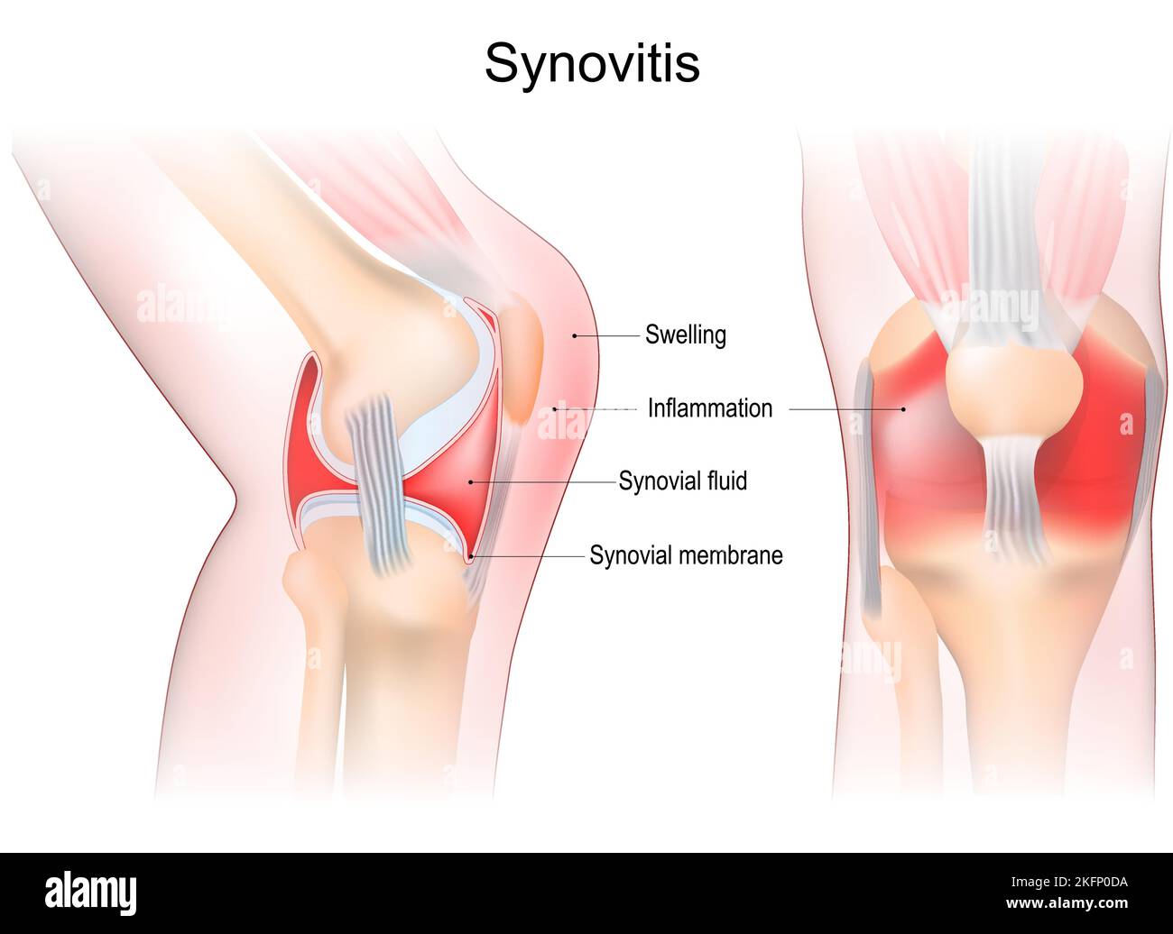 synovitis of a Knee. Close-up of joint with inflammation of the synovial membrane. Signs and symptoms of the disease. Synovial joint anatomy. Stock Vectorhttps://www.alamy.com/image-license-details/?v=1https://www.alamy.com/synovitis-of-a-knee-close-up-of-joint-with-inflammation-of-the-synovial-membrane-signs-and-symptoms-of-the-disease-synovial-joint-anatomy-image491593462.html
RF2KFP0DA–synovitis of a Knee. Close-up of joint with inflammation of the synovial membrane. Signs and symptoms of the disease. Synovial joint anatomy.
 vector illustration of a puncture synovitis of the knee joint Stock Vectorhttps://www.alamy.com/image-license-details/?v=1https://www.alamy.com/vector-illustration-of-a-puncture-synovitis-of-the-knee-joint-image229174799.html
RFR8RPP7–vector illustration of a puncture synovitis of the knee joint
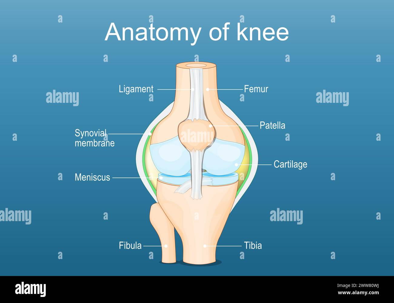 Knee joint anatomy. Labeled of all bones. Isometric Flat vector illustration Stock Vectorhttps://www.alamy.com/image-license-details/?v=1https://www.alamy.com/knee-joint-anatomy-labeled-of-all-bones-isometric-flat-vector-illustration-image600695246.html
RF2WW80WJ–Knee joint anatomy. Labeled of all bones. Isometric Flat vector illustration
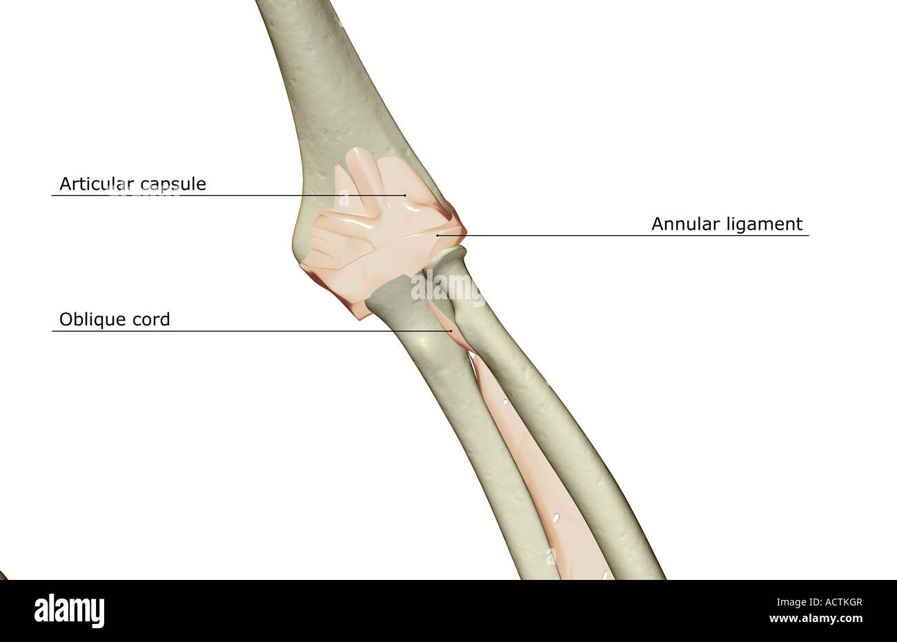 Ligaments of the elbow joint Stock Photohttps://www.alamy.com/image-license-details/?v=1https://www.alamy.com/stock-photo-ligaments-of-the-elbow-joint-13227958.html
RFACTKGR–Ligaments of the elbow joint
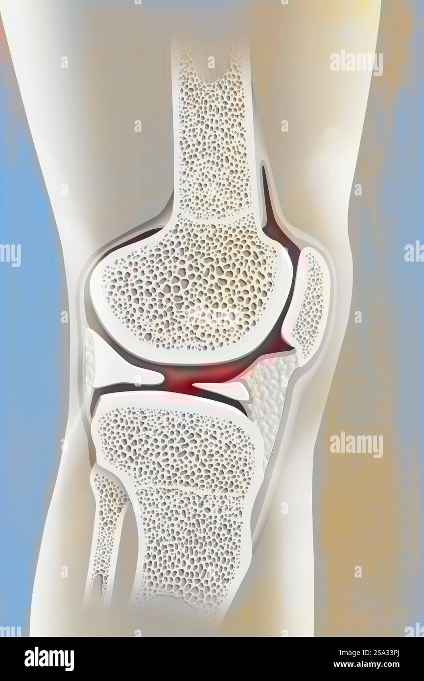 Anatomy of the knee profile. Knee drawing 016238 056 Stock Photohttps://www.alamy.com/image-license-details/?v=1https://www.alamy.com/anatomy-of-the-knee-profile-knee-drawing-016238-056-image642999018.html
RM2SA33PJ–Anatomy of the knee profile. Knee drawing 016238 056
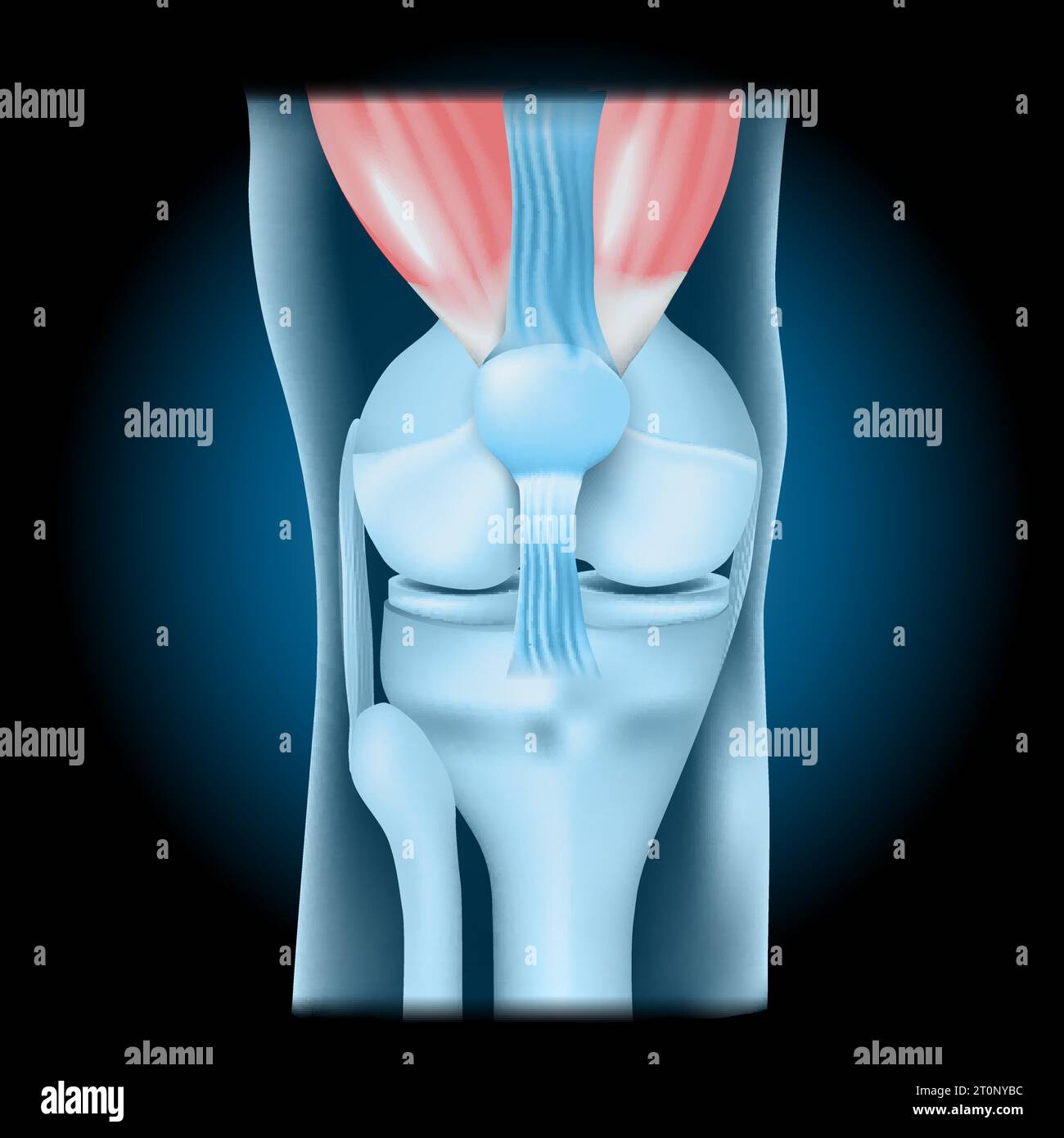 Knee joint with Quadriceps. Front view of human knee with glowing effect. Realistic transparent blue joint on dark background. vector illustration lik Stock Vectorhttps://www.alamy.com/image-license-details/?v=1https://www.alamy.com/knee-joint-with-quadriceps-front-view-of-human-knee-with-glowing-effect-realistic-transparent-blue-joint-on-dark-background-vector-illustration-lik-image568424624.html
RF2T0NYBC–Knee joint with Quadriceps. Front view of human knee with glowing effect. Realistic transparent blue joint on dark background. vector illustration lik
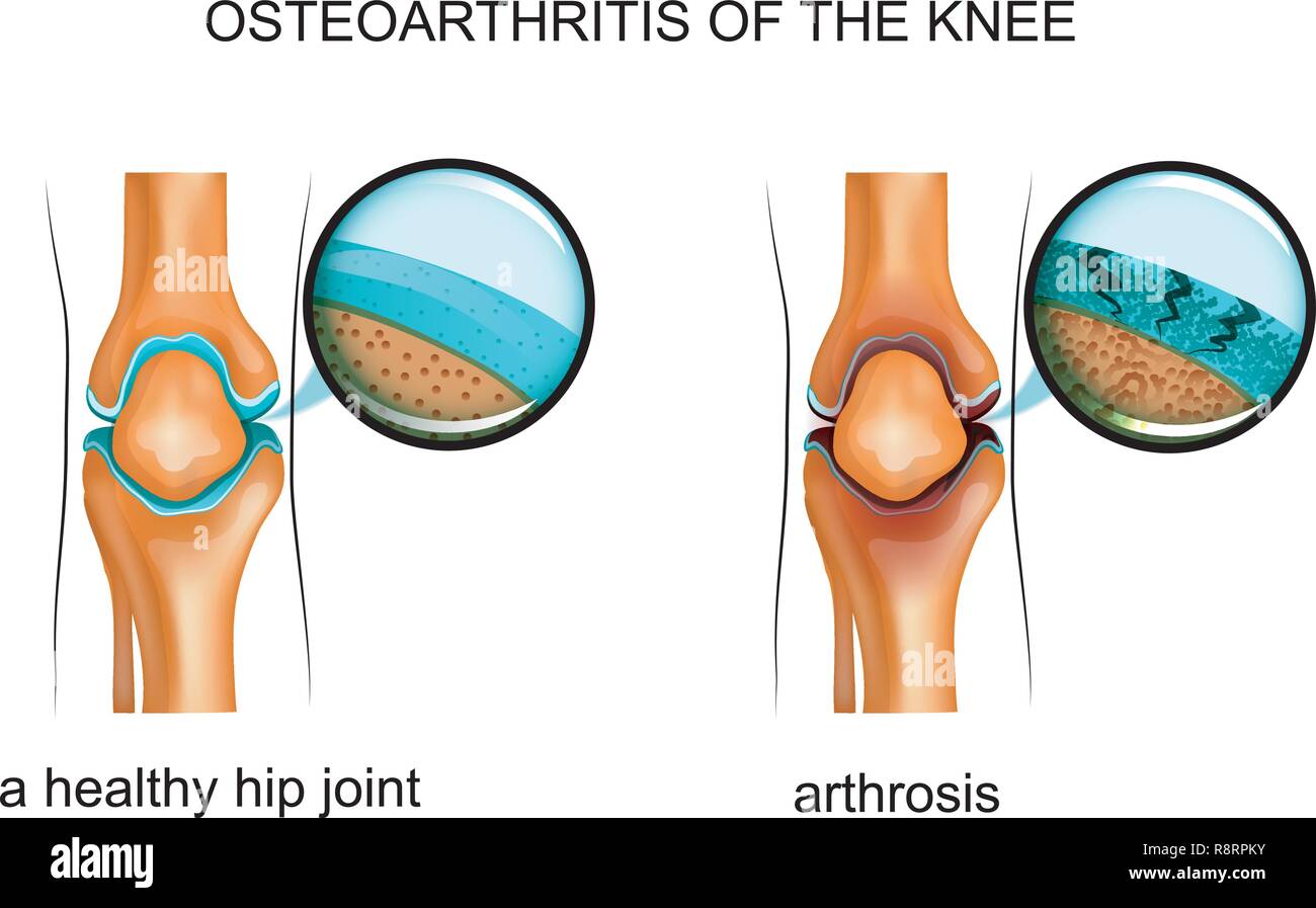 vector illustration of osteoarthritis of the knee Stock Vectorhttps://www.alamy.com/image-license-details/?v=1https://www.alamy.com/vector-illustration-of-osteoarthritis-of-the-knee-image229174735.html
RFR8RPKY–vector illustration of osteoarthritis of the knee
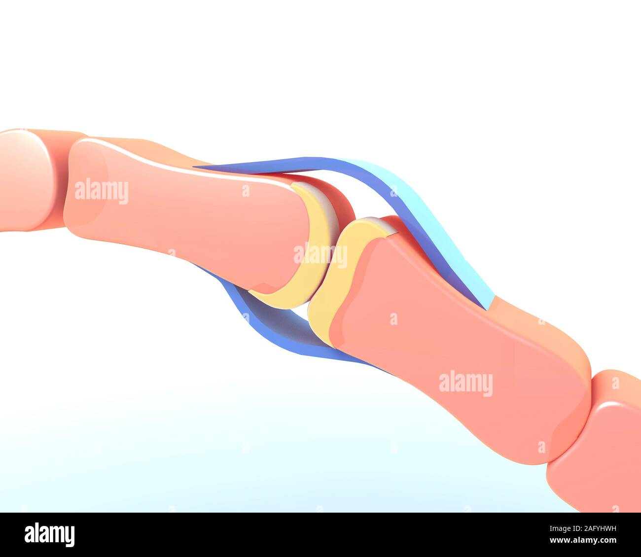 3d illustration of the synovial joint of the bone of a hand. Schematic and symbolic graphic representation. Stock Photohttps://www.alamy.com/image-license-details/?v=1https://www.alamy.com/3d-illustration-of-the-synovial-joint-of-the-bone-of-a-hand-schematic-and-symbolic-graphic-representation-image336823581.html
RF2AFYHWH–3d illustration of the synovial joint of the bone of a hand. Schematic and symbolic graphic representation.
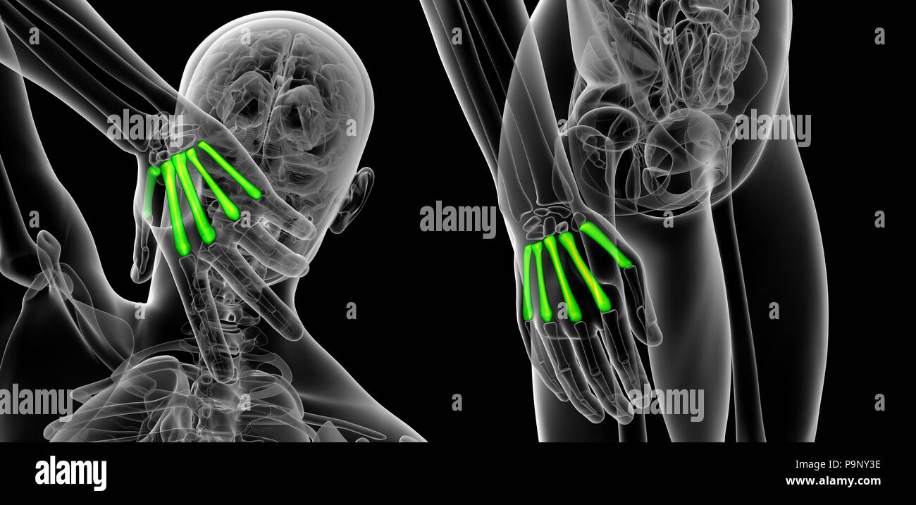 3d rendering illustration of metacarpal Stock Photohttps://www.alamy.com/image-license-details/?v=1https://www.alamy.com/3d-rendering-illustration-of-metacarpal-image212538578.html
RFP9NY3E–3d rendering illustration of metacarpal
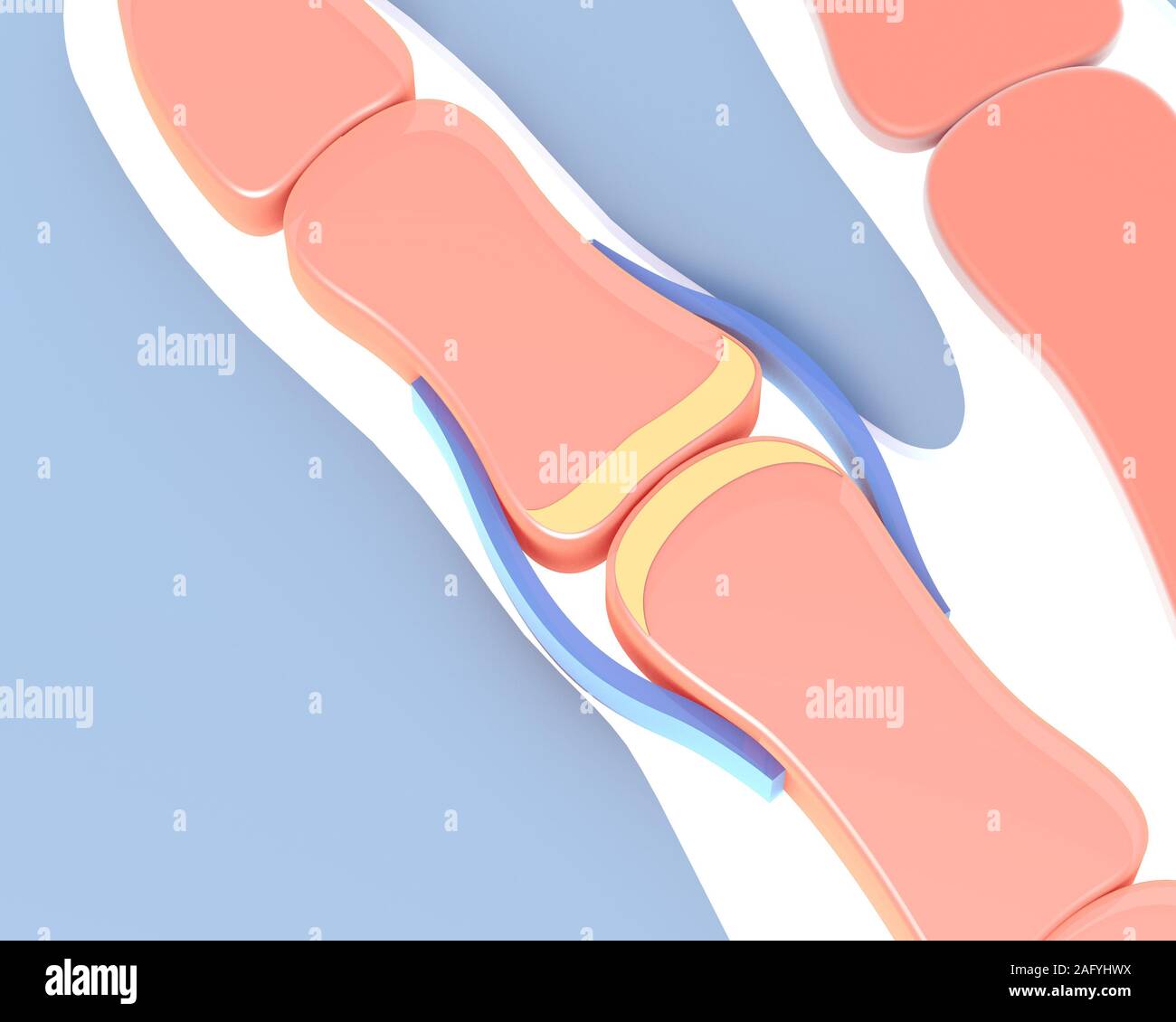 3d illustration of the synovial joint of the bone of a hand. Schematic and symbolic graphic representation. Stock Photohttps://www.alamy.com/image-license-details/?v=1https://www.alamy.com/3d-illustration-of-the-synovial-joint-of-the-bone-of-a-hand-schematic-and-symbolic-graphic-representation-image336823590.html
RF2AFYHWX–3d illustration of the synovial joint of the bone of a hand. Schematic and symbolic graphic representation.
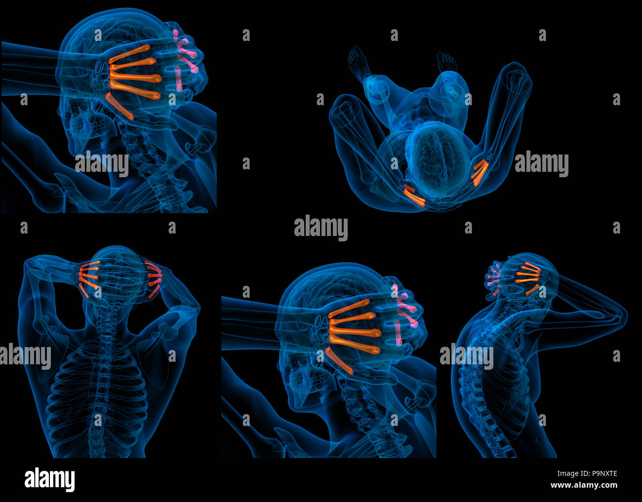 3d rendering of metacarpal Stock Photohttps://www.alamy.com/image-license-details/?v=1https://www.alamy.com/3d-rendering-of-metacarpal-image212538382.html
RFP9NXTE–3d rendering of metacarpal
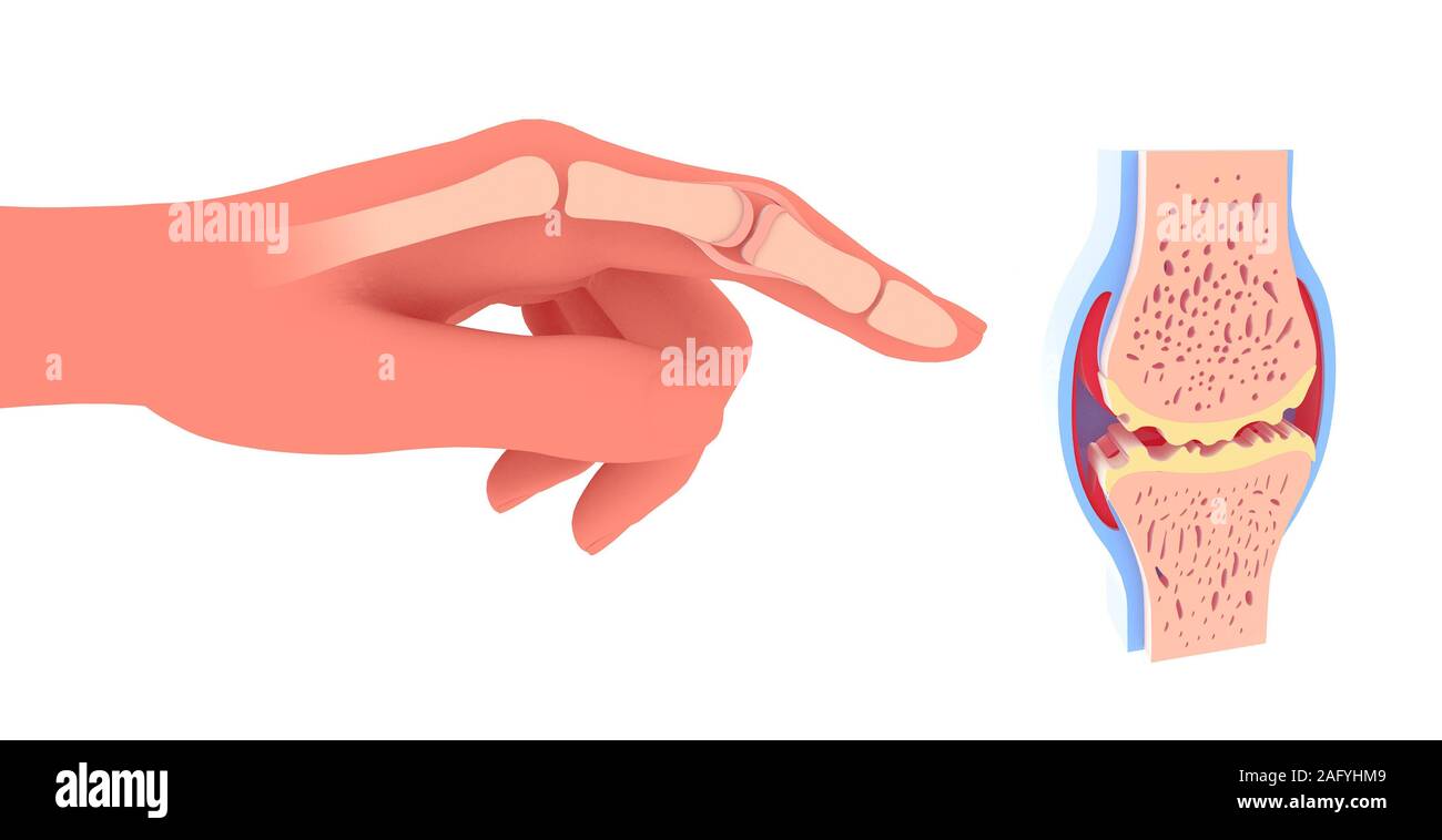 3d illustration of synovial joint with osteoarthritis. Next to the graphic representation of a hand placing the joint. Stock Photohttps://www.alamy.com/image-license-details/?v=1https://www.alamy.com/3d-illustration-of-synovial-joint-with-osteoarthritis-next-to-the-graphic-representation-of-a-hand-placing-the-joint-image336823433.html
RF2AFYHM9–3d illustration of synovial joint with osteoarthritis. Next to the graphic representation of a hand placing the joint.
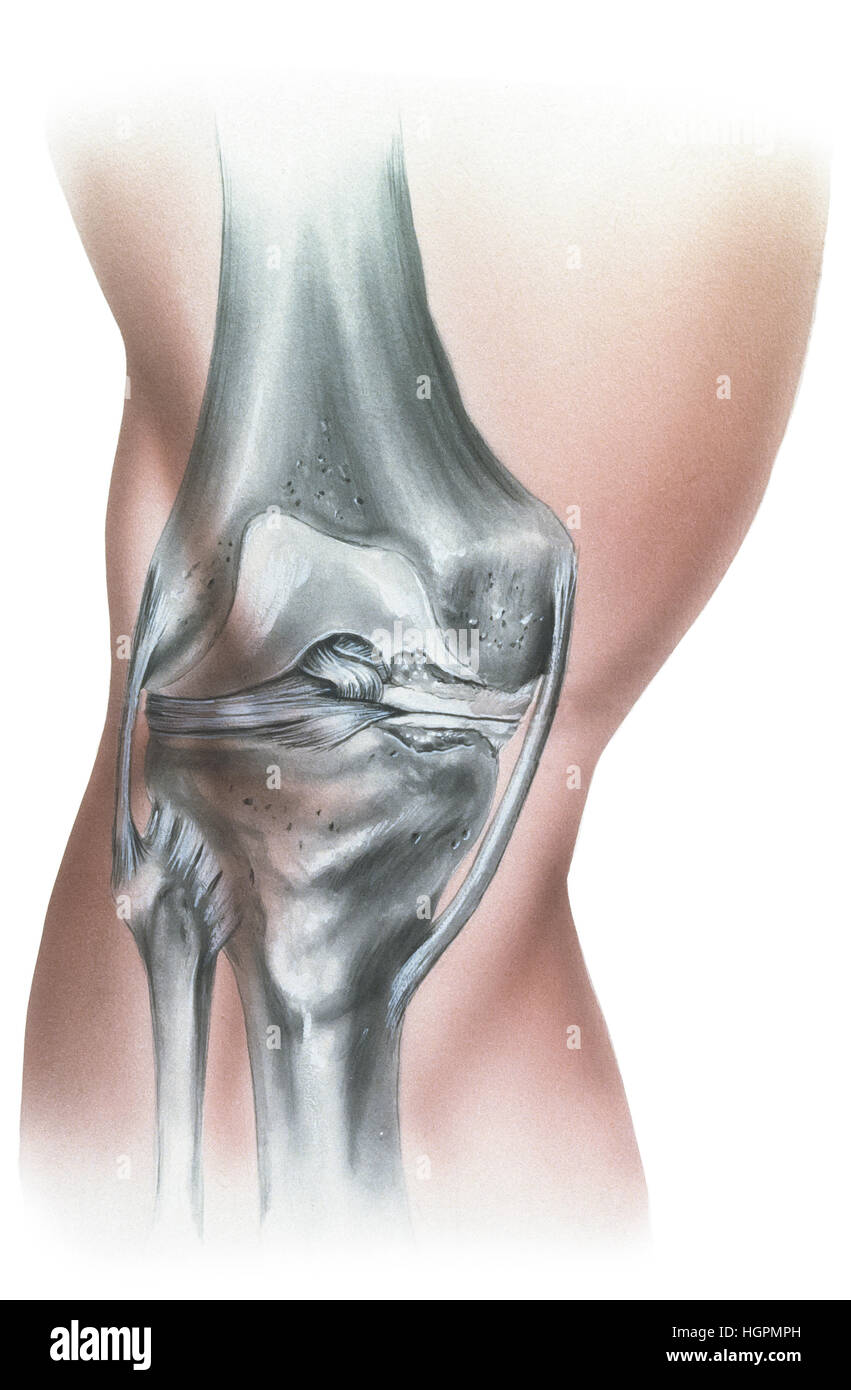 Shown is a knee with erosion of the cartilage causing meniscus damage. There is also hypertrphyof the knee bone and lining. Stock Photohttps://www.alamy.com/image-license-details/?v=1https://www.alamy.com/stock-photo-shown-is-a-knee-with-erosion-of-the-cartilage-causing-meniscus-damage-130806329.html
RFHGPMPH–Shown is a knee with erosion of the cartilage causing meniscus damage. There is also hypertrphyof the knee bone and lining.
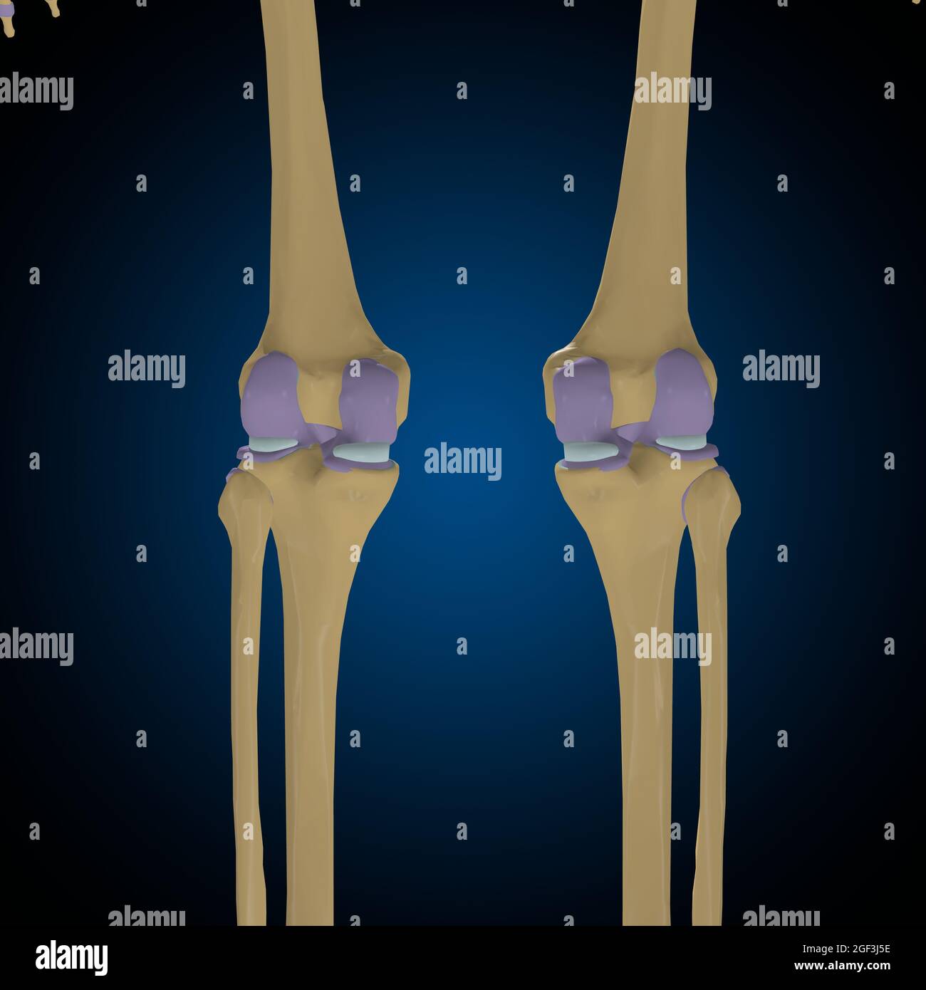 Articular capsule Anatomy For Medical Concept 3D Illustration Stock Photohttps://www.alamy.com/image-license-details/?v=1https://www.alamy.com/articular-capsule-anatomy-for-medical-concept-3d-illustration-image439559162.html
RF2GF3J5E–Articular capsule Anatomy For Medical Concept 3D Illustration
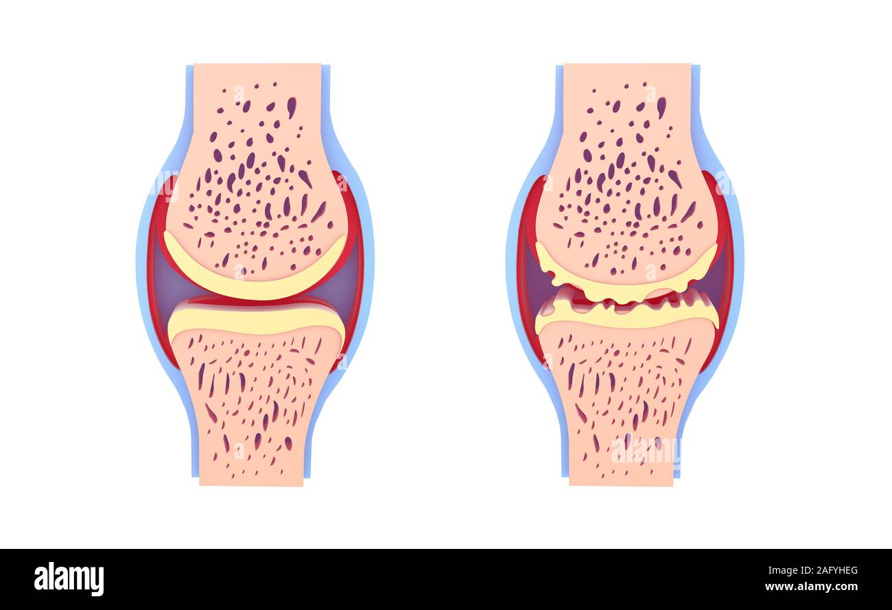 3d illustration of healthy synovial joint and with osteoarthritis. Images isolated on white background front view. Vivid colors. Stock Photohttps://www.alamy.com/image-license-details/?v=1https://www.alamy.com/3d-illustration-of-healthy-synovial-joint-and-with-osteoarthritis-images-isolated-on-white-background-front-view-vivid-colors-image336823272.html
RF2AFYHEG–3d illustration of healthy synovial joint and with osteoarthritis. Images isolated on white background front view. Vivid colors.
 Radius and ulna (bones of the forearm) seen by their front face, vintage engraved illustration. Usual Medicine Dictionary by Dr Stock Vectorhttps://www.alamy.com/image-license-details/?v=1https://www.alamy.com/stock-photo-radius-and-ulna-bones-of-the-forearm-seen-by-their-front-face-vintage-84408204.html
RFEW93EM–Radius and ulna (bones of the forearm) seen by their front face, vintage engraved illustration. Usual Medicine Dictionary by Dr
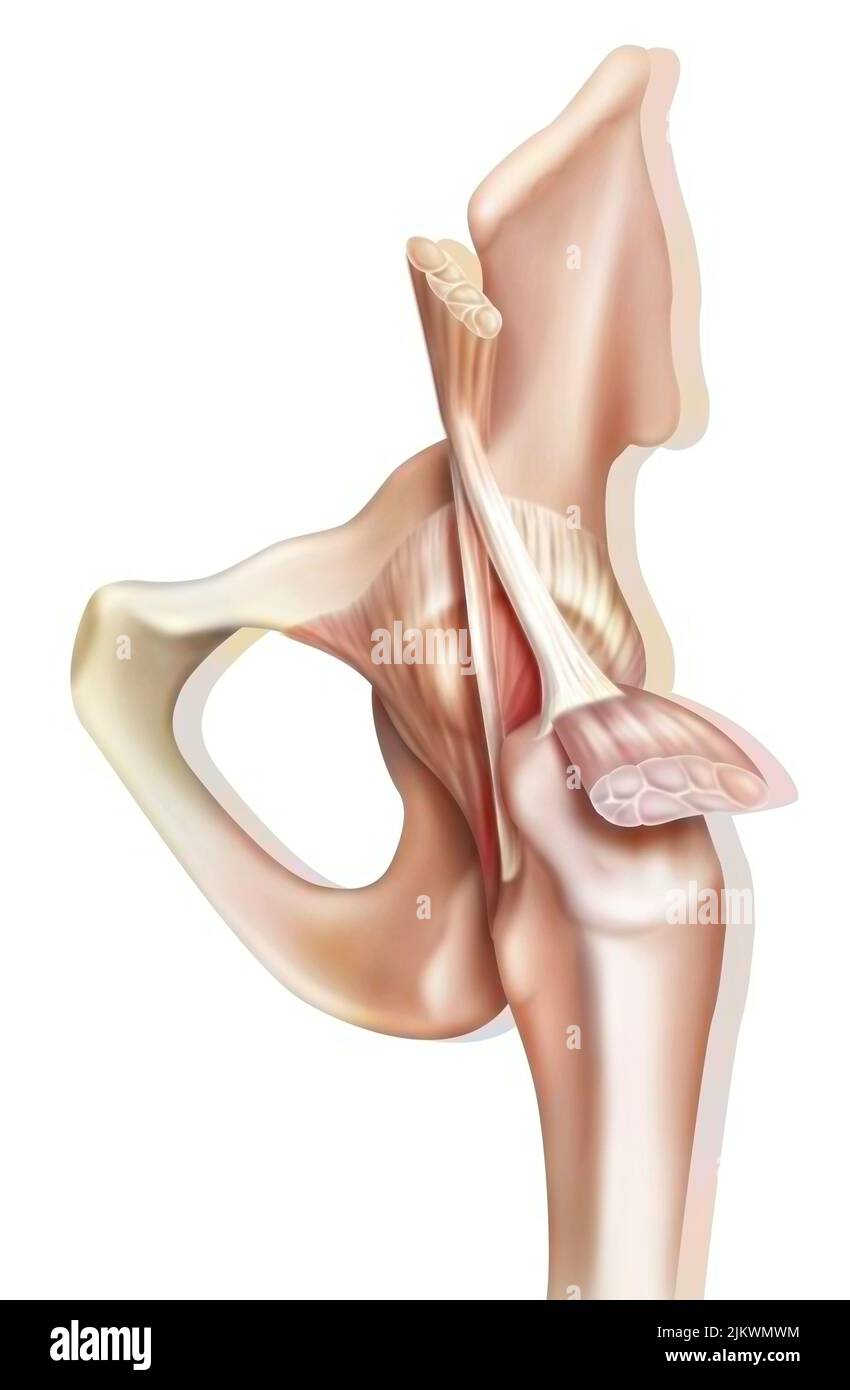 Anatomy of the coxofemoral (hip) joint with muscles, tendons. Stock Photohttps://www.alamy.com/image-license-details/?v=1https://www.alamy.com/anatomy-of-the-coxofemoral-hip-joint-with-muscles-tendons-image476923600.html
RF2JKWMWM–Anatomy of the coxofemoral (hip) joint with muscles, tendons.
 3D illustration showing knee joint with transparent femur and articular capsule, menisci and ligaments Stock Photohttps://www.alamy.com/image-license-details/?v=1https://www.alamy.com/3d-illustration-showing-knee-joint-with-transparent-femur-and-articular-capsule-menisci-and-ligaments-image357782791.html
RF2BP2BGR–3D illustration showing knee joint with transparent femur and articular capsule, menisci and ligaments
 Scanogram is a Full-length standing AP radiograph of both lower extremities including the hip, knee, and ankle with 3D rendering. Stock Photohttps://www.alamy.com/image-license-details/?v=1https://www.alamy.com/scanogram-is-a-full-length-standing-ap-radiograph-of-both-lower-extremities-including-the-hip-knee-and-ankle-with-3d-rendering-image518160197.html
RF2N306H9–Scanogram is a Full-length standing AP radiograph of both lower extremities including the hip, knee, and ankle with 3D rendering.
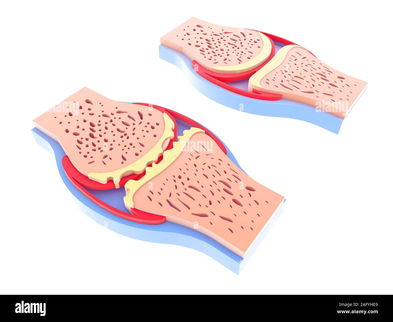 3d illustration of healthy synovial joint and with osteoarthritis. Images isolated on white background leaning on the floor. Vivid colors. Stock Photohttps://www.alamy.com/image-license-details/?v=1https://www.alamy.com/3d-illustration-of-healthy-synovial-joint-and-with-osteoarthritis-images-isolated-on-white-background-leaning-on-the-floor-vivid-colors-image336823265.html
RF2AFYHE9–3d illustration of healthy synovial joint and with osteoarthritis. Images isolated on white background leaning on the floor. Vivid colors.
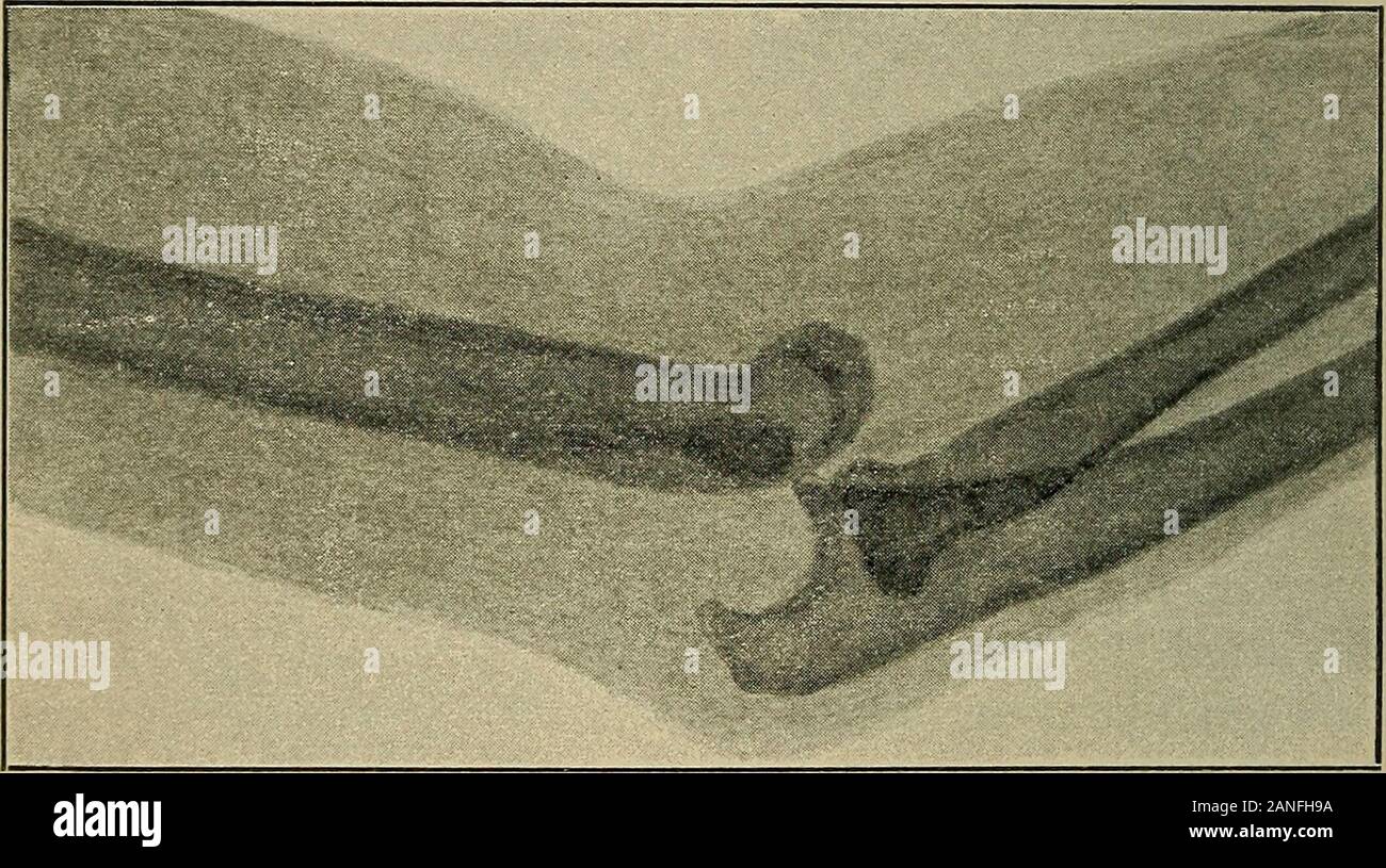 Atlas and epitome of traumatic fractures and dislocations . Fig. 81.—Eecent backward dislo-cation of the left forearm in a boyfourteen years of age (Kriiger, 1896).Swelling, prominence of the olecra-non, shortening of the forearm areseen. The dislocation was reducedand perfect recovery ensued. 188 FRACTURES AND DISLOCATIONS. cadaver. The arm need only be overextended to producea tear in the anterior segment of the articular capsule ; theolecranon during this movement is braced against the pos-terior supratrochlear fossa, and after the bones have beensufficiently forced apart, the forearm is su Stock Photohttps://www.alamy.com/image-license-details/?v=1https://www.alamy.com/atlas-and-epitome-of-traumatic-fractures-and-dislocations-fig-81eecent-backward-dislo-cation-of-the-left-forearm-in-a-boyfourteen-years-of-age-kriiger-1896swelling-prominence-of-the-olecra-non-shortening-of-the-forearm-areseen-the-dislocation-was-reducedand-perfect-recovery-ensued-188-fractures-and-dislocations-cadaver-the-arm-need-only-be-overextended-to-producea-tear-in-the-anterior-segment-of-the-articular-capsule-theolecranon-during-this-movement-is-braced-against-the-pos-terior-supratrochlear-fossa-and-after-the-bones-have-beensufficiently-forced-apart-the-forearm-is-su-image340247638.html
RM2ANFH9A–Atlas and epitome of traumatic fractures and dislocations . Fig. 81.—Eecent backward dislo-cation of the left forearm in a boyfourteen years of age (Kriiger, 1896).Swelling, prominence of the olecra-non, shortening of the forearm areseen. The dislocation was reducedand perfect recovery ensued. 188 FRACTURES AND DISLOCATIONS. cadaver. The arm need only be overextended to producea tear in the anterior segment of the articular capsule ; theolecranon during this movement is braced against the pos-terior supratrochlear fossa, and after the bones have beensufficiently forced apart, the forearm is su
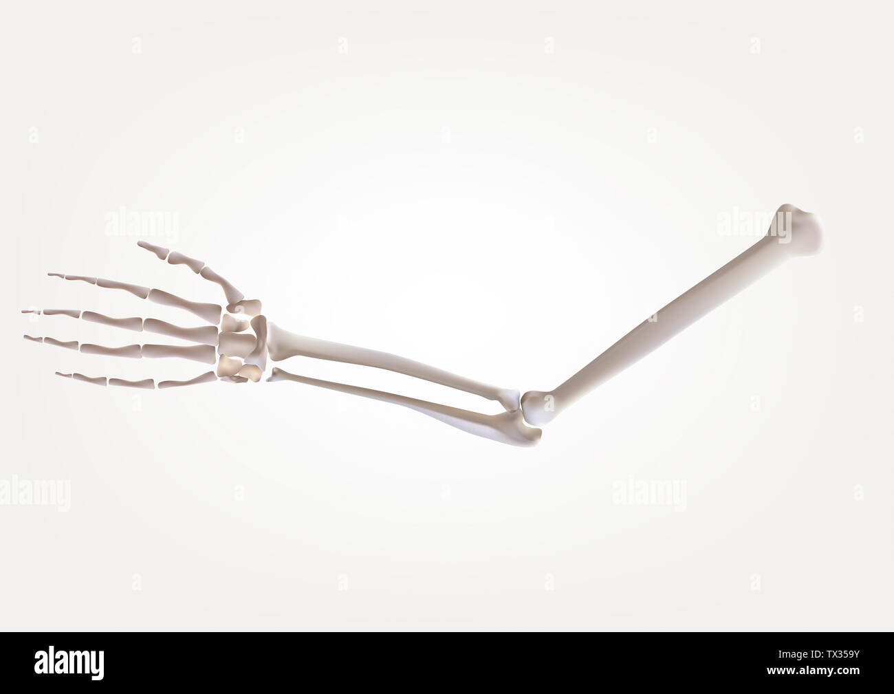 Skeleton in human hands, isolated on a white background Medical illustration, 3D illustration Stock Photohttps://www.alamy.com/image-license-details/?v=1https://www.alamy.com/skeleton-in-human-hands-isolated-on-a-white-background-medical-illustration-3d-illustration-image256996263.html
RFTX359Y–Skeleton in human hands, isolated on a white background Medical illustration, 3D illustration
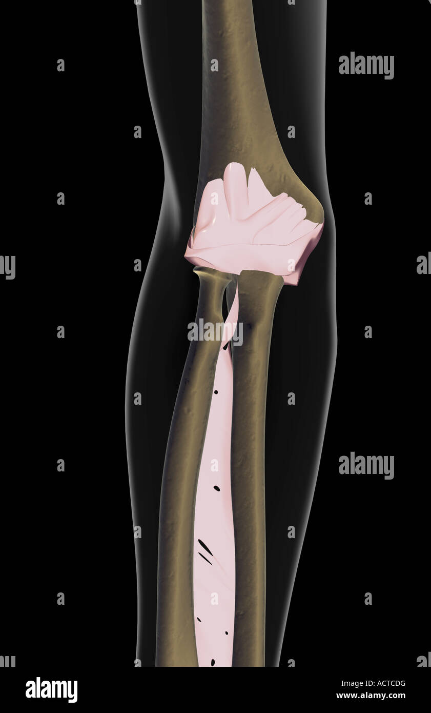 The ligaments of the elbow Stock Photohttps://www.alamy.com/image-license-details/?v=1https://www.alamy.com/stock-photo-the-ligaments-of-the-elbow-13225563.html
RFACTCDG–The ligaments of the elbow
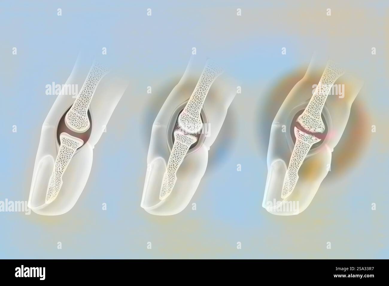 Finger osteoarthritis. Left: healthy joint. Middle: moderate osteoarthritis. The cartilage is worn, the cartilaginous debris causes inflammation of th Stock Photohttps://www.alamy.com/image-license-details/?v=1https://www.alamy.com/finger-osteoarthritis-left-healthy-joint-middle-moderate-osteoarthritis-the-cartilage-is-worn-the-cartilaginous-debris-causes-inflammation-of-th-image642999035.html
RM2SA33R7–Finger osteoarthritis. Left: healthy joint. Middle: moderate osteoarthritis. The cartilage is worn, the cartilaginous debris causes inflammation of th
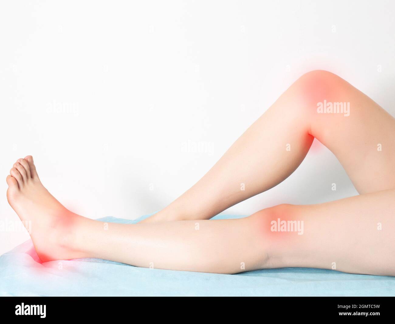 female legs on a white background with sore reddened knee joints and ankle joints with heels. Concept of disease and treatment of arthritis and joint Stock Photohttps://www.alamy.com/image-license-details/?v=1https://www.alamy.com/female-legs-on-a-white-background-with-sore-reddened-knee-joints-and-ankle-joints-with-heels-concept-of-disease-and-treatment-of-arthritis-and-joint-image443088741.html
RF2GMTC5W–female legs on a white background with sore reddened knee joints and ankle joints with heels. Concept of disease and treatment of arthritis and joint
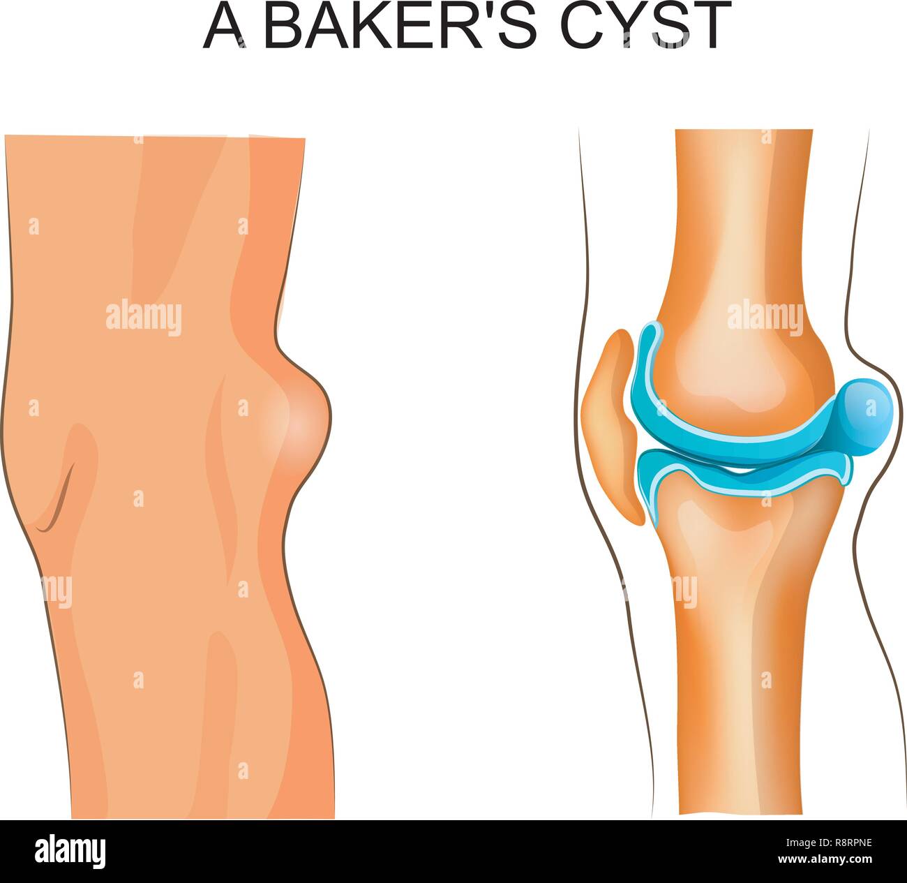 vector illustration of Baker's cyst. traumatology and orthopedics Stock Vectorhttps://www.alamy.com/image-license-details/?v=1https://www.alamy.com/vector-illustration-of-bakers-cyst-traumatology-and-orthopedics-image229174778.html
RFR8RPNE–vector illustration of Baker's cyst. traumatology and orthopedics
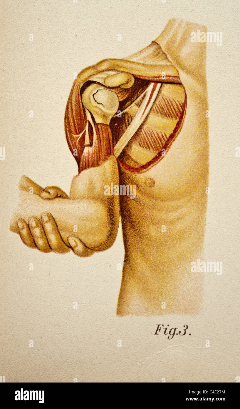 Illustration of the Human Shoulder copyright 1902 Stock Photohttps://www.alamy.com/image-license-details/?v=1https://www.alamy.com/stock-photo-illustration-of-the-human-shoulder-copyright-1902-37188472.html
RFC4E27M–Illustration of the Human Shoulder copyright 1902
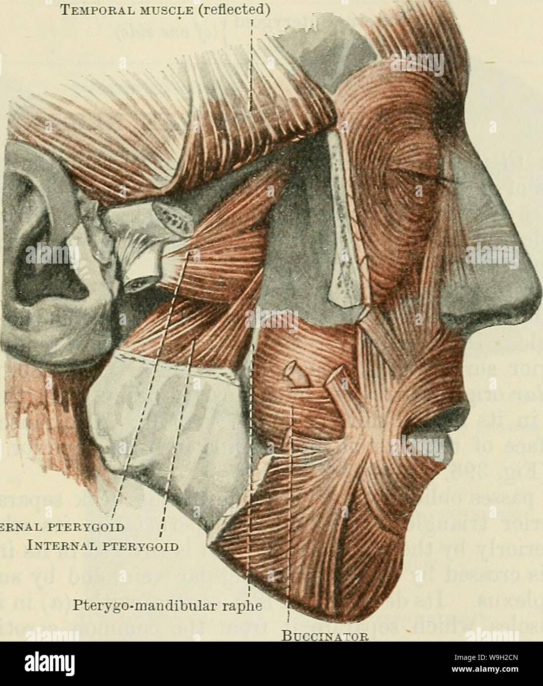 Archive image from page 490 of Cunningham's Text-book of anatomy (1914). Cunningham's Text-book of anatomy cunninghamstextb00cunn Year: 1914 ( MUSCLES OF MASTICATION. 457 fovea pterygoidea on the anterior aspect of the neck of the mandible (Figs. 403 and 404, p. 455), and (2) the articular disc and capsule of the mandibular articulation. This muscle is covered by the insertion of the temporal muscle and the coronoid process of the mandible, and is usually crossed by the internal maxillary artery. It conceals the mandibular branch of the trigeminal nerve, and the pterygoid origin of the intern Stock Photohttps://www.alamy.com/image-license-details/?v=1https://www.alamy.com/archive-image-from-page-490-of-cunninghams-text-book-of-anatomy-1914-cunninghams-text-book-of-anatomy-cunninghamstextb00cunn-year-1914-muscles-of-mastication-457-fovea-pterygoidea-on-the-anterior-aspect-of-the-neck-of-the-mandible-figs-403-and-404-p-455-and-2-the-articular-disc-and-capsule-of-the-mandibular-articulation-this-muscle-is-covered-by-the-insertion-of-the-temporal-muscle-and-the-coronoid-process-of-the-mandible-and-is-usually-crossed-by-the-internal-maxillary-artery-it-conceals-the-mandibular-branch-of-the-trigeminal-nerve-and-the-pterygoid-origin-of-the-intern-image264062533.html
RMW9H2CN–Archive image from page 490 of Cunningham's Text-book of anatomy (1914). Cunningham's Text-book of anatomy cunninghamstextb00cunn Year: 1914 ( MUSCLES OF MASTICATION. 457 fovea pterygoidea on the anterior aspect of the neck of the mandible (Figs. 403 and 404, p. 455), and (2) the articular disc and capsule of the mandibular articulation. This muscle is covered by the insertion of the temporal muscle and the coronoid process of the mandible, and is usually crossed by the internal maxillary artery. It conceals the mandibular branch of the trigeminal nerve, and the pterygoid origin of the intern
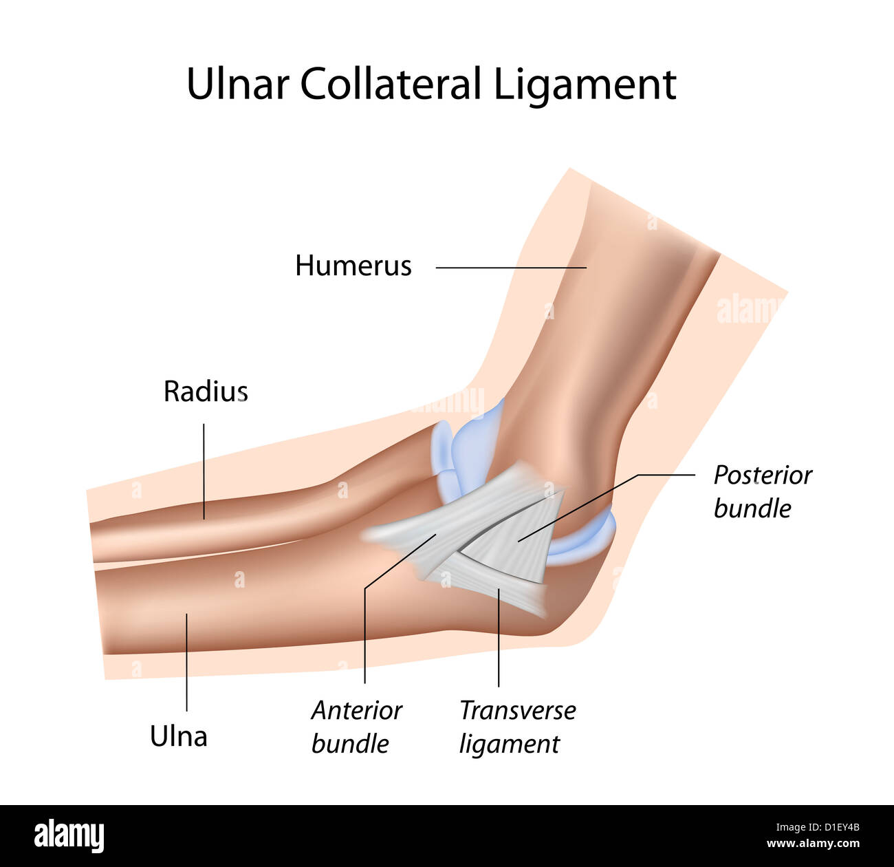 Ulnar collateral ligament of elbow Stock Photohttps://www.alamy.com/image-license-details/?v=1https://www.alamy.com/stock-photo-ulnar-collateral-ligament-of-elbow-52574379.html
RFD1EY4B–Ulnar collateral ligament of elbow
 Symbolic for arthralgia Stock Photohttps://www.alamy.com/image-license-details/?v=1https://www.alamy.com/stock-photo-symbolic-for-arthralgia-132740315.html
RFHKXRHF–Symbolic for arthralgia
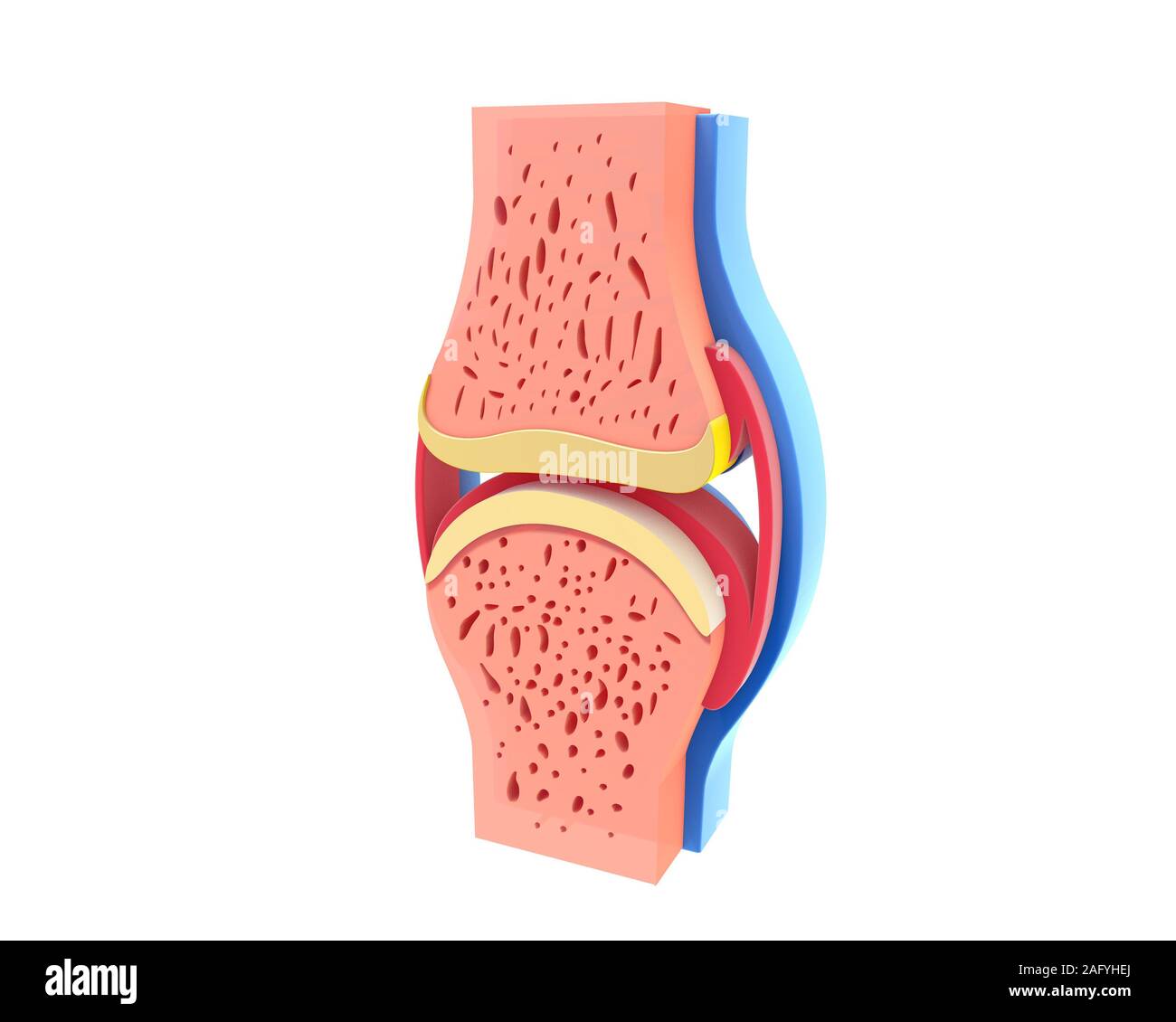 3d illustration of synovial joint lateral view. Image isolated on white background. Vivid colors. Stock Photohttps://www.alamy.com/image-license-details/?v=1https://www.alamy.com/3d-illustration-of-synovial-joint-lateral-view-image-isolated-on-white-background-vivid-colors-image336823274.html
RF2AFYHEJ–3d illustration of synovial joint lateral view. Image isolated on white background. Vivid colors.
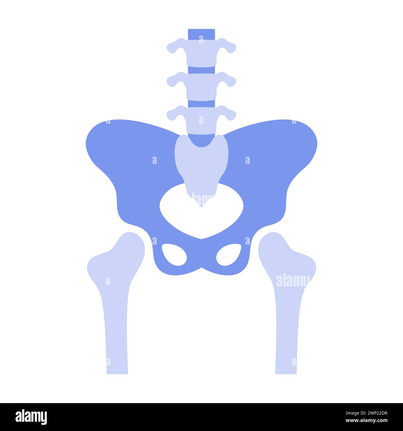 Human hip joint, simple anatomical presentation of pelvic bones vector illustration Stock Vectorhttps://www.alamy.com/image-license-details/?v=1https://www.alamy.com/human-hip-joint-simple-anatomical-presentation-of-pelvic-bones-vector-illustration-image598720803.html
RF2WP22DR–Human hip joint, simple anatomical presentation of pelvic bones vector illustration
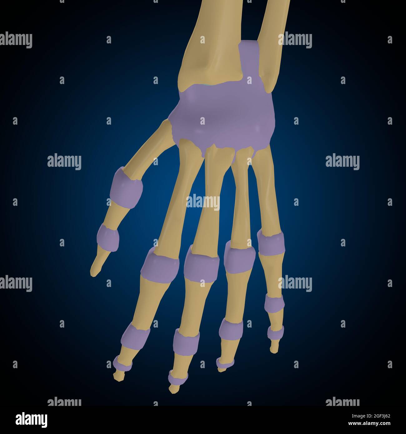 Articular capsule Anatomy For Medical Concept 3D Illustration Stock Photohttps://www.alamy.com/image-license-details/?v=1https://www.alamy.com/articular-capsule-anatomy-for-medical-concept-3d-illustration-image439559178.html
RF2GF3J62–Articular capsule Anatomy For Medical Concept 3D Illustration
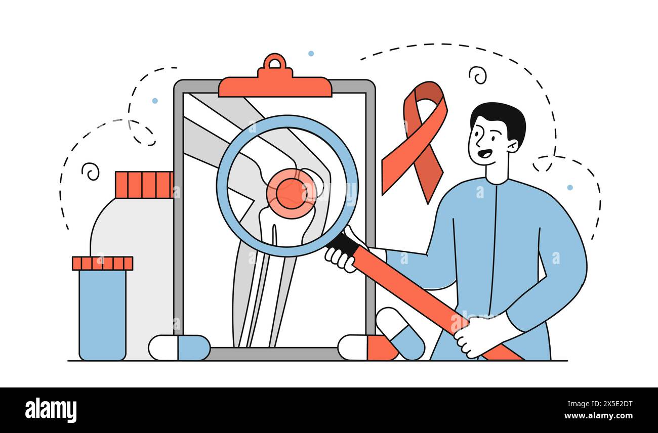 Doctor studying arthritis vector simple Stock Vectorhttps://www.alamy.com/image-license-details/?v=1https://www.alamy.com/doctor-studying-arthritis-vector-simple-image605745444.html
RF2X5E2DT–Doctor studying arthritis vector simple
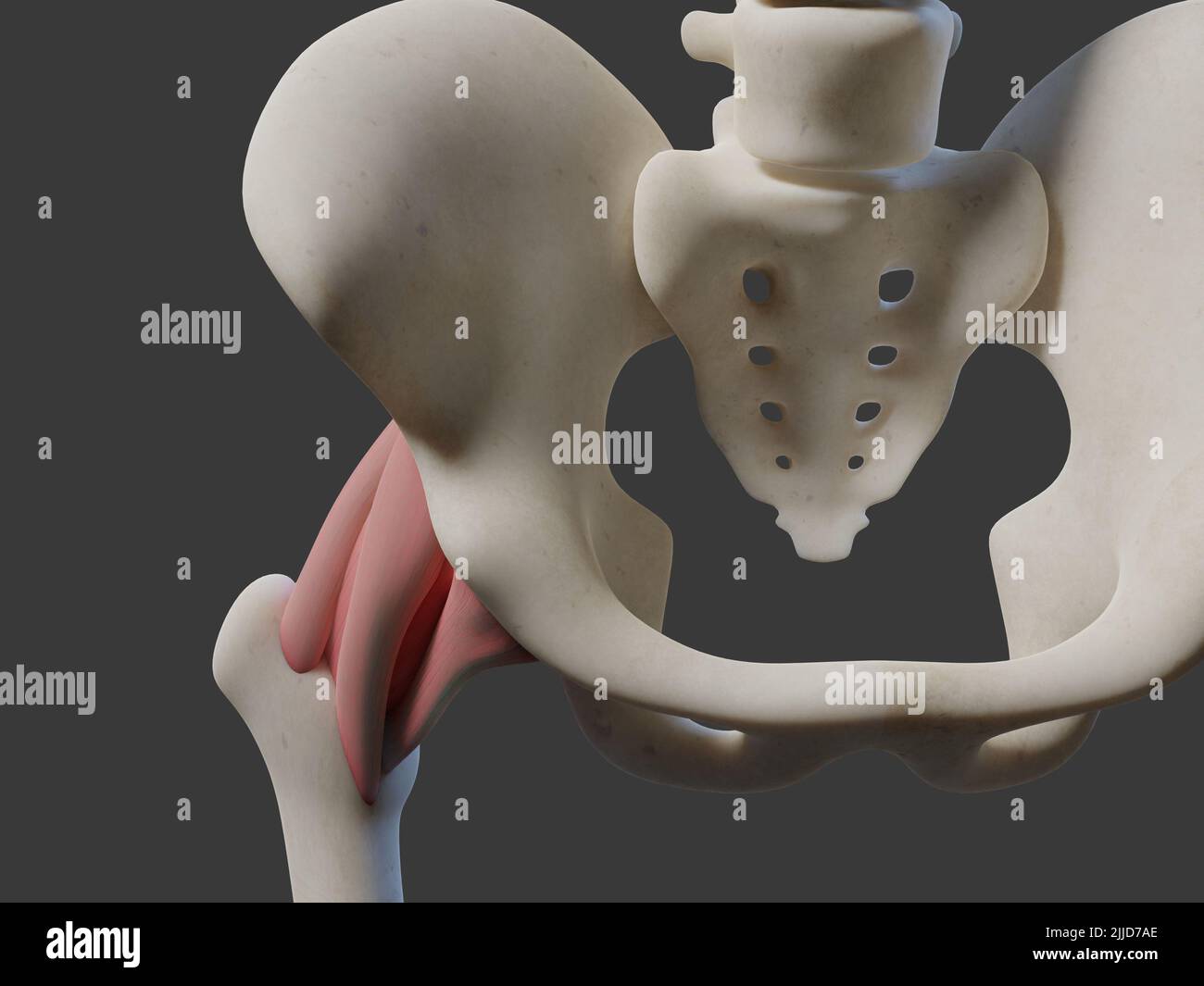 3D illustration of human joint. Includes Stock Photohttps://www.alamy.com/image-license-details/?v=1https://www.alamy.com/3d-illustration-of-human-joint-includes-image476034902.html
RF2JJD7AE–3D illustration of human joint. Includes
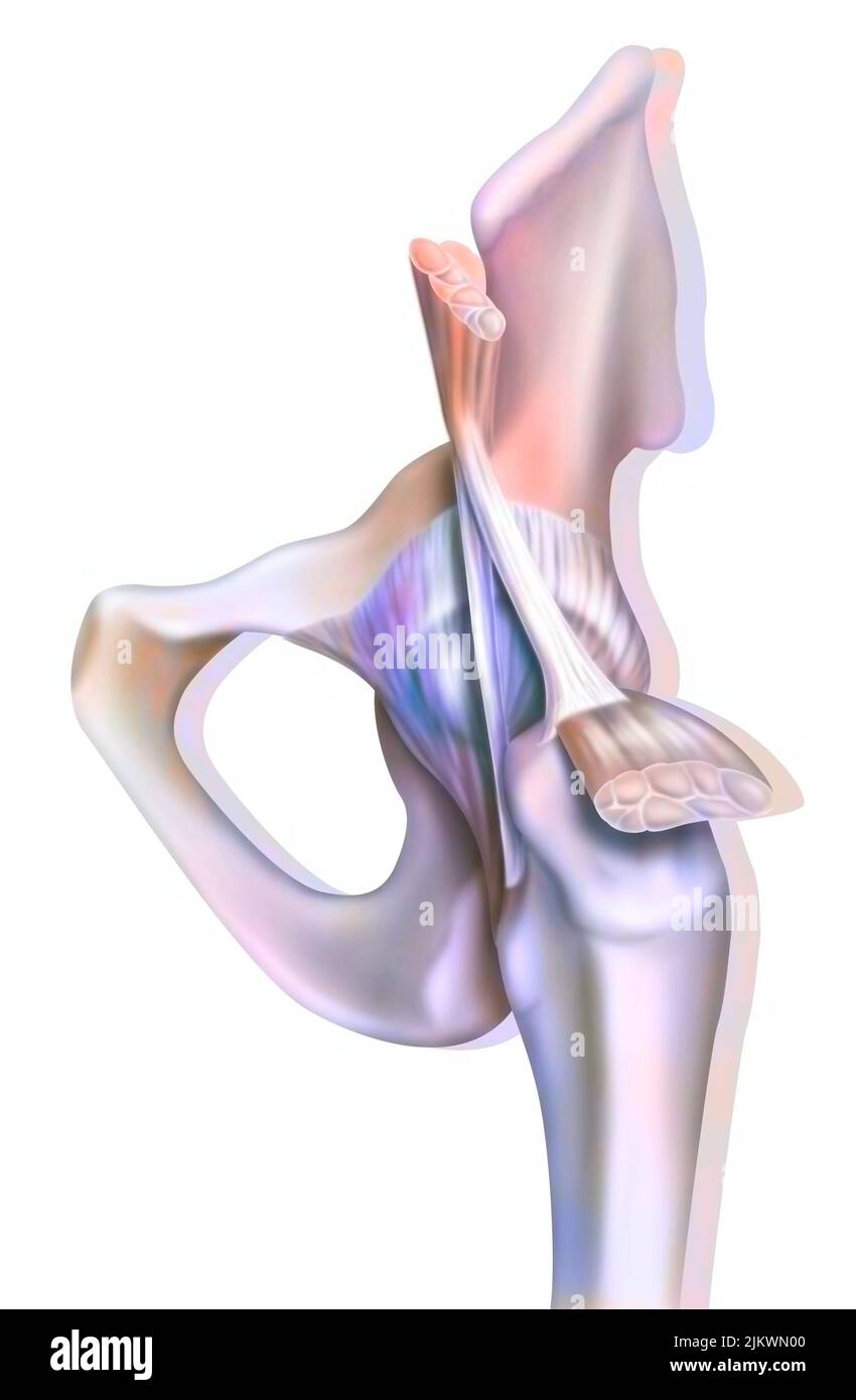 Anatomy of the coxofemoral (hip) joint with muscles, tendons. Stock Photohttps://www.alamy.com/image-license-details/?v=1https://www.alamy.com/anatomy-of-the-coxofemoral-hip-joint-with-muscles-tendons-image476923664.html
RF2JKWN00–Anatomy of the coxofemoral (hip) joint with muscles, tendons.
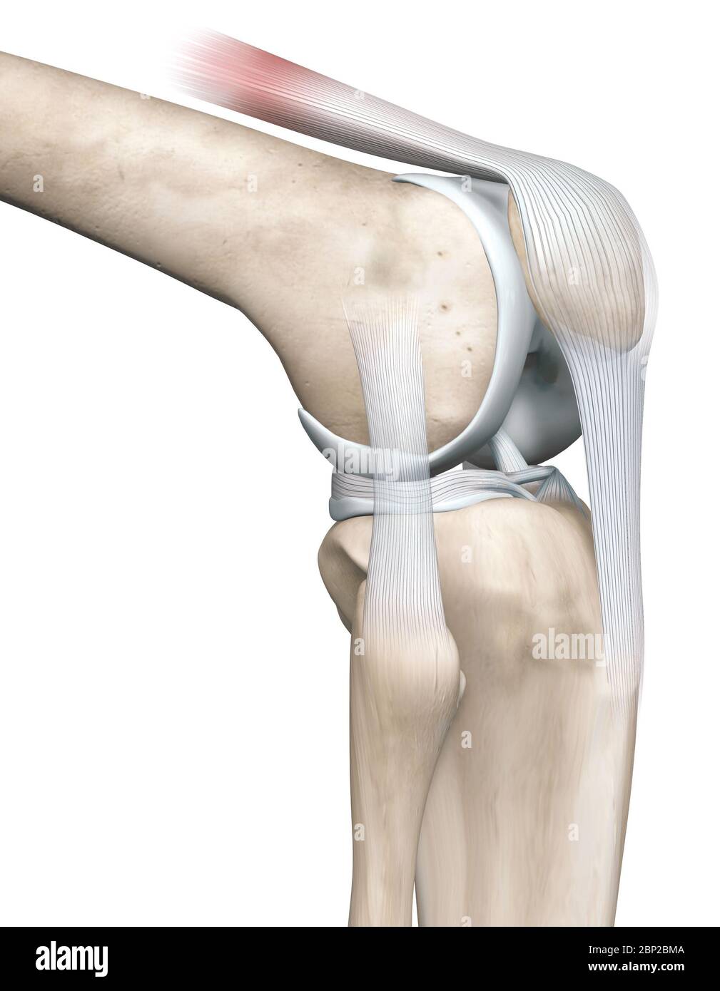 3D illustration showing knee joint with femur, fibula and articular capsule, menisci and ligaments Stock Photohttps://www.alamy.com/image-license-details/?v=1https://www.alamy.com/3d-illustration-showing-knee-joint-with-femur-fibula-and-articular-capsule-menisci-and-ligaments-image357782890.html
RF2BP2BMA–3D illustration showing knee joint with femur, fibula and articular capsule, menisci and ligaments
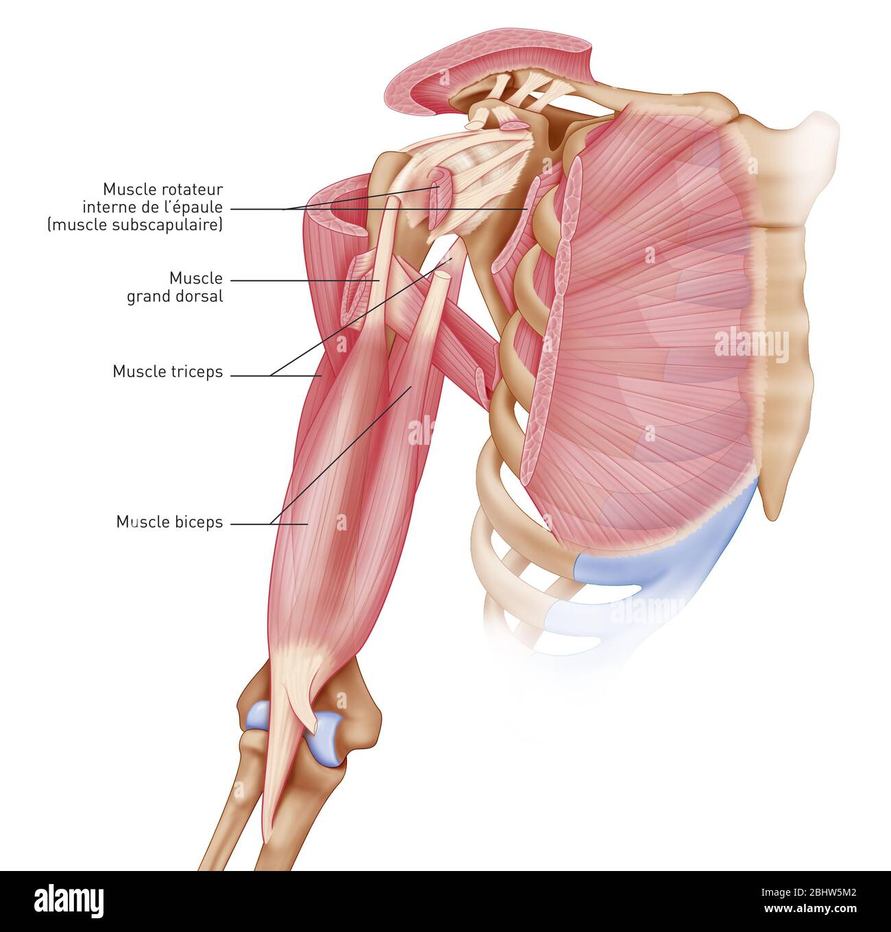 Medical illustration representing the muscles of the shoulder, joint and shoulder muscles. The articular capsule, the ligaments, the small pectoralis Stock Photohttps://www.alamy.com/image-license-details/?v=1https://www.alamy.com/medical-illustration-representing-the-muscles-of-the-shoulder-joint-and-shoulder-muscles-the-articular-capsule-the-ligaments-the-small-pectoralis-image355209794.html
RM2BHW5M2–Medical illustration representing the muscles of the shoulder, joint and shoulder muscles. The articular capsule, the ligaments, the small pectoralis
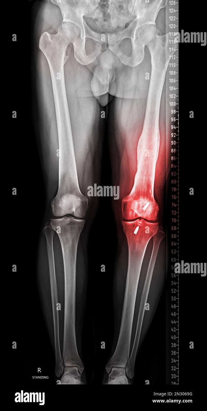 Scanogram is a Full-length standing AP radiograph of both lower extremities including the hip, knee, and ankle. Stock Photohttps://www.alamy.com/image-license-details/?v=1https://www.alamy.com/scanogram-is-a-full-length-standing-ap-radiograph-of-both-lower-extremities-including-the-hip-knee-and-ankle-image518159980.html
RF2N3069G–Scanogram is a Full-length standing AP radiograph of both lower extremities including the hip, knee, and ankle.
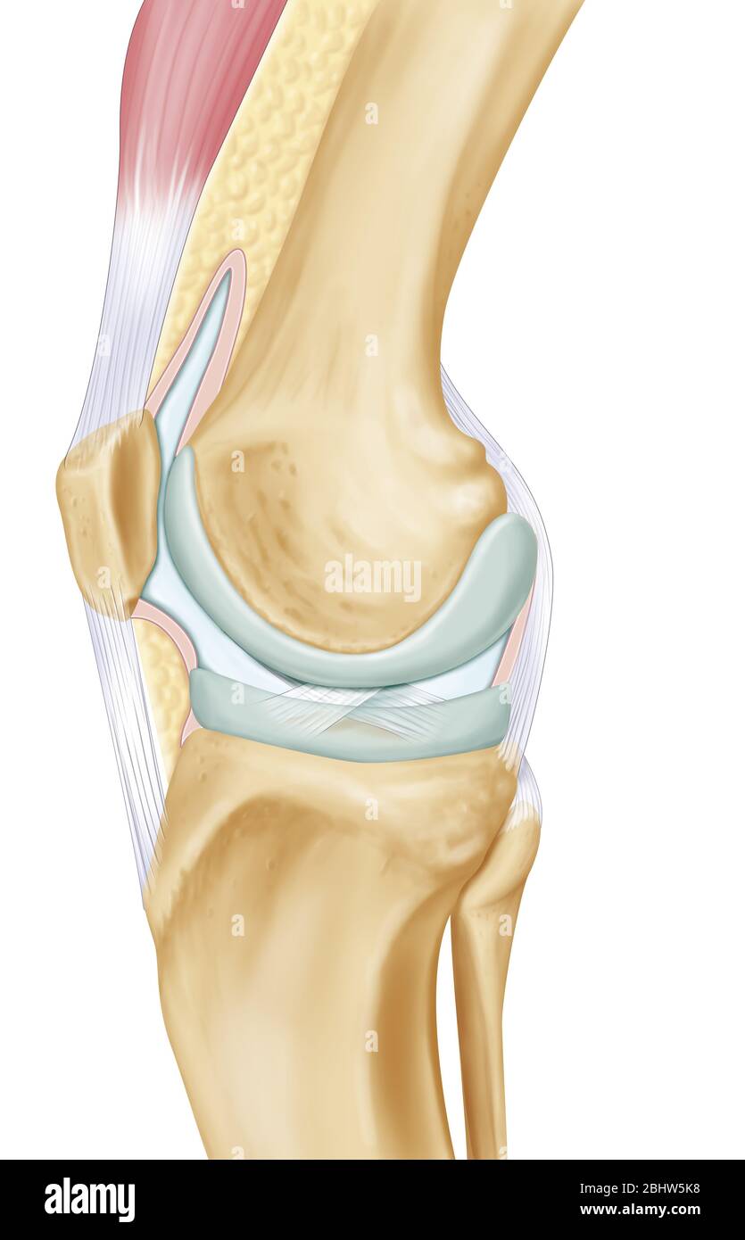 Representation of the knee joint on an internal lateral view of the right leg. At the center of the illustration are the two articular surfaces in gra Stock Photohttps://www.alamy.com/image-license-details/?v=1https://www.alamy.com/representation-of-the-knee-joint-on-an-internal-lateral-view-of-the-right-leg-at-the-center-of-the-illustration-are-the-two-articular-surfaces-in-gra-image355209772.html
RM2BHW5K8–Representation of the knee joint on an internal lateral view of the right leg. At the center of the illustration are the two articular surfaces in gra
 . Manual of operative surgery. Fig. 1376.—Arthroplasty,authors method. ARTHROPLASTY ELBOW III9 4. Resect the olecranon at its base. Fashion the bone as shown in Figs.1378 and 1379. Resect that portion of the head of the radius which projectsabove the sawn surface of the ulna. If anchylosis exists between the radiusand ulna separate these bones with a fine chisel. 5. Remove the elastic constrictor. Attend to hemostasis with usual care. 6. Interposition of Muscle.—(a) Flex the forearm acutely. Divide theanterior articular capsule transversely at its ulnar insertion; continue thisincision into th Stock Photohttps://www.alamy.com/image-license-details/?v=1https://www.alamy.com/manual-of-operative-surgery-fig-1376arthroplastyauthors-method-arthroplasty-elbow-iii9-4-resect-the-olecranon-at-its-base-fashion-the-bone-as-shown-in-figs1378-and-1379-resect-that-portion-of-the-head-of-the-radius-which-projectsabove-the-sawn-surface-of-the-ulna-if-anchylosis-exists-between-the-radiusand-ulna-separate-these-bones-with-a-fine-chisel-5-remove-the-elastic-constrictor-attend-to-hemostasis-with-usual-care-6-interposition-of-musclea-flex-the-forearm-acutely-divide-theanterior-articular-capsule-transversely-at-its-ulnar-insertion-continue-thisincision-into-th-image336741929.html
RM2AFRWND–. Manual of operative surgery. Fig. 1376.—Arthroplasty,authors method. ARTHROPLASTY ELBOW III9 4. Resect the olecranon at its base. Fashion the bone as shown in Figs.1378 and 1379. Resect that portion of the head of the radius which projectsabove the sawn surface of the ulna. If anchylosis exists between the radiusand ulna separate these bones with a fine chisel. 5. Remove the elastic constrictor. Attend to hemostasis with usual care. 6. Interposition of Muscle.—(a) Flex the forearm acutely. Divide theanterior articular capsule transversely at its ulnar insertion; continue thisincision into th
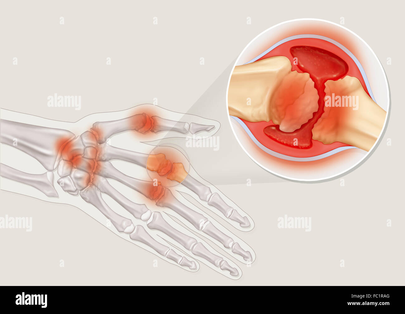 RHEUMATOID ARTHRITIS, DRAWING Stock Photohttps://www.alamy.com/image-license-details/?v=1https://www.alamy.com/stock-photo-rheumatoid-arthritis-drawing-93467992.html
RMFC1RAG–RHEUMATOID ARTHRITIS, DRAWING
 The ligaments of the elbow Stock Photohttps://www.alamy.com/image-license-details/?v=1https://www.alamy.com/stock-photo-the-ligaments-of-the-elbow-13170982.html
RFACJJ0R–The ligaments of the elbow
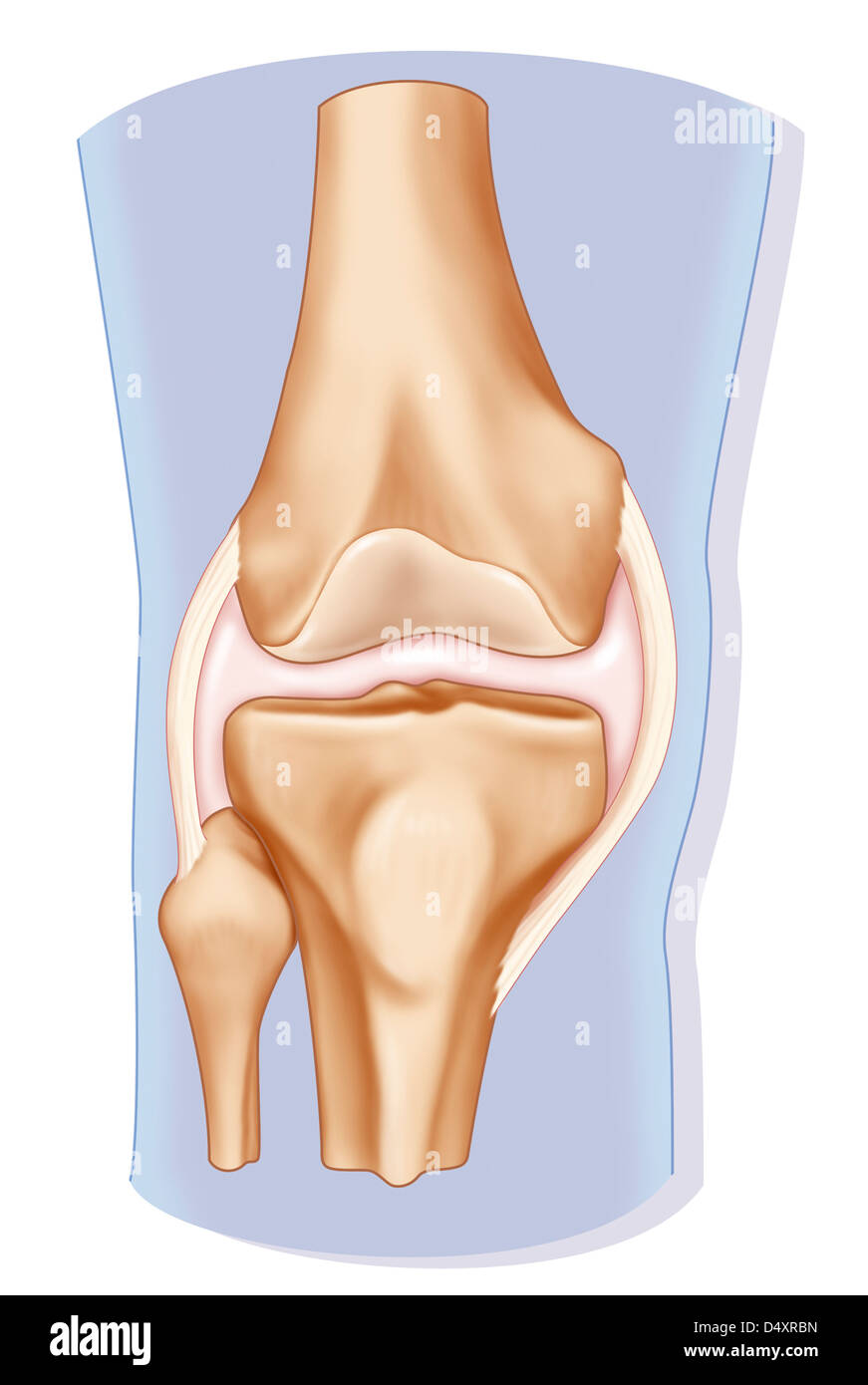 KNEE, DRAWING Stock Photohttps://www.alamy.com/image-license-details/?v=1https://www.alamy.com/stock-photo-knee-drawing-54678841.html
RMD4XRBN–KNEE, DRAWING
 vector illustration of a healthy knee joint Stock Vectorhttps://www.alamy.com/image-license-details/?v=1https://www.alamy.com/vector-illustration-of-a-healthy-knee-joint-image229174772.html
RFR8RPN8–vector illustration of a healthy knee joint
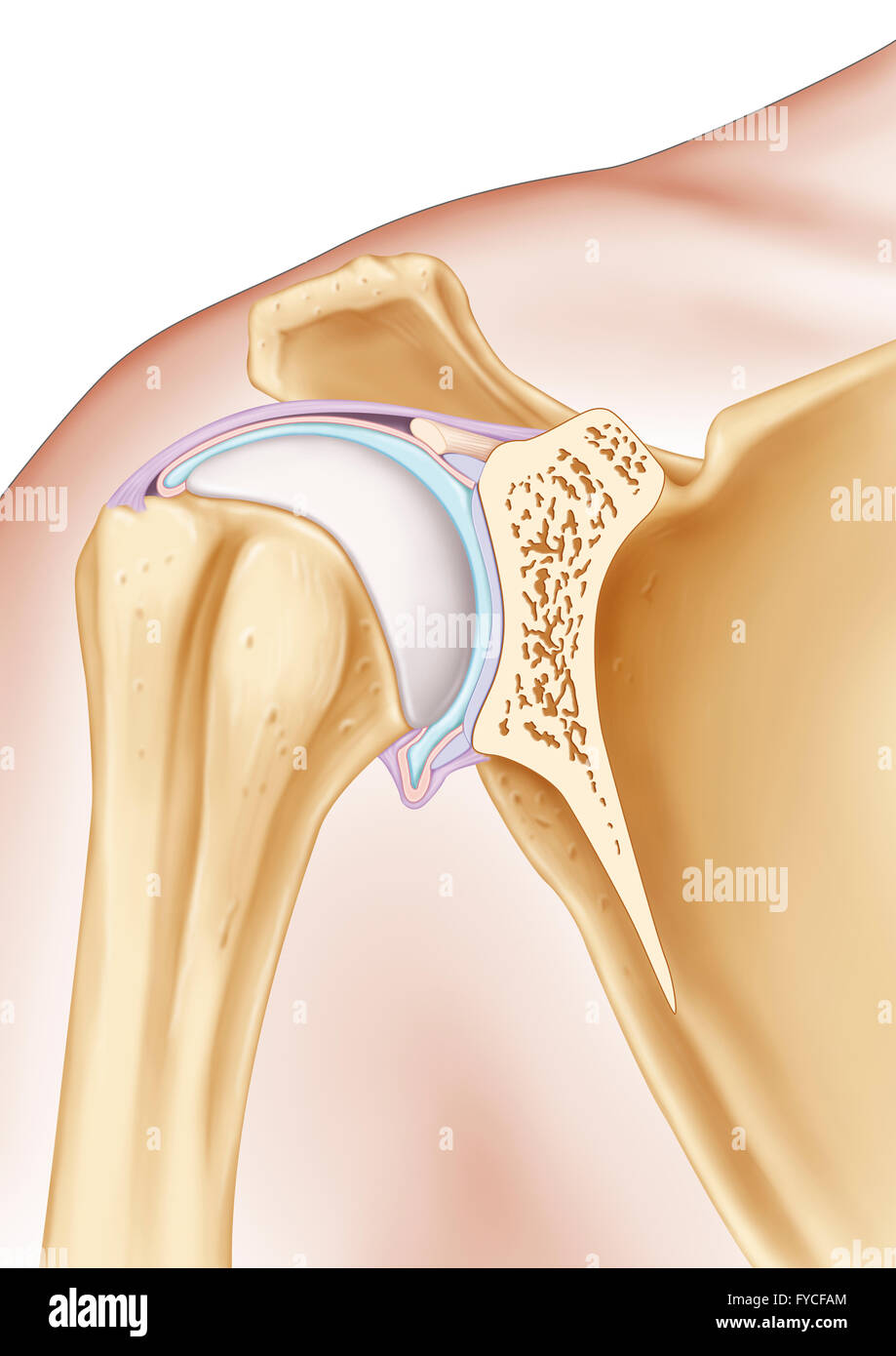 SHOULDER, ILLUSTRATION Stock Photohttps://www.alamy.com/image-license-details/?v=1https://www.alamy.com/stock-photo-shoulder-illustration-102923036.html
RMFYCFAM–SHOULDER, ILLUSTRATION
 vector illustration of osteoarthritis of the knee Stock Vectorhttps://www.alamy.com/image-license-details/?v=1https://www.alamy.com/vector-illustration-of-osteoarthritis-of-the-knee-image221007865.html
RFPRFNP1–vector illustration of osteoarthritis of the knee
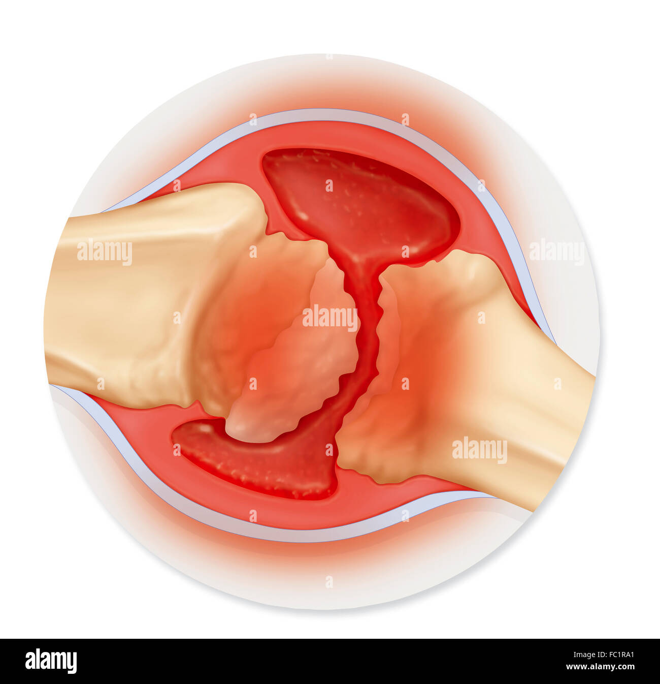 RHEUMATOID ARTHRITIS, DRAWING Stock Photohttps://www.alamy.com/image-license-details/?v=1https://www.alamy.com/stock-photo-rheumatoid-arthritis-drawing-93467977.html
RMFC1RA1–RHEUMATOID ARTHRITIS, DRAWING
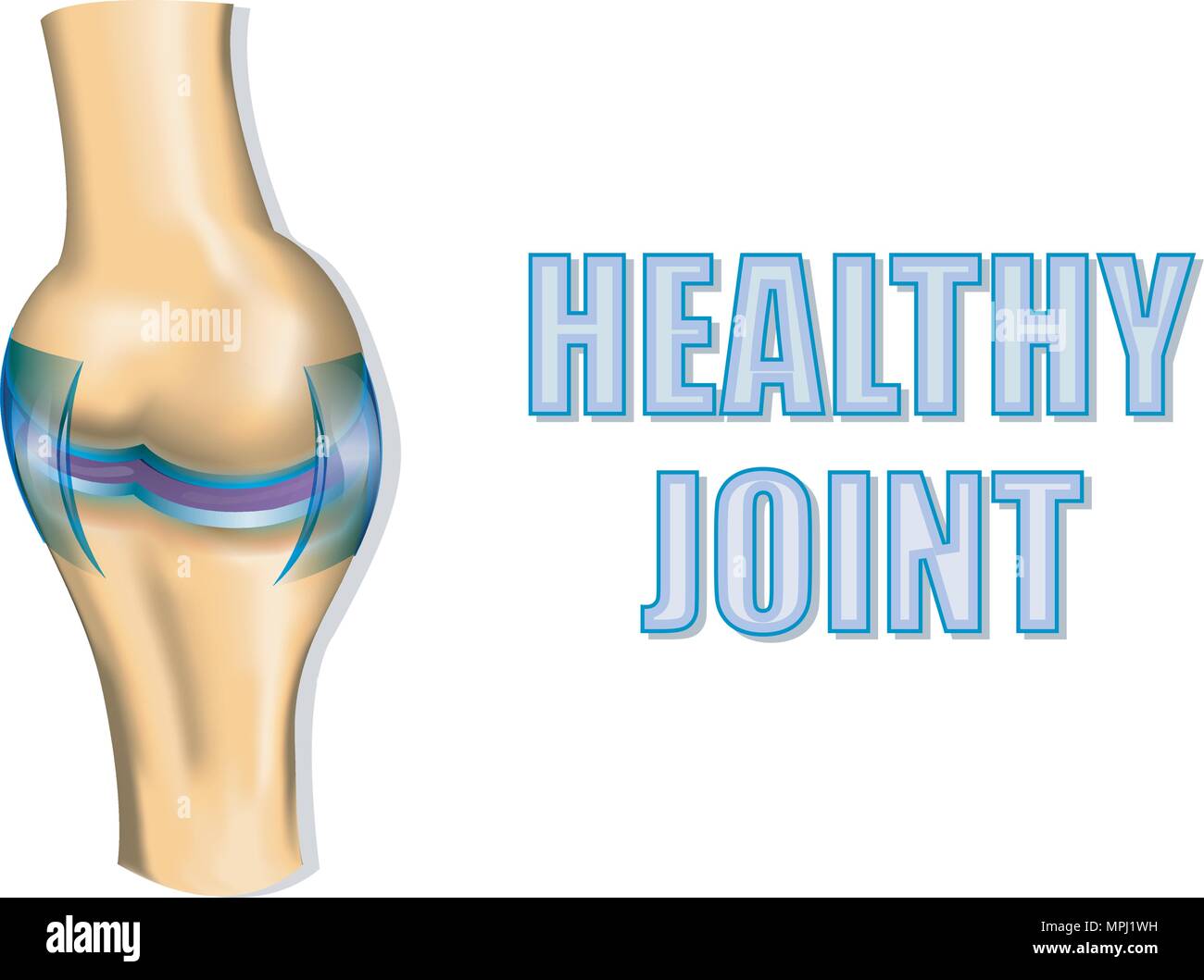 ILLUSTRATION OF THE HEALTHY JOINT. CUTAWAY VIEW. Stock Vectorhttps://www.alamy.com/image-license-details/?v=1https://www.alamy.com/illustration-of-the-healthy-joint-cutaway-view-image186022749.html
RFMPJ1WH–ILLUSTRATION OF THE HEALTHY JOINT. CUTAWAY VIEW.
 A woman's hand on a blue table with redness in the joints of the hand and fingers. Concept of arthritis disease, inflammation of small susavs on the h Stock Photohttps://www.alamy.com/image-license-details/?v=1https://www.alamy.com/a-womans-hand-on-a-blue-table-with-redness-in-the-joints-of-the-hand-and-fingers-concept-of-arthritis-disease-inflammation-of-small-susavs-on-the-h-image443089411.html
RF2GMTD1R–A woman's hand on a blue table with redness in the joints of the hand and fingers. Concept of arthritis disease, inflammation of small susavs on the h
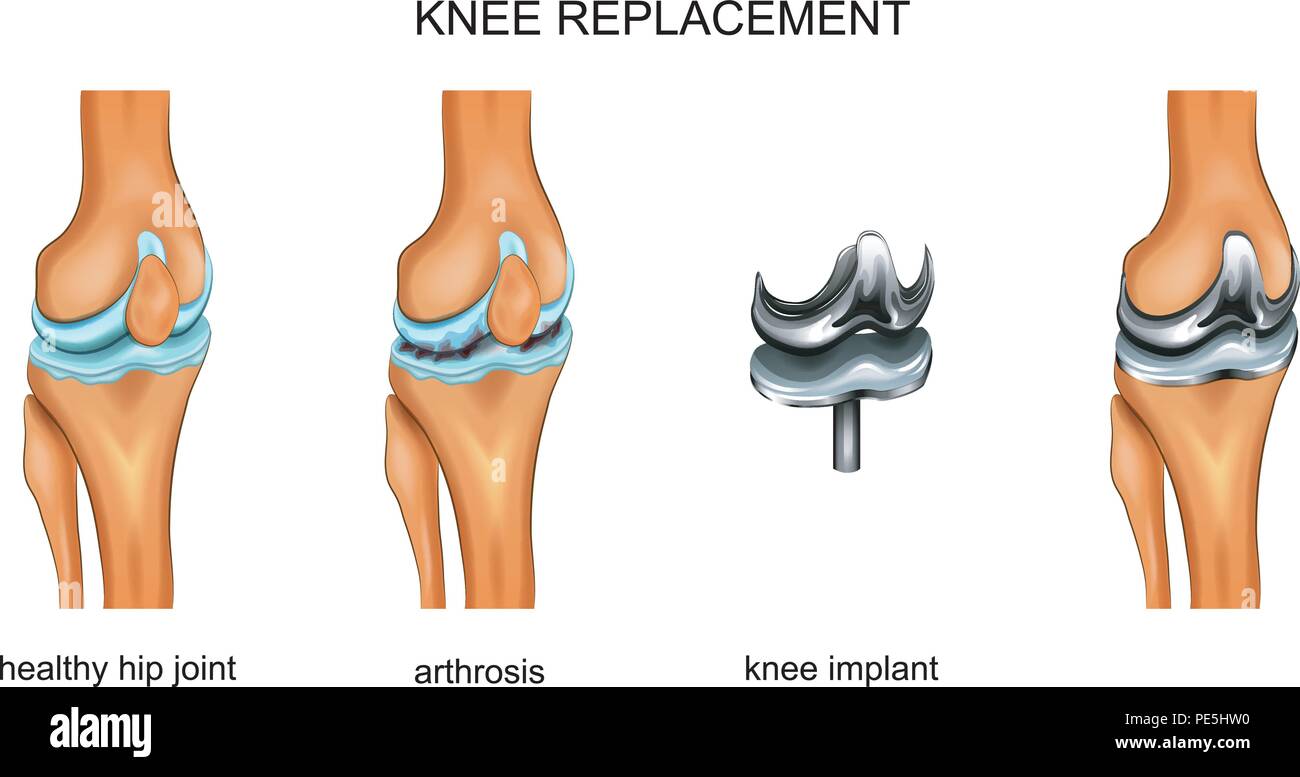 vector illustration of a total knee replacement Stock Vectorhttps://www.alamy.com/image-license-details/?v=1https://www.alamy.com/vector-illustration-of-a-total-knee-replacement-image215253388.html
RFPE5HW0–vector illustration of a total knee replacement
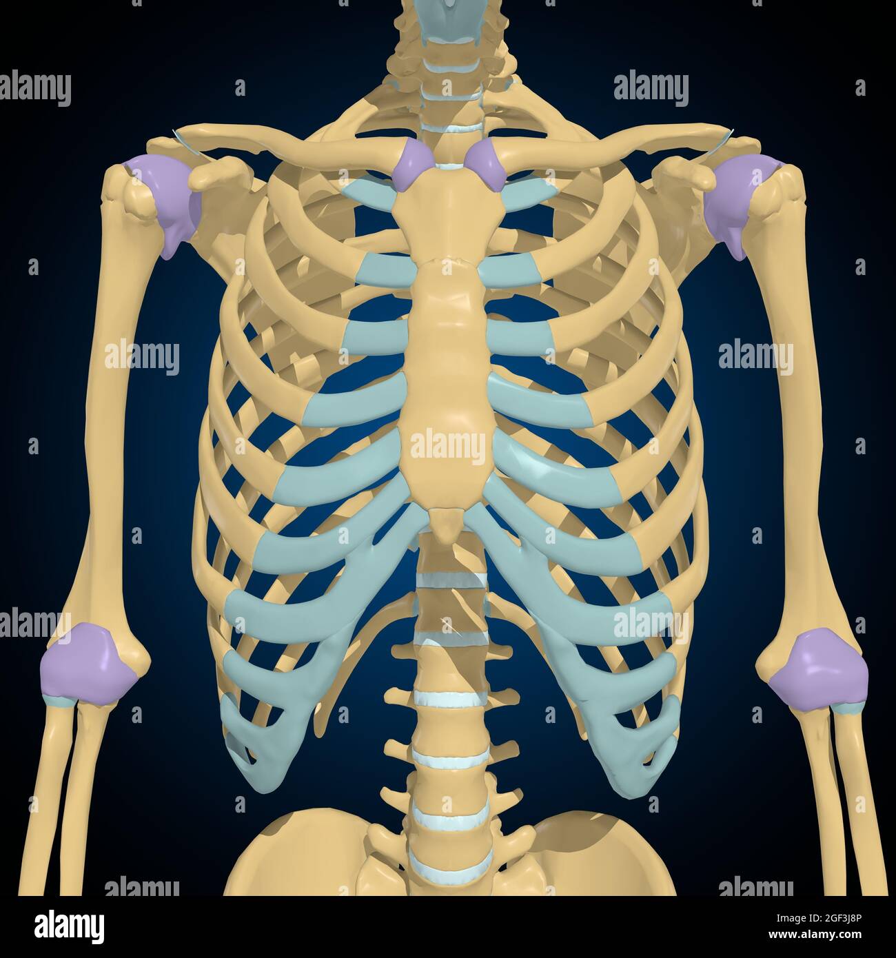 Articular capsule Anatomy For Medical Concept 3D Illustration Stock Photohttps://www.alamy.com/image-license-details/?v=1https://www.alamy.com/articular-capsule-anatomy-for-medical-concept-3d-illustration-image439559254.html
RF2GF3J8P–Articular capsule Anatomy For Medical Concept 3D Illustration
 illustration of the joint anatomy. bone. vector Stock Vectorhttps://www.alamy.com/image-license-details/?v=1https://www.alamy.com/illustration-of-the-joint-anatomy-bone-vector-image186022767.html
RFMPJ1X7–illustration of the joint anatomy. bone. vector
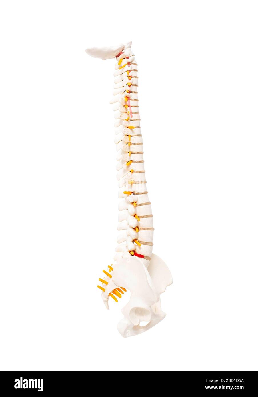 Mock up human spine on a white background. The concept of segments and divisions of the spine, the structure and anatomy of the bone marrow, nerves an Stock Photohttps://www.alamy.com/image-license-details/?v=1https://www.alamy.com/mock-up-human-spine-on-a-white-background-the-concept-of-segments-and-divisions-of-the-spine-the-structure-and-anatomy-of-the-bone-marrow-nerves-an-image352230182.html
RF2BD1D5A–Mock up human spine on a white background. The concept of segments and divisions of the spine, the structure and anatomy of the bone marrow, nerves an