. The anatomy of the domestic fowl . Domestic animals; Veterinary medicine; Poultry. THE URO-GENITAL SYSTEM 179 active state are developed, one by one, into yolks with their blasto-- derms. From the blastoderm the fetus may later be developed. In the active ovary of the laying hen the ovarian mass is of consider- able size, as it contains ova in different stages of development. Only one ovum is completely developed at a time, though occasion- ally there may be only a few hours between the maturity of succes-. FiG. 56.—Functionating female generative organs of a hen. i, Ova in process of format
Image details
Contributor:
Central Historic Books / Alamy Stock PhotoImage ID:
PG1AGAFile size:
7.1 MB (473.3 KB Compressed download)Releases:
Model - no | Property - noDo I need a release?Dimensions:
1751 x 1427 px | 29.7 x 24.2 cm | 11.7 x 9.5 inches | 150dpiMore information:
This image is a public domain image, which means either that copyright has expired in the image or the copyright holder has waived their copyright. Alamy charges you a fee for access to the high resolution copy of the image.
This image could have imperfections as it’s either historical or reportage.
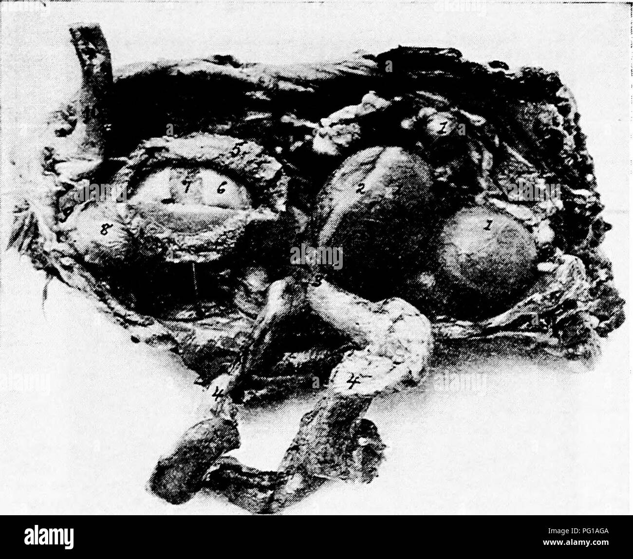
. The anatomy of the domestic fowl . Domestic animals; Veterinary medicine; Poultry. THE URO-GENITAL SYSTEM 179 active state are developed, one by one, into yolks with their blasto-- derms. From the blastoderm the fetus may later be developed. In the active ovary of the laying hen the ovarian mass is of consider- able size, as it contains ova in different stages of development. Only one ovum is completely developed at a time, though occasion- ally there may be only a few hours between the maturity of succes-. FiG. 56.—Functionating female generative organs of a hen. i, Ova in process of formation of yolk. 2, Stigmal line at -which point the capsule ruptures when ovum is mature. 3, The funnel end of the oviduct. 4, The oviduct torn loose and laid to one side, the albumin-secreting portion. 5, The shell membrane secreting portion. 6, The albumin. 7, The yolk. 8, The shell-secreting por- tion. 9, The cloaca. 10, The rectum. sive ova. The ova receives nourishment from the blood-vessels of the capsule, which vessels are branches of the ovarian artery. Structure.—The ovary contains very vascular cellulofibrous tissue. The ovum as it develops is attached to the ovarian body by means of a delicate white fibrous pedicle. When the yolk is mature it escapes from the enveloping fibrous capsule by a cleavage of the" capsule. The cleavage line is called the stigmen (Fig. 56, No.. Please note that these images are extracted from scanned page images that may have been digitally enhanced for readability - coloration and appearance of these illustrations may not perfectly resemble the original work.. Kaupp, Benjamin Franklyn, 1874-. Philadelphia ; London : W. B. Saunders Company