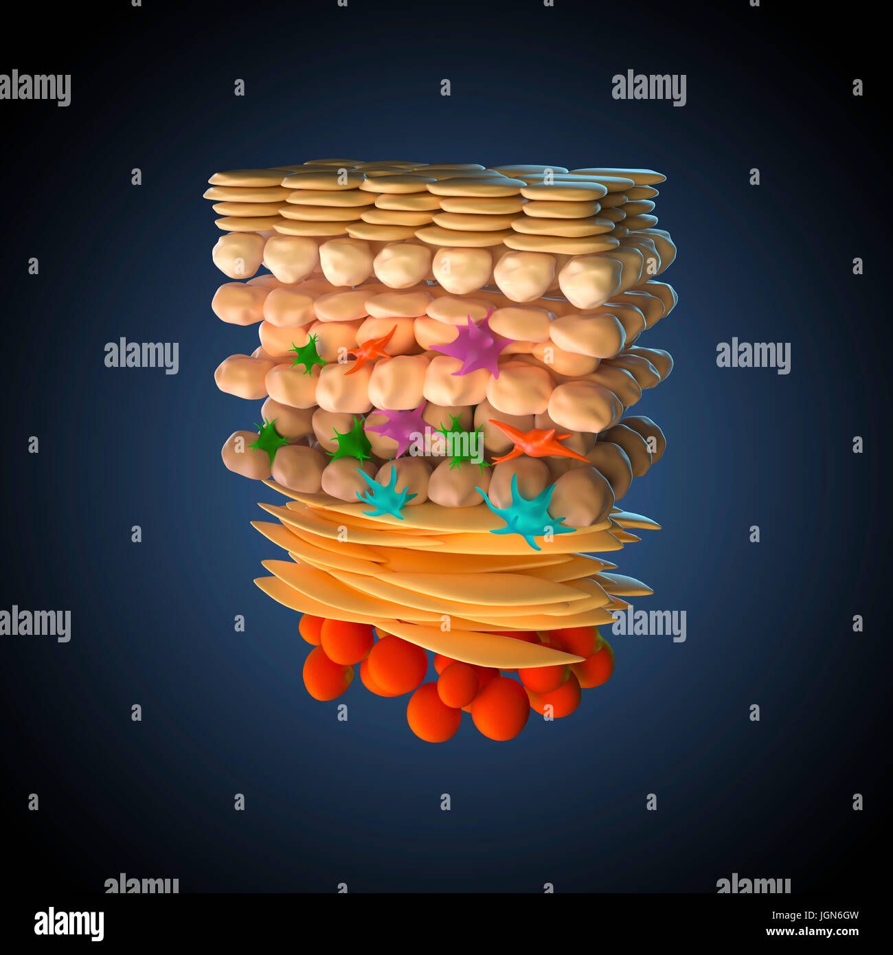
Illustration of a cross-section through human skin. At bottom are adipocyte (fat) cells (red). Above this is the dermis (flattened yellow cells), and then the basal, spinous and granular layers of the epidermis and keratinised dead cells at the surface. Also shown are fibroblasts (orange), melanocytes (green), langerhans cells (pink) and merkel cells (blue).
RFID:Image ID:JGN6GW
Image details
Contributor:
Science Photo Library / Alamy Stock PhotoImage ID:
JGN6GWFile size:
74.4 MB (947.5 KB Compressed download)Releases:
Model - no | Property - noDo I need a release?Dimensions:
5100 x 5100 px | 43.2 x 43.2 cm | 17 x 17 inches | 300dpiDate taken:
18 May 2017Photographer:
ROGER HARRIS/SCIENCE PHOTO LIBRARY