Brain of Sheep Embryo, Transverse Section
Image details
Contributor:
Science History Images / Alamy Stock PhotoImage ID:
HRH464File size:
39.2 MB (2.2 MB Compressed download)Releases:
Model - no | Property - noDo I need a release?Dimensions:
3656 x 3750 px | 31 x 31.8 cm | 12.2 x 12.5 inches | 300dpiPhotographer:
Photo ResearchersMore information:
This image could have imperfections as it’s either historical or reportage.
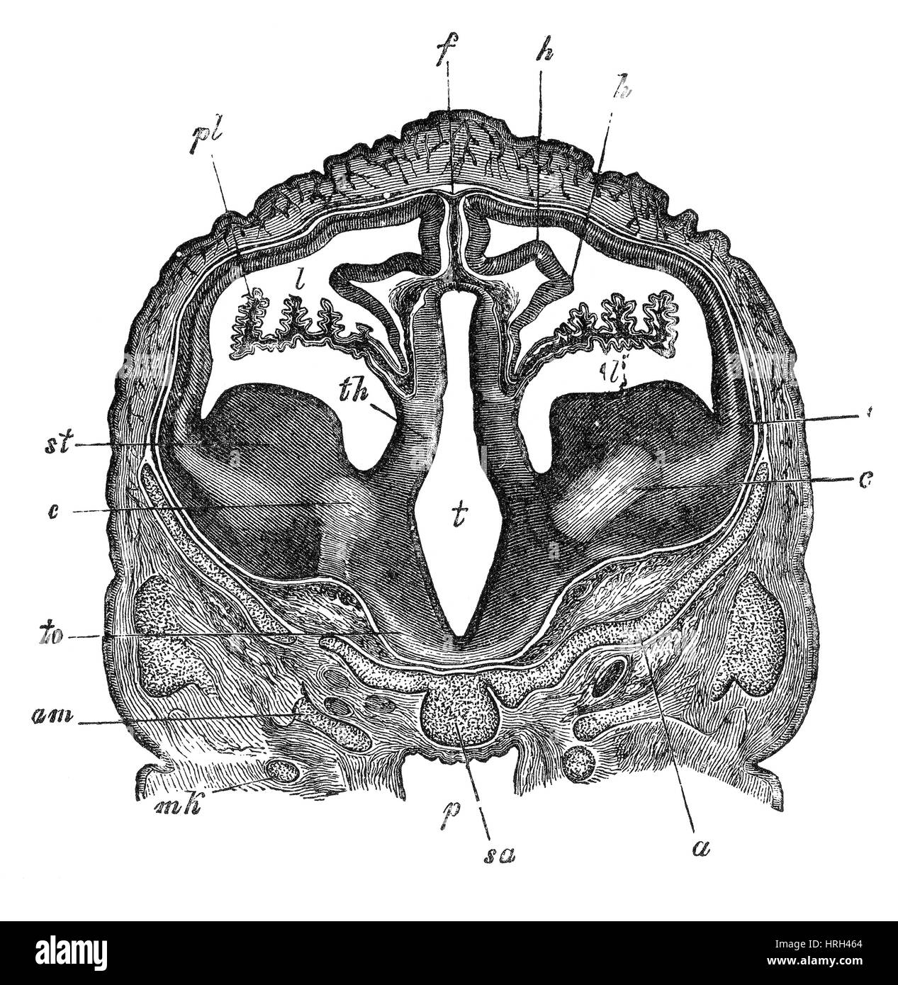
Transverse section through the brain of sheep embryo passing through the third and lateral ventricles. st, corpus striatum; th, optic thalamus; t, third ventricle; c, c1, rudiment of internal capsule and corona radiate; l, lateral ventricle with choroid plexus, pl; h, hippocampus major; f, primitive falx; am orbito-spenoid; ch, chiasma; o, optic nerve; m, m, foramina of Monro; to, optic tract; mk, Meckels's cartilage. Jones Quain (November, 1796 - January 31, 1865) was an Irish anatomist, professor of Anatomy and Physiology in the University of London, and author of Elements of Anatomy. The first edition was published in 1828 and it quickly became a standard text-book in English-speaking countries. 10th edition, 1896.