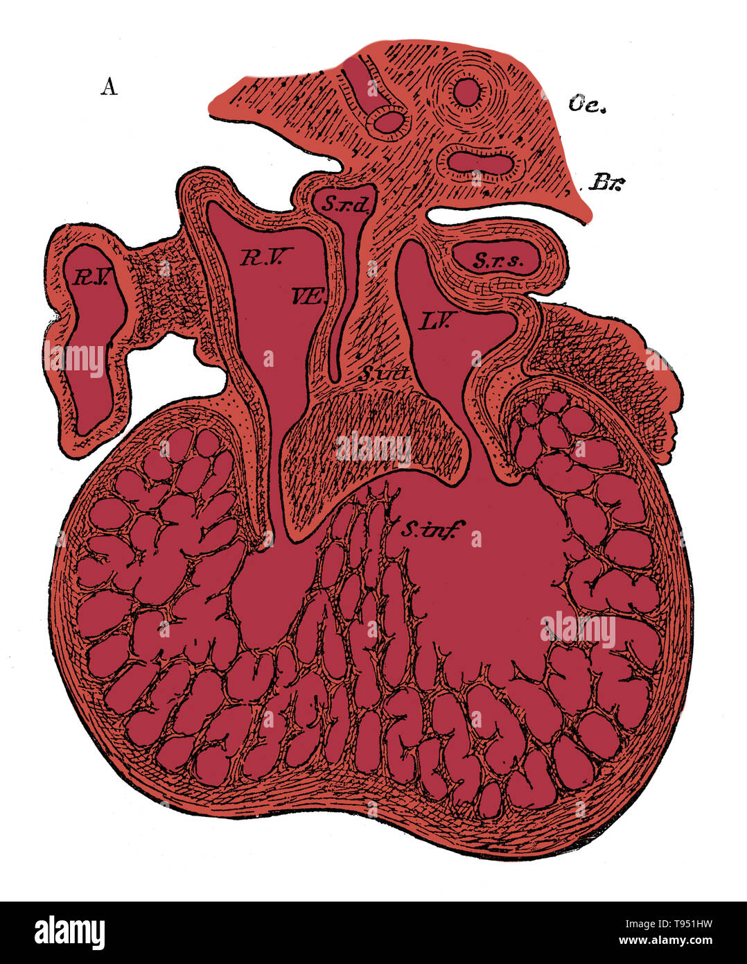
Section through the heart of human embryo showing the formation of the cardiac septa and the auriculo-ventricular valves, 5 to 6 weeks. R.V, right auricle; L.V, left auricle; S.r.d, right horn of sinus; Sr.s, left horn of sinus; s. int, septum superior and endocardial cushion (septum intermedium); s. inf, septum infers ventriculorum; This septum, as well as the bulk of the ventricle, is a muscular sponge at this stage. Oc, esophagus; Br, bronchus.
RMID:Image ID:T951HW
Image details
Contributor:
Science History Images / Alamy Stock PhotoImage ID:
T951HWFile size:
38.9 MB (1.9 MB Compressed download)Releases:
Model - no | Property - noDo I need a release?Dimensions:
3359 x 4050 px | 28.4 x 34.3 cm | 11.2 x 13.5 inches | 300dpiDate taken:
28 August 2017Photographer:
Science History ImagesMore information:
This image could have imperfections as it’s either historical or reportage.
Human Embryo, Heart Development