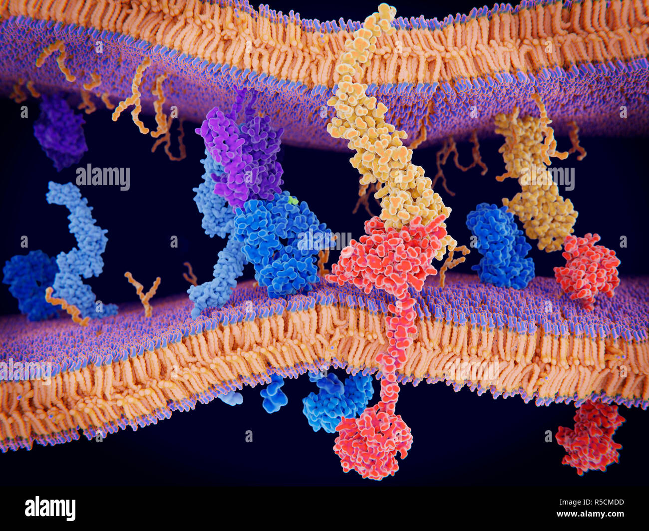
Programmed cell death in immune system, illustration. The protein PD-1 (programmed cell death protein 1, red) is seen extending from the surface membrane of a T-cell (across bottom), interacting with the ligand protein PD-L1 (orange) from an antigen presenting cell (APC). Although the T-cell has been activated through the interaction at left of a T-cell receptor (blue) and a MHC protein (purple), PD-1 regulates and reduces this activation. It acts to slow down the effects of T-cells.
RFID:Image ID:R5CMDD
Image details
Contributor:
Science Photo Library / Alamy Stock PhotoImage ID:
R5CMDDFile size:
100 MB (3.9 MB Compressed download)Releases:
Model - no | Property - noDo I need a release?Dimensions:
6827 x 5120 px | 57.8 x 43.3 cm | 22.8 x 17.1 inches | 300dpiDate taken:
26 November 2018Photographer:
JUAN GAERTNER/SCIENCE PHOTO LIBRARY