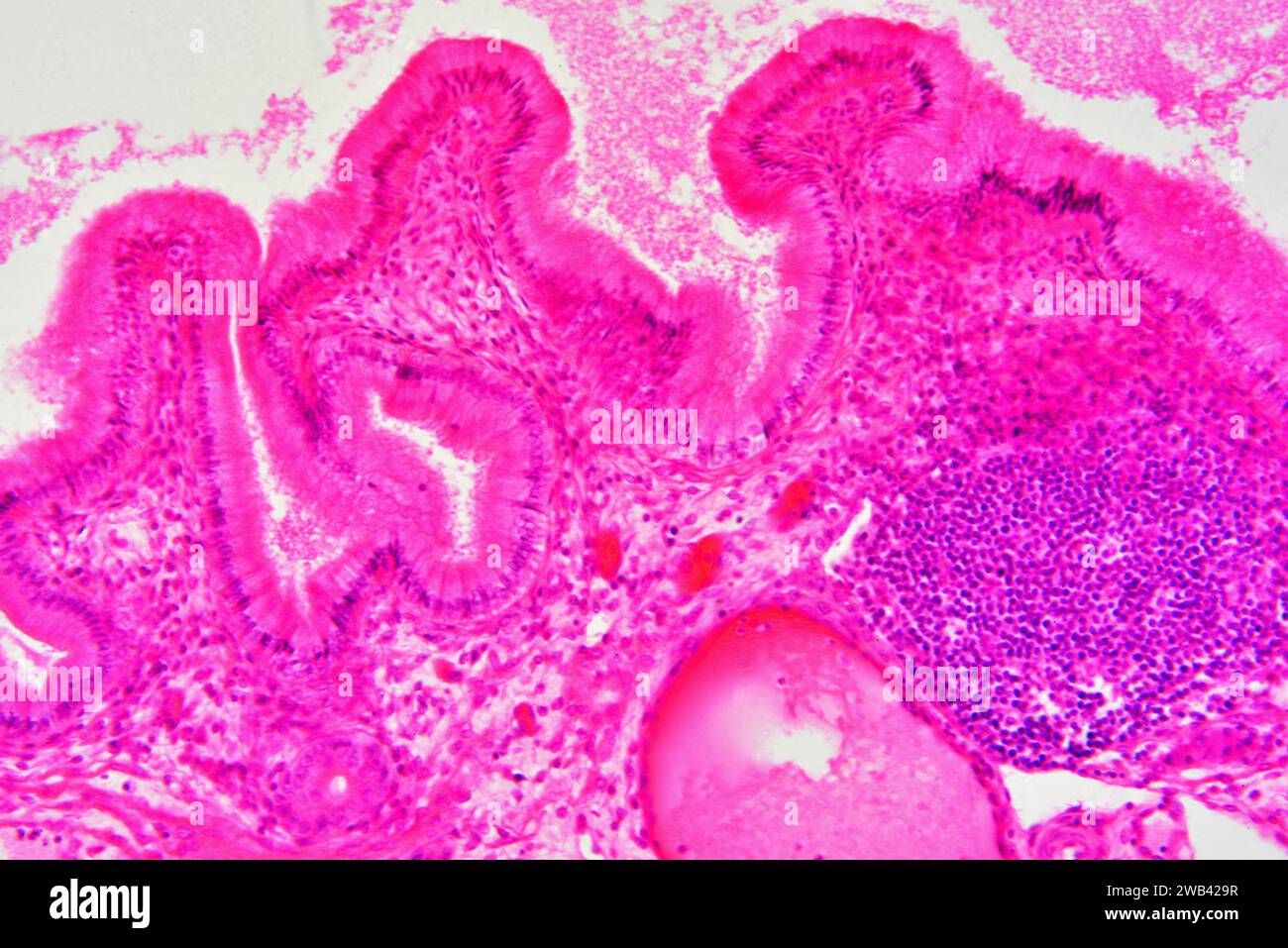
Gallbladder wall showing columnar epithelium with mucosal folds, connective tissue, blood vessels and smooth muscle fibers. Photomicrograph X150 at 10
RFID:Image ID:2WB429R
Image details
Contributor:
José María Barres Manuel / Alamy Stock PhotoImage ID:
2WB429RFile size:
103.4 MB (6.9 MB Compressed download)Releases:
Model - no | Property - noDo I need a release?Dimensions:
7360 x 4912 px | 62.3 x 41.6 cm | 24.5 x 16.4 inches | 300dpiDate taken:
6 December 2020More information:
Gallbladder wall showing columnar epithelium with mucosal folds, connective tissue, blood vessels and smooth muscle fibers. Photomicrograph X150 at 10 cm wide.