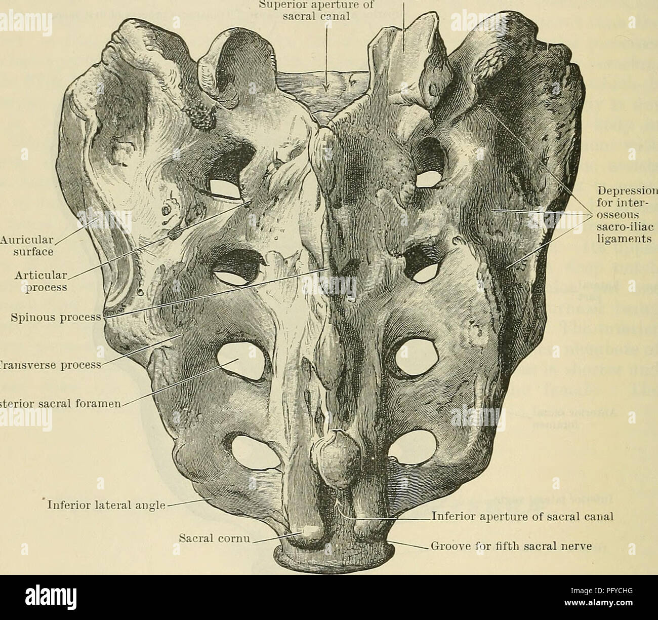
. Cunningham's Text-book of anatomy. Anatomy. 08 OSTEOLOGY. laterales), and which are serially homologous with the true transverse processes of the lumbar vertebrae. The posterior surface of the bone furnishes an extensive surface for the origin of the sacro - spinalis, whilst the edge of the bone lateral to the third and fourth foramen gives attachment to the glutaeus maximus. The base of the bone displays features more in accordance with a typical vertebra. Centrally, and in front, is placed the body, the superior surface of which articulates with the last lumbar vertebra through the medium
Image details
Contributor:
Central Historic Books / Alamy Stock PhotoImage ID:
PFYCHGFile size:
7.1 MB (515.3 KB Compressed download)Releases:
Model - no | Property - noDo I need a release?Dimensions:
1692 x 1477 px | 28.7 x 25 cm | 11.3 x 9.8 inches | 150dpiMore information:
This image is a public domain image, which means either that copyright has expired in the image or the copyright holder has waived their copyright. Alamy charges you a fee for access to the high resolution copy of the image.
This image could have imperfections as it’s either historical or reportage.
. Cunningham's Text-book of anatomy. Anatomy. 08 OSTEOLOGY. laterales), and which are serially homologous with the true transverse processes of the lumbar vertebrae. The posterior surface of the bone furnishes an extensive surface for the origin of the sacro - spinalis, whilst the edge of the bone lateral to the third and fourth foramen gives attachment to the glutaeus maximus. The base of the bone displays features more in accordance with a typical vertebra. Centrally, and in front, is placed the body, the superior surface of which articulates with the last lumbar vertebra through the medium of an intervertebral fibro-cartilage. The anterior margin is thin and projecting, overhanging the general concavity of the pelvic surface of the bone, and forming what is called the promontory. Posterior to the body, the sacral canal, of triangular form but slightly compressed dorso-ventrally, is seen, whilst still more posteriorly is the short spinous Superior articular process. Transverse proces: Posterior sacral foramen^^^ w Inferior lateral angle Sacral cornu Inferior aperture of sacral canal Groove for fifth sacral nerve Coccygeal articular surface Fig. 113.—The Sacrum (posterior view). process, forming the highest tubercle of the median crest. Spreading out from the sides, and partly from the back of the body on either side, is a fan-shaped mass of bone, the upper surface of which is slightly concave from side to side, and convex from above and behind downwards and forwards. This, the ala sacralis, corresponds to the thick upper border of the lateral part, and is formed, as will be explained hereafter, by elements which correspond to the roots of the vertebral arches (O.T. pedicles) and the transverse processes of the sacral vertebras, together with superadded structures—the sacral ribs. The lateral margin of the lateral part, as seen from above, is sharp and laterally convex, terminating posteriorly in a prominent tubercle— the highest of the series of elevations s