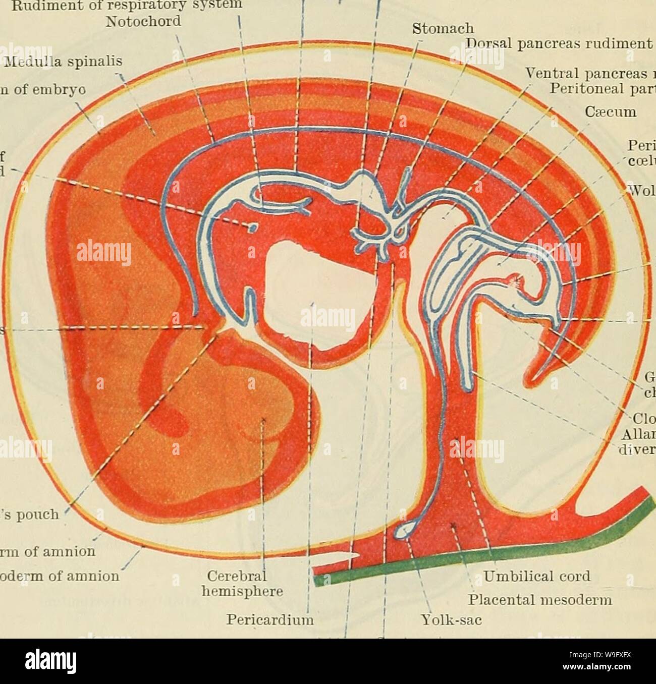
Archive image from page 79 of Cunningham's Text-book of anatomy (1914). Cunningham's Text-book of anatomy cunninghamstextb00cunn Year: 1914 ( Ftg. 61.—Schema showing stages in the development of the tongue. are developed. The tuberculum impar either disappears or it forms the median part of the anterior two-thirds of the organ. The posterior or dorsal third of the tongue, which lies in the ventral or anterior wall 'of the permanent pharynx, is formed from the copula of the second arches. It follows from what has been said Oesophagus Rudiment of respiratory system Xotochord | Medulla spinalis
Image details
Contributor:
Bookive / Alamy Stock PhotoImage ID:
W9FXFXFile size:
5.7 MB (265.2 KB Compressed download)Releases:
Model - no | Property - noDo I need a release?Dimensions:
1433 x 1396 px | 24.3 x 23.6 cm | 9.6 x 9.3 inches | 150dpiMore information:
This image is a public domain image, which means either that copyright has expired in the image or the copyright holder has waived their copyright. Alamy charges you a fee for access to the high resolution copy of the image.
This image could have imperfections as it’s either historical or reportage.
Archive image from page 79 of Cunningham's Text-book of anatomy (1914). Cunningham's Text-book of anatomy cunninghamstextb00cunn Year: 1914 ( Ftg. 61.—Schema showing stages in the development of the tongue. are developed. The tuberculum impar either disappears or it forms the median part of the anterior two-thirds of the organ. The posterior or dorsal third of the tongue, which lies in the ventral or anterior wall 'of the permanent pharynx, is formed from the copula of the second arches. It follows from what has been said Oesophagus Rudiment of respiratory system Xotochord | Medulla spinalis Ectoderm of embryo Rudiment of thyreoid gland - Liver diverticulum branching in septum transversum f I Stomach Dorsal pancreas rudiment Ventral pancreas rudiment •/ Peritoneal part of caelum N. Ca;cum Peritoneal part of 'ccelum Wolffian duct Rectum Hypophysi Rathke's pouch ' Ectoderm of amnion Mesoderm of amnion Genito-urinary chamber Cloacal membrane Allantoic diverticulum horion Pericardium Umbilical cord Placental mesoderm 'Solk-sac Septum transversum Fig. 62.—Schema showing further stages in the development of the diverticula from the primitive gut and modifications of the mid-gut and the mid-gut regions. The heart is not shown. (After Mall, modified.) that the commencement of the thyreoid rudiment, which persists in the adult as the foramen csecuni of the tongue, must he at the junction of the dorsal third with the ventral two-thirds. In many cases it appears to He in the dorsal end of the ventral two-thirds, a position which may be associated with the fact that in some cases the rudiment of the thyreoid passes through the substance of the tuberculum