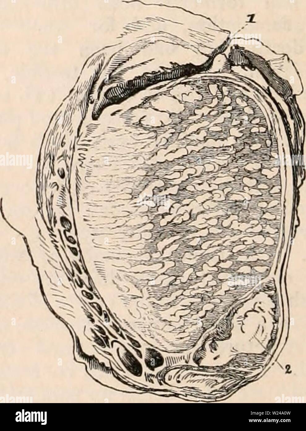
Archive image from page 221 of The cyclopædia of anatomy and. The cyclopædia of anatomy and physiology cyclopdiaofana0402todd Year: 1849 several are interspersed throughout the tes- ticle, portions of sound gland intervening. In a case of chronic enlargement of both testicles taken from a patient who died of ramollissement of the medulla spinalis, I found six or seven separate deposits of this yellow matter in the substance of the right testicle, and a single one only in the body of the left. The small masses as they enlarge coalesce, or the single one increases, until the whole testicle pre
Image details
Contributor:
Bookive / Alamy Stock PhotoImage ID:
W24A0WFile size:
5.7 MB (284.4 KB Compressed download)Releases:
Model - no | Property - noDo I need a release?Dimensions:
1234 x 1621 px | 20.9 x 27.4 cm | 8.2 x 10.8 inches | 150dpiMore information:
This image is a public domain image, which means either that copyright has expired in the image or the copyright holder has waived their copyright. Alamy charges you a fee for access to the high resolution copy of the image.
This image could have imperfections as it’s either historical or reportage.
Archive image from page 221 of The cyclopædia of anatomy and. The cyclopædia of anatomy and physiology cyclopdiaofana0402todd Year: 1849 several are interspersed throughout the tes- ticle, portions of sound gland intervening. In a case of chronic enlargement of both testicles taken from a patient who died of ramollissement of the medulla spinalis, I found six or seven separate deposits of this yellow matter in the substance of the right testicle, and a single one only in the body of the left. The small masses as they enlarge coalesce, or the single one increases, until the whole testicle presents an uniform yel- lowish-white appearance. The epididymis is frequently invaded at the same time by a similar kind of morbid deposit, which also tends to obliterate its tubular structure. This, however, is not, as some pathologists suppose, a constant occurrence; for in the majority of testicles thus diseased that 1 have examined, the epididymis had entirely escaped. I have never succeeded in injecting this deposit, or in tracing vessels into it. But the vessels of the testicle generally are enlarged. Patholo- gists have differed as to the particular tissue in which this yellow matter is deposited. Sir A. Cooper and Cruveilhier describes it to be seated in the areolar tissue between the tubuli; whilst Sir B. Brodie is of opinion, that it is secreted from their inner surface, as he discovered the yellow substance in the canal of the epididymis and also in the vas deferens which are continuous with the tubuli. I have had the opportunity of in- specting a testicle affected with this disease, in what seems to me to be its early stage from which examination I think I have been able satisfactorily to confirm this opinion. The testicle was injected with red size, and a section then made of it. The surfaces of the tunica vaginalis were partly adherent, and about three drachms of serum were collected in one part of the sac. The body of the tes- ticle was not much enlarged : it con