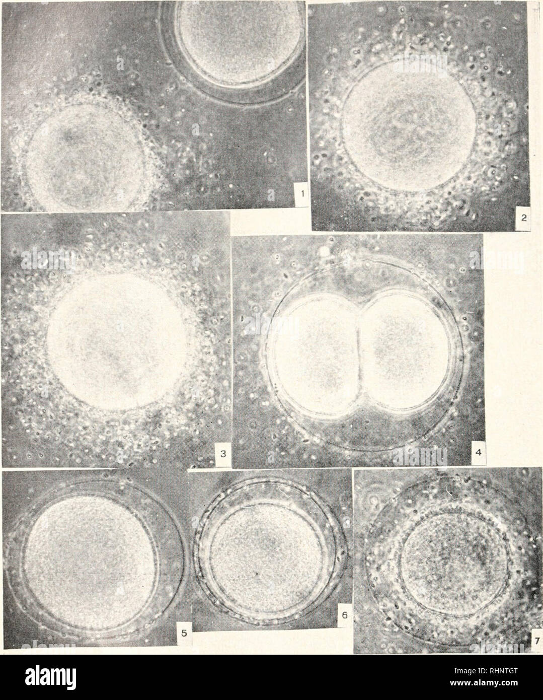
. The Biological bulletin. Biology; Zoology; Biology; Marine Biology. 1. An oocyte and a ripe egg of Paraccutrotus liruins; 30 minutes after insemination. 310 X. l'"i(.ri<K 2. Oocyte ni I'lit-iiccutrolus In-idits. c<i. 25 minutes after insemination. 330 X. l-'iGi/RE 3. Oocyte of I'Miinnit'cliini/x microtuberculatus in meiotic division. Penetration of M vend Nperniato/oa lias provoked the formation of mitotic figure*. 325 X. FIGURE 4. I'^« of I'siiiiniiccliiniis microtuberculatus about 70 minutes after insemination. 325 X. FIGURE 5. KKK of Paracentrotus li-riihis. four minutes after
Image details
Contributor:
Library Book Collection / Alamy Stock PhotoImage ID:
RHNTGTFile size:
7.2 MB (554.7 KB Compressed download)Releases:
Model - no | Property - noDo I need a release?Dimensions:
1441 x 1735 px | 24.4 x 29.4 cm | 9.6 x 11.6 inches | 150dpiMore information:
This image is a public domain image, which means either that copyright has expired in the image or the copyright holder has waived their copyright. Alamy charges you a fee for access to the high resolution copy of the image.
This image could have imperfections as it’s either historical or reportage.
. The Biological bulletin. Biology; Zoology; Biology; Marine Biology. 1. An oocyte and a ripe egg of Paraccutrotus liruins; 30 minutes after insemination. 310 X. l'"i(.ri<K 2. Oocyte ni I'lit-iiccutrolus In-idits. c<i. 25 minutes after insemination. 330 X. l-'iGi/RE 3. Oocyte of I'Miinnit'cliini/x microtuberculatus in meiotic division. Penetration of M vend Nperniato/oa lias provoked the formation of mitotic figure*. 325 X. FIGURE 4. I'^« of I'siiiiniiccliiniis microtuberculatus about 70 minutes after insemination. 325 X. FIGURE 5. KKK of Paracentrotus li-riihis. four minutes after fertilization. 350 X. I-'K.I in, o. I-'.^KS of I'dnifcntrotus liridus, tvo minutes after insemination, exposed to 0.01% brilliant cresyl blue, reception cone visible in the proximal region of the egg. 310 X. FIGURE 7. Oocyte of 1'araccntrotus lii'ulux, tvo minutes after insemination, exposed to <l.0lr; brilliant cresyl blue: photographed 35 minutes later. 310 X. 136. Please note that these images are extracted from scanned page images that may have been digitally enhanced for readability - coloration and appearance of these illustrations may not perfectly resemble the original work.. Marine Biological Laboratory (Woods Hole, Mass. ); Marine Biological Laboratory (Woods Hole, Mass. ). Annual report 1907/08-1952; Lillie, Frank Rattray, 1870-1947; Moore, Carl Richard, 1892-; Redfield, Alfred Clarence, 1890-1983. Woods Hole, Mass. : Marine Biological Laboratory