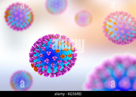
 Flu viruses, computer illustration. Each virus consists of a core of RNA (ribonucleic acid) genetic material surrounded by a protein coat (blue). Embedded in the coat are surface proteins (spikes). There are two types of surface protein, hemagglutinin (purple) and neuraminidase (orange), and each exists in several subtypes. Both surface proteins are associated with the pathogenicity of a virus. Hemagglutinin binds to host cells, allowing the virus to enter them and replicate. Neuraminidase allows the new particles to exit the host after replication. Stock Photohttps://www.alamy.com/licenses-and-pricing/?v=1https://www.alamy.com/stock-image-flu-viruses-computer-illustration-each-virus-consists-of-a-core-of-164842929.html
RFKG56RD–Flu viruses, computer illustration. Each virus consists of a core of RNA (ribonucleic acid) genetic material surrounded by a protein coat (blue). Embedded in the coat are surface proteins (spikes). There are two types of surface protein, hemagglutinin (purple) and neuraminidase (orange), and each exists in several subtypes. Both surface proteins are associated with the pathogenicity of a virus. Hemagglutinin binds to host cells, allowing the virus to enter them and replicate. Neuraminidase allows the new particles to exit the host after replication.
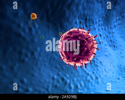 macrophage kills the viruses, 3d rendered macrophage and virus, inside human body, Medical video background, viruses in the human body Stock Photohttps://www.alamy.com/licenses-and-pricing/?v=1https://www.alamy.com/macrophage-kills-the-viruses-3d-rendered-macrophage-and-virus-inside-human-body-medical-video-background-viruses-in-the-human-body-image332580182.html
RF2A929BJ–macrophage kills the viruses, 3d rendered macrophage and virus, inside human body, Medical video background, viruses in the human body
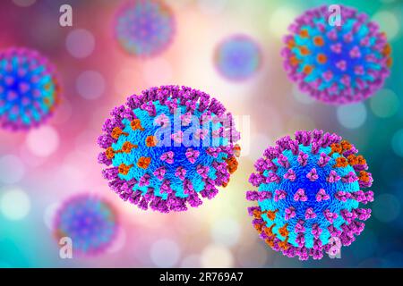 Flu viruses, computer illustration. Each virus consists of a core of RNA (ribonucleic acid) genetic material surrounded by a protein coat (blue). Embe Stock Photohttps://www.alamy.com/licenses-and-pricing/?v=1https://www.alamy.com/flu-viruses-computer-illustration-each-virus-consists-of-a-core-of-rna-ribonucleic-acid-genetic-material-surrounded-by-a-protein-coat-blue-embe-image555173423.html
RF2R769A7–Flu viruses, computer illustration. Each virus consists of a core of RNA (ribonucleic acid) genetic material surrounded by a protein coat (blue). Embe
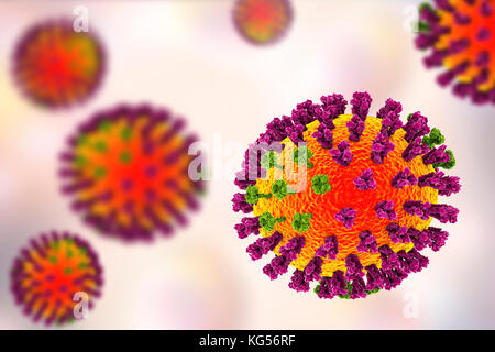 Flu viruses, computer illustration. Each virus consists of a core of RNA (ribonucleic acid) genetic material surrounded by a protein coat (orange). Embedded in the coat are surface proteins (spikes). There are two types of surface protein, hemagglutinin (purple) and neuraminidase (green), and each exists in several subtypes. Both surface proteins are associated with the pathogenicity of a virus. Hemagglutinin binds to host cells, allowing the virus to enter them and replicate. Neuraminidase allows the new particles to exit the host after replication. Stock Photohttps://www.alamy.com/licenses-and-pricing/?v=1https://www.alamy.com/stock-image-flu-viruses-computer-illustration-each-virus-consists-of-a-core-of-164842931.html
RFKG56RF–Flu viruses, computer illustration. Each virus consists of a core of RNA (ribonucleic acid) genetic material surrounded by a protein coat (orange). Embedded in the coat are surface proteins (spikes). There are two types of surface protein, hemagglutinin (purple) and neuraminidase (green), and each exists in several subtypes. Both surface proteins are associated with the pathogenicity of a virus. Hemagglutinin binds to host cells, allowing the virus to enter them and replicate. Neuraminidase allows the new particles to exit the host after replication.
 word LOVE with viruses 3D rendering Stock Photohttps://www.alamy.com/licenses-and-pricing/?v=1https://www.alamy.com/word-love-with-viruses-3d-rendering-image383913836.html
RF2D8GP0C–word LOVE with viruses 3D rendering
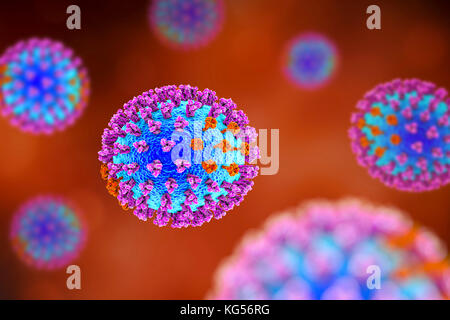 Flu viruses, computer illustration. Each virus consists of a core of RNA (ribonucleic acid) genetic material surrounded by a protein coat (blue). Embedded in the coat are surface proteins (spikes). There are two types of surface protein, hemagglutinin (purple) and neuraminidase (orange), and each exists in several subtypes. Both surface proteins are associated with the pathogenicity of a virus. Hemagglutinin binds to host cells, allowing the virus to enter them and replicate. Neuraminidase allows the new particles to exit the host after replication. Stock Photohttps://www.alamy.com/licenses-and-pricing/?v=1https://www.alamy.com/stock-image-flu-viruses-computer-illustration-each-virus-consists-of-a-core-of-164842932.html
RFKG56RG–Flu viruses, computer illustration. Each virus consists of a core of RNA (ribonucleic acid) genetic material surrounded by a protein coat (blue). Embedded in the coat are surface proteins (spikes). There are two types of surface protein, hemagglutinin (purple) and neuraminidase (orange), and each exists in several subtypes. Both surface proteins are associated with the pathogenicity of a virus. Hemagglutinin binds to host cells, allowing the virus to enter them and replicate. Neuraminidase allows the new particles to exit the host after replication.
 Mutated Omicron SARS-CoV-2 virus. Electron microscope enlargement size computer model, 3D rendering Stock Photohttps://www.alamy.com/licenses-and-pricing/?v=1https://www.alamy.com/mutated-omicron-sars-cov-2-virus-electron-microscope-enlargement-size-computer-model-3d-rendering-image453041706.html
RF2H91R8X–Mutated Omicron SARS-CoV-2 virus. Electron microscope enlargement size computer model, 3D rendering
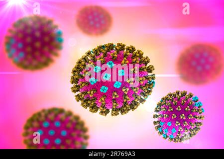 Flu viruses, computer illustration. Each virus consists of a core of RNA (ribonucleic acid) genetic material surrounded by a protein coat (purple). Em Stock Photohttps://www.alamy.com/licenses-and-pricing/?v=1https://www.alamy.com/flu-viruses-computer-illustration-each-virus-consists-of-a-core-of-rna-ribonucleic-acid-genetic-material-surrounded-by-a-protein-coat-purple-em-image555173419.html
RF2R769A3–Flu viruses, computer illustration. Each virus consists of a core of RNA (ribonucleic acid) genetic material surrounded by a protein coat (purple). Em
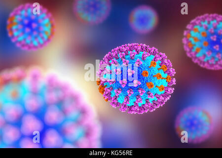 Flu viruses, computer illustration. Each virus consists of a core of RNA (ribonucleic acid) genetic material surrounded by a protein coat (blue). Embedded in the coat are surface proteins (spikes). There are two types of surface protein, hemagglutinin (purple) and neuraminidase (orange), and each exists in several subtypes. Both surface proteins are associated with the pathogenicity of a virus. Hemagglutinin binds to host cells, allowing the virus to enter them and replicate. Neuraminidase allows the new particles to exit the host after replication. Stock Photohttps://www.alamy.com/licenses-and-pricing/?v=1https://www.alamy.com/stock-image-flu-viruses-computer-illustration-each-virus-consists-of-a-core-of-164842930.html
RFKG56RE–Flu viruses, computer illustration. Each virus consists of a core of RNA (ribonucleic acid) genetic material surrounded by a protein coat (blue). Embedded in the coat are surface proteins (spikes). There are two types of surface protein, hemagglutinin (purple) and neuraminidase (orange), and each exists in several subtypes. Both surface proteins are associated with the pathogenicity of a virus. Hemagglutinin binds to host cells, allowing the virus to enter them and replicate. Neuraminidase allows the new particles to exit the host after replication.
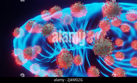 Mutated Omicron SARS-CoV-2 viruses. Electron microscope transparent enlargement size computer model, 3D rendering. Stock Photohttps://www.alamy.com/licenses-and-pricing/?v=1https://www.alamy.com/mutated-omicron-sars-cov-2-viruses-electron-microscope-transparent-enlargement-size-computer-model-3d-rendering-image467577672.html
RF2J4M02G–Mutated Omicron SARS-CoV-2 viruses. Electron microscope transparent enlargement size computer model, 3D rendering.
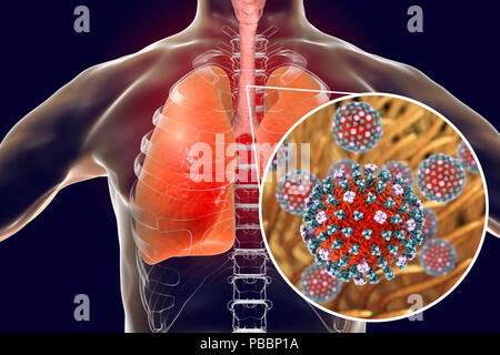 Flu viruses in trachea, computer illustration. Influenza viruses have tropism to tracheal epithelium, they decrease tracheal mucociliary velocity thus contributing to the development of secondary infections of the lower respiratory tract. Due to this, pneumonia is one of the more common complications of flu infection. Stock Photohttps://www.alamy.com/licenses-and-pricing/?v=1https://www.alamy.com/flu-viruses-in-trachea-computer-illustration-influenza-viruses-have-tropism-to-tracheal-epithelium-they-decrease-tracheal-mucociliary-velocity-thus-contributing-to-the-development-of-secondary-infections-of-the-lower-respiratory-tract-due-to-this-pneumonia-is-one-of-the-more-common-complications-of-flu-infection-image213544390.html
RFPBBP1A–Flu viruses in trachea, computer illustration. Influenza viruses have tropism to tracheal epithelium, they decrease tracheal mucociliary velocity thus contributing to the development of secondary infections of the lower respiratory tract. Due to this, pneumonia is one of the more common complications of flu infection.
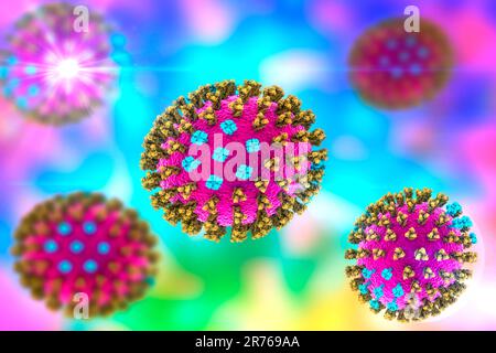 Flu viruses, computer illustration. Each virus consists of a core of RNA (ribonucleic acid) genetic material surrounded by a protein coat (purple). Em Stock Photohttps://www.alamy.com/licenses-and-pricing/?v=1https://www.alamy.com/flu-viruses-computer-illustration-each-virus-consists-of-a-core-of-rna-ribonucleic-acid-genetic-material-surrounded-by-a-protein-coat-purple-em-image555173426.html
RF2R769AA–Flu viruses, computer illustration. Each virus consists of a core of RNA (ribonucleic acid) genetic material surrounded by a protein coat (purple). Em