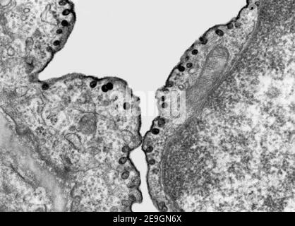
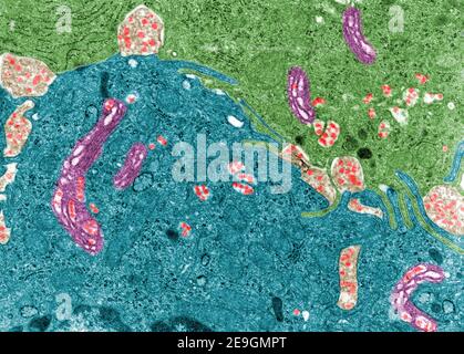
Transmission electron microscope (TEM) micrograph showing the labelling of vesicles of pinocytosis with an electron dense marker, the ruthenium red. Stock Photo
RF2E9GN6X–Transmission electron microscope (TEM) micrograph showing the labelling of vesicles of pinocytosis with an electron dense marker, the ruthenium red.
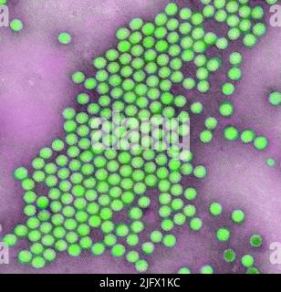
This transmission electron microscopic (TEM), negative stain image, reveals some of the ultrastructural features exhibited by a grouping of icosahedral-shaped polio virus particle. Digitally colourised /false colour image.  An optimised and enhanced version of an image produced by the US Centers for Disease Control and Prevention / credit CDC / J.J.Esposito; F.A.Murphy Stock Photo
RM2JFX1KC–This transmission electron microscopic (TEM), negative stain image, reveals some of the ultrastructural features exhibited by a grouping of icosahedral-shaped polio virus particle. Digitally colourised /false colour image. An optimised and enhanced version of an image produced by the US Centers for Disease Control and Prevention / credit CDC / J.J.Esposito; F.A.Murphy
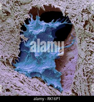
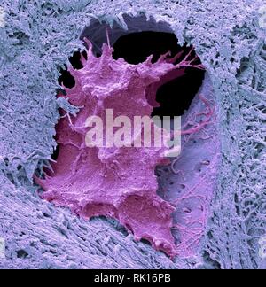
Osteocyte bone cell, SEM Stock Photo
RFRK16PB–Osteocyte bone cell, SEM
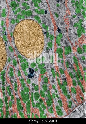
False colour transmission electron microscope (TEM) micrograph showing chylomicrons (red) in enterocytes of small intestine. They appear in the Golgi Stock Photo
RF2E9GMPT–False colour transmission electron microscope (TEM) micrograph showing chylomicrons (red) in enterocytes of small intestine. They appear in the Golgi
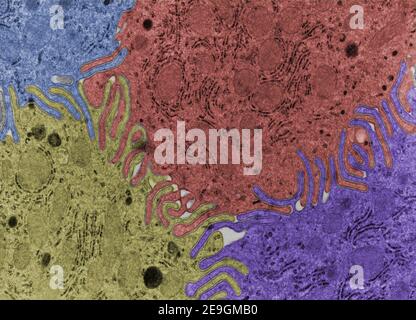
False colour transmission electron microscope (TEM) micrograph showing very complex cellular interdigitations joining the lateral surface of four epit Stock Photo
RF2E9GMB0–False colour transmission electron microscope (TEM) micrograph showing very complex cellular interdigitations joining the lateral surface of four epit
Osteocyte bone cell, SEM Stock Photo
RFRK16P1–Osteocyte bone cell, SEM
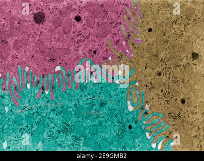
False colour transmission electron microscope (TEM) micrograph showing very complex cellular interdigitations joining the lateral surface of four epit Stock Photo
RF2E9GMB2–False colour transmission electron microscope (TEM) micrograph showing very complex cellular interdigitations joining the lateral surface of four epit
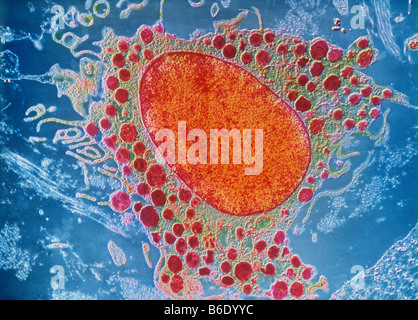
Mast cell coloured transmission electronmicrograph (TEM). Mast cells are a type of whiteblood cell found in connective tissue. Stock Photo
RFB6DYYC–Mast cell coloured transmission electronmicrograph (TEM). Mast cells are a type of whiteblood cell found in connective tissue.
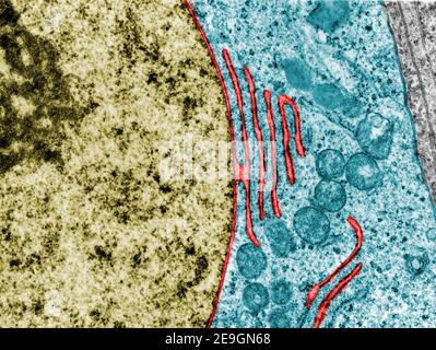
False colour transmission electron microscope micrograph showing a continuity between the nuclear envelope and a cistern of the rough endoplasmic reti Stock Photo
RF2E9GN68–False colour transmission electron microscope micrograph showing a continuity between the nuclear envelope and a cistern of the rough endoplasmic reti
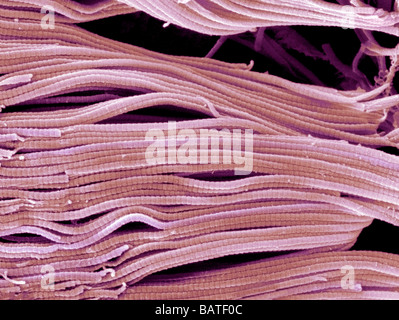
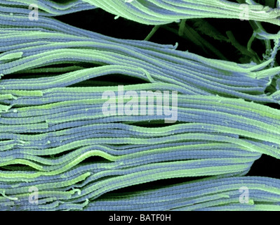
Collagen. Scanning electron micrograph (SEM) of collagen bundles from the delicate connectivetissue endoneurium. Stock Photo
RFBATF0C–Collagen. Scanning electron micrograph (SEM) of collagen bundles from the delicate connectivetissue endoneurium.
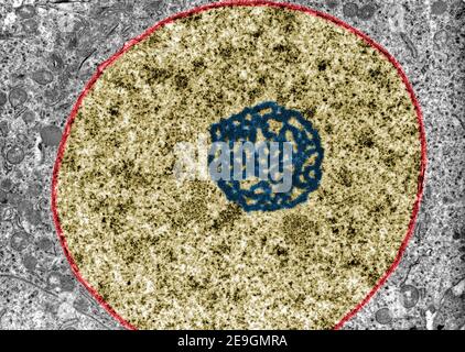
False colour transmission electron microscope (TEM) micrograph showing the ultrastructure of a nucleus (gold) with a very prominent nucleolus (blue) a Stock Photo
RF2E9GMRA–False colour transmission electron microscope (TEM) micrograph showing the ultrastructure of a nucleus (gold) with a very prominent nucleolus (blue) a
Collagen. Scanning electron micrograph (SEM) of collagen bundles from the delicate connectivetissue endoneurium. Stock Photo
RFBATF0H–Collagen. Scanning electron micrograph (SEM) of collagen bundles from the delicate connectivetissue endoneurium.
False colour transmission electron microscope (TEM) micrograph of a kidney distal convoluted tube. Basal interdigitations or infoldings are labelled i Stock Photo
RF2E9GMCX–False colour transmission electron microscope (TEM) micrograph of a kidney distal convoluted tube. Basal interdigitations or infoldings are labelled i