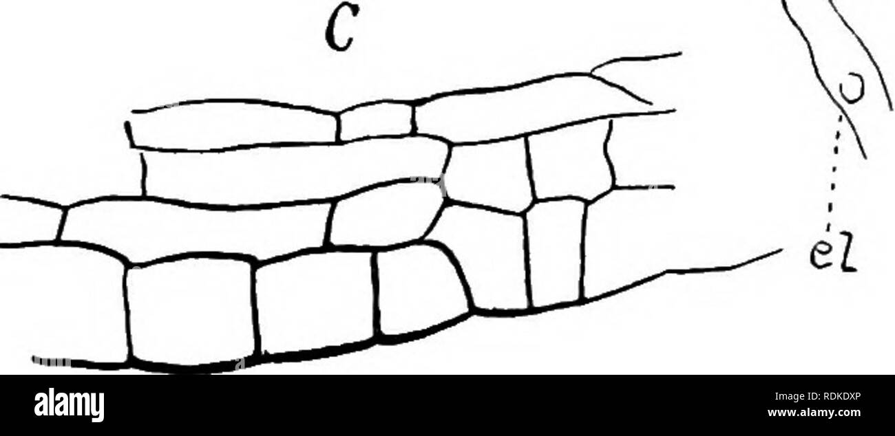
. Dudley memorial volume, containing a paper by William Russel Dudley and appreciations and contributions in his memory by friends and colleagues ... Dudley, William Russel, 1849-1911; Botany. Fig. 10. A, median section of a sporogonium at the time of the mitosis of the spore mother cells, x 15. B, apical region of a younger sporogonium, sp. spore mother cells, el. young elator, x 225. C, part of the lateral wall of the sporogonium. The capsule (Fig. 10) has a relatively thick wall which is better de- veloped at the apex than at the sides, this difference becoming still more marked in the late
Image details
Contributor:
The Book Worm / Alamy Stock PhotoImage ID:
RDKDXPFile size:
7.1 MB (124.3 KB Compressed download)Releases:
Model - no | Property - noDo I need a release?Dimensions:
2441 x 1023 px | 41.3 x 17.3 cm | 16.3 x 6.8 inches | 150dpiMore information:
This image is a public domain image, which means either that copyright has expired in the image or the copyright holder has waived their copyright. Alamy charges you a fee for access to the high resolution copy of the image.
This image could have imperfections as it’s either historical or reportage.
. Dudley memorial volume, containing a paper by William Russel Dudley and appreciations and contributions in his memory by friends and colleagues ... Dudley, William Russel, 1849-1911; Botany. Fig. 10. A, median section of a sporogonium at the time of the mitosis of the spore mother cells, x 15. B, apical region of a younger sporogonium, sp. spore mother cells, el. young elator, x 225. C, part of the lateral wall of the sporogonium. The capsule (Fig. 10) has a relatively thick wall which is better de- veloped at the apex than at the sides, this difference becoming still more marked in the later stages. The inner tissue now shows a separation into the roundish spore mother cells and the elongated young elaters. Long before the division of the spore mother cells begins, they show the first indi- cations of the lobing which later becomes so conspicuous. At this stage the walls of both the spore mother cells and the elaters are very delicate, but can be readily demonstrated by suitable stains, e. g. Bismarck brown.. Please note that these images are extracted from scanned page images that may have been digitally enhanced for readability - coloration and appearance of these illustrations may not perfectly resemble the original work.. Stanford University, Cal. , The University