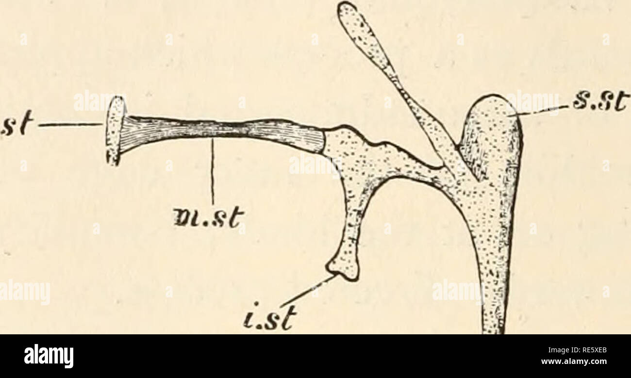
. A course of instruction in zootomy (vertebrata). Anatomy, Comparative. H2 ZOOTOMY. 64. The epipterygoid (e.pt.) (so-called columella), a slender rod of bone, lying just in front of and external to the anterior edge of the prootic : below it articulates with the pterygoid, above with the prootic. 65. The columella auris (Fig. 36), a small rod of combined bone and cartilage, lying in the dorsal wall of the tympanic recess : its inner end is inserted into the fenestra ovalis, a small aperture between the prootic and opisthotic, while its outer end is, in the entire head, fixed to the inner surf
Image details
Contributor:
The Book Worm / Alamy Stock PhotoImage ID:
RE5XEBFile size:
7.1 MB (148.8 KB Compressed download)Releases:
Model - no | Property - noDo I need a release?Dimensions:
2171 x 1151 px | 36.8 x 19.5 cm | 14.5 x 7.7 inches | 150dpiMore information:
This image is a public domain image, which means either that copyright has expired in the image or the copyright holder has waived their copyright. Alamy charges you a fee for access to the high resolution copy of the image.
This image could have imperfections as it’s either historical or reportage.
. A course of instruction in zootomy (vertebrata). Anatomy, Comparative. H2 ZOOTOMY. 64. The epipterygoid (e.pt.) (so-called columella), a slender rod of bone, lying just in front of and external to the anterior edge of the prootic : below it articulates with the pterygoid, above with the prootic. 65. The columella auris (Fig. 36), a small rod of combined bone and cartilage, lying in the dorsal wall of the tympanic recess : its inner end is inserted into the fenestra ovalis, a small aperture between the prootic and opisthotic, while its outer end is, in the entire head, fixed to the inner surface of the tympanic membrane.. i.at jf CM FIG. 36.—Lacerta agilis. The columella auris (after W. K. Parke?) x 14. The cartilaginous parts are dotted. e.st, extra-stapedial: i.st, infra-stapedial: m.st, medio-stapedial: s.st, supra-stapedial: st. stapes. The columella auris consists of the following distinct parts which are only to be made out by careful dissection of an entire head : (a) the stapes, (st), a small cartilaginous nodule in the fenestra ovalis : (b) the medio-stapedial (m.sf), a bony bar connected with the stapes, the ossification from it extending into the latter : (c) a. cartilaginous rod continuous with the distal end of the medio-stapedial, sending off a downwardly directed process, the infra-stapedial (i.st), and expanding at its outer or free extremity into a bar set transversely to the rest of the columella like the head of a hammer : the lower somewhat pointed end of this bar is the extra-stapedial (e.st) : its dorsal extremity, the supra- stapedial (s.st) is blunt and rounded, and gives off a process which becomes connected with the auditory capsule. 66. The vagus foramen (Fig. 35, IX, X, XI, ), a small aperture in the combined exoccipital and opisthotic, behind. Please note that these images are extracted from scanned page images that may have been digitally enhanced for readability - coloration and appearance of these illustrations may not perfectly res